


Ⅰ. Introduction
There are many reasons for tooth discoloration, which can be classified into extrinsic and intrinsic according to the inducing factor. Extrinsic tooth discoloration is caused by the accumulation of causative materials on the tooth surface, and so it can be removed. Such causes include beverage (e.g., coffee and tea) food, oral hygiene, bacteria, smoking, oral rinses (chlorhexidine), and metals (e.g., copper, nickel)[1-4]. In contrast, intrinsic tooth discoloration is caused by metabolic disorders, anomalies of the developing dental follicle, genetic factors, drug administration, and fluorosis. Because these causative materials infiltrate into the tooth structure, their removal is impossible[5-8]. Tooth discoloration can be an esthetic problem, and it is one of main reasons why patients visit dentists.
Jaundice of the newborn infant is common due to immature kidney metabolism resulting in elevated blood bilirubin. Jaundice appears clinically when the level of blood bilirubin exceeds over 7.0 mg/dL. Treatment for physiologic jaundice is necessary only in underweight premature babies[9-11]. An increase in the bilirubin level may cause tooth discolorations[5]. Such cases are rare, but most involve tooth discoloration with a greenish hue[12,13].
The aim of this report is to describe 2 pediatric patients with tooth discoloration associated with hyperbilirubinemia who visited the Yonsei University Dental Hospital having green teeth as the chief complaint.
Ⅱ. Case Reports
Case 1
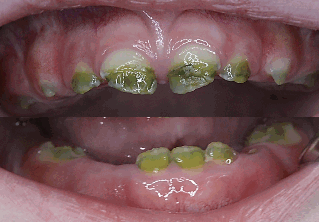
A 22-month old male visited the Department of Pediatric Dentistry, Yonsei University Dental Hospital due to tooth discoloration. A clinical oral examination revealed that the primary central and lateral incisors (with the exception of the lower right lateral incisor) were erupted, and that the primary lower first molars were partially erupted. These teeth showed green discoloration, and the upper central teeth exhibited enamel hypoplasia (Fig. 1).
Owing to chorioamnionitis, the patient was born prematurely after 25 weeks and 6 days of gestation, weighing only 750 g. The patient received intubation in an incubator due to perinatal asphyxia. At birth the patient suffered from a neonatal seizure and was diagnosed with periventricular leukomalacia and cerebral palsy. The patient also received phototherapy for 1 week, due to neonatal hyperbilirubinemia.
After 3 month, primary upper canines and first molar and lower left canine could be partially observed. These teeth exhibited varying degrees of general green discoloration with the central and lateral incisors being most affected, followed by the first molars and canines. The various tooth areas exhibiting discoloration and normal hue could be easily as certainly (Fig. 2).
Case 2
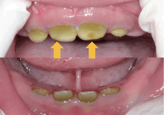
A 16-month-old boy who had congenital green teeth visited the Department of Pediatric Dentistry, Yonsei University Dental Hospital. An intraoral examination revealed that the primary central and lateral incisors were partially erupted in the maxilla and mandible. These teeth appeared greenish due to intrinsic factors. The upper central incisors exhibited enamel hypoplasia bilaterally (Fig. 3).
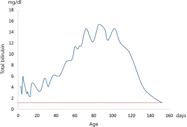
The patient was born by Cesarean section due to an amnion rupture after 26 weeks and 6 days of gestation, weighing 930 g. The patient suffered from severe bronchial pulmonary dysplasia. The patient received ligation of patent ductus arteriosus because of an atrial septal defect and patent ductus arteriosus. Similar to case 1, the patient had neonatal hyperbilirubinemia and exchange transfusion histories. The serum bilirubin level peaked at 15.3 mg/dL when he was 84 days old, but had returned to within the normal range at 152 days old (Fig. 4).
Ⅲ. Discussion
Tooth discoloration may manifest as different colors, with green discoloration being uncommon. Its etiology is classified into extrinsic and intrinsic causative factors. The known extrinsic causes include chromogenic bacteria, systemic antibiotics (e.g., minocycline), copper salts contained in mouth rinse, and copper and nickel metal ions[3,4,14]. Intrinsic causes include hyperbilirubinemia, and drugs such as minocycline and ciprofloxacin[8,15-17]. Tooth discoloration had resulted from intrinsic causes in the present cases, and so it was not physically removed. In addition, they had no history of minocycline or ciprofloxacin administration.
According to Guimaraes and Silva[5], there had been 48 case reports on green teeth up to 2003. The prevalence of green teeth has not been measured officially, but it can be described as rare. The prevalence was similar in women (51.3%) and men (48.7%), with the age at onset most commonly ranging from birth to 11 years, and being more common in primary dentition than permanent dentition.
Hepatobiliary disorders and hematologic disease have been reported as systemic diseases that cause green teeth. The hematologic diseases include Rh incompatibility, ABO incompatibility disease, hematologic anemia and erythroblastosis fetalis[15,18-21]. Biliary atresia has been reported as the most frequent disease, related to green teeth[5]. Both of the present premature infants were diagnosed as hyperbilirubinemia, which is high likely to cause green teeth, due to immature metabolism in the liver.
There are various phenotypes of tooth discoloration by bilirubin[5]. Green is the most frequently reported, followed by brown, yellow and gray. The degree of discoloration is known to be proportional to the serum bilirubin level[19]; for example, Tank[22] reported that the degree of green discoloration was positively correlated with the jaundice index. On the other hand, it is considered that there is no direct relation between the degree of green discoloration and the duration of hyperbilirubinemia. Marsland and Gerrard[23] reported the green teeth in a patient who had suffered the jaundice for only 1 week, while in the present cases, the jaundice period were 1 week and 5 months.
The primary dentition develops during the prenatal period. Bilirubin accumulates at dental follicles via the blood-stream. The calcification process is initiated at 4 months prenatally and finishes after 11 months. It is known that the primary central incisors form at 1 month and that the primary canine and molars are formed completely at 6 months[24]. In addition, the crown portion with a normal hue that developed when the bilirubin level was normal and the green portion that formed during the hyperbilirubieneimia period can be clearly distinguished by lines on the teeth. The color of the tooth returns to normal after the hyperbilirubinemia is treated[18,19], and so it is considered that the degree and location of pigmentation is determined by the timing of hyperbilirubinemia.
Green discoloration of primary teeth was observed in both of the present cases. However, the discoloration portion ranged from the incisal to the middle of the primary central incisor in case 1, while in second case 2 the discoloration started from the middle to cervical portion. This indicates that there was a difference in the times of dental follicle formation when the patients suffered hyperbilirubinemia. In both cases the primary central incisors had enamel hypoplasia, and both patients had an intubation history due to being born prematurely. It has been reported that intubation in the neonatal periods can cause enamel hypoplasia[25], and so it can be considered as a cause of enamel hypoplasia in the maxillary primary central incisor in the present cases.
For asthetic needs, there are several treatment options, such as tooth bleaching, direct composite restoration and full veneer crown. Full veneer crown is more suitable for restoring extensive discoloration area because of its durability and esthetics[26]. In these cases, patients showed enamel hypoplasia on upper central incisors, so retention of direct composite could be reduced. On the other hand, some adverse effects of tooth bleaching in children and adolescents have been reported. So peroxides in lower concentration are recommended, but bleaching leads to limited clinical satisfaction[27]. In addition, there is a possibility that neonatal hyperbilirubinemia affected tooth development of permanent molars and incisors, so regular check-ups should be performed until eruption.
Ⅳ. Summary
2 patients aged a 16 and 22-months visited in the Department of Pediatric Dentistry, Yonsei University Dental Hospital having a chief complaint of erupting teeth with abnormal color. Both patients were diagnosed as green intrinsic discoloration in oral examinations, and they had a systemic history of being born prematurely and suffering neonatal hyperbilirubinemia.
Tooth development is affected by systemic disease that can lead to tooth discoloration. Pediatric dentists may be the first people to observe patients with discoloration in their primary dentition, and they can explain the possibility of systemic disease to their parents. In addition, they should perform regular check-ups to preserve their oral health and resolve the esthetic demands of patients and parents regarding discoloration.

 PDF Links
 PubReader
 ePub Link
 Full text via DOI
 Download Citation
 Print
Print


