Short root anomaly (SRA) 환아의 교정적 처치 증례
Orthodontic Treatment of a Child with Short Root Anomaly : a Case Report
Article information
Abstract
Short root anomaly (SRA)는 매우 드문 질환으로, 교정력에 의한 치근흡수에 있어서 정상적인 치아보다 더 취약하므로 처치시에 어려움이 있다. 또한, 혼합치열기 시기에 나타나는 전반적인 SRA의 경우는 진단 또한 쉽지 않다. 초기 혼합치열기 환아에서 방사선 사진만으로 전반적인 SRA를 예측하기 어렵지만, 가족력이나 관련 전신질환, 또는 구강 내 다른 치아이상 (tooth anomaly) 등의 요소가 있는 경우, SRA를 염두에 두어야 한다.
본 증례는 전악에 걸쳐SRA를 보이는 초기 혼합치열기 환아의 교정적 처치를 다루었다. 비록 국소적인 치아 상실이 있었으나 적절한 교정적 배열을 통해 더 이상의 과도한 치근흡수 없이 치료를 마무리 하였으며, 비교적 양호한 치료 결과를 얻었기에 이를 보고하고자 한다. 결론적으로, SRA 환자에서도 교정 치료가 가능하며, SRA가 예측되는 혼합치열기 환아에서 치근흡수나 치아상실과 같은 합병증이 나타나지 않도록 특히 주의를 기울여야 한다.
Trans Abstract
Short root anomaly (SRA) is very rare, but can be problematic for physicians because patients with SRA are more vulnerable to root resorption with orthodontic forces. During the mixed dentition period, it may be difficult to diagnose generalized SRA. This article reports the treatment of an orthodontic patient with SRA at the early mixed dentition stage. Despite local tooth loss, a relatively favorable outcome was obtained without excessive root resorption. Ultimately, orthodontic therapy is possible for patients with generalized SRA, but precautions should be taken to avoid complications, such as tooth loss or root resorption.
Ⅰ. Introduction
In 1972, Lind [1] discovered short root anomaly (SRA) in a Swedish population, and defined the condition as being characterized by an abnormally short and blunt root shape, affecting mainly the maxillary central incisors. SRA is typically diagnosed based on the crown-to-root ratio when the root length is equal to or smaller than the crown length [1,2]. The estimated prevalence of SRA is 1.3~2.7% [2]. It shows familial occurrence, and hereditary characteristics have been reported in studies of parents and siblings [2,3]. Apajalahti et al. [2] suggested that SRA affected only the permanent teeth, based on the absence of reports on SRA in the primary teeth. SRA typically affects the maxillary central incisors, but sometimes the premolars [1]. Rarely, generalized SRA may affect the entire dentition [4-6].
Orthodontic forces can occasionally result in root resorption, and the risk increases with a short root [1,7-9]. Orthodontic therapy often begins in the mixed dentition period in growing children. Unfortunately, root formation is incomplete at this stage, which makes diagnosis difficult with radiographs alone.
Here, we present a case involving orthodontic treatment performed in a patient at the early mixed dentition stage, when the identification of SRA is most challenging. This report highlights the importance of the early detection of SRA and precautions needed during orthodontic procedures. Informed consent was obtained from the patient’s parents for publication of this report.
Ⅱ. Case report
A boy, age 9 years, 5 months, visited the pediatric dental clinic at Wonkwang University Dental Hospital. His parents reported that the permanent lateral incisors had not erupted since he had lost the primary teeth 2 years previously. There was sufficient space for the successors. The four erupted mandibular incisors seemed to complete apical closure with relatively short roots. The mandibular primary second molars formed an infraocclusion due to ankylosis. The ectopic eruption of the first molars bilaterally had resulted in space deficiency for the second premolars (Fig. 1). His primary second molars had been extracted bilaterally 1 - 2 years previously. After oral screening, it was decided that orthodontic intervention would be initiated on eruption of the maxillary lateral incisors because there had been no space to arrange permanent maxillary premolars.

Panoramic radiograph at first examination.
At the patient’s next visit, 8 months later, we found that the maxillary left lateral incisor had erupted, but the right one had not, showing no significant change from the initial radiograph (Fig. 2). A treatment plan was established using a removable appliance to move the first molars distally, followed by a fixed appliance. The patient had early mixed dentition, and root development of the permanent teeth appeared incomplete overall. Consequently, the treatment was performed with great care to avoid an openbite, injury, or anchorage loss in the anterior teeth.
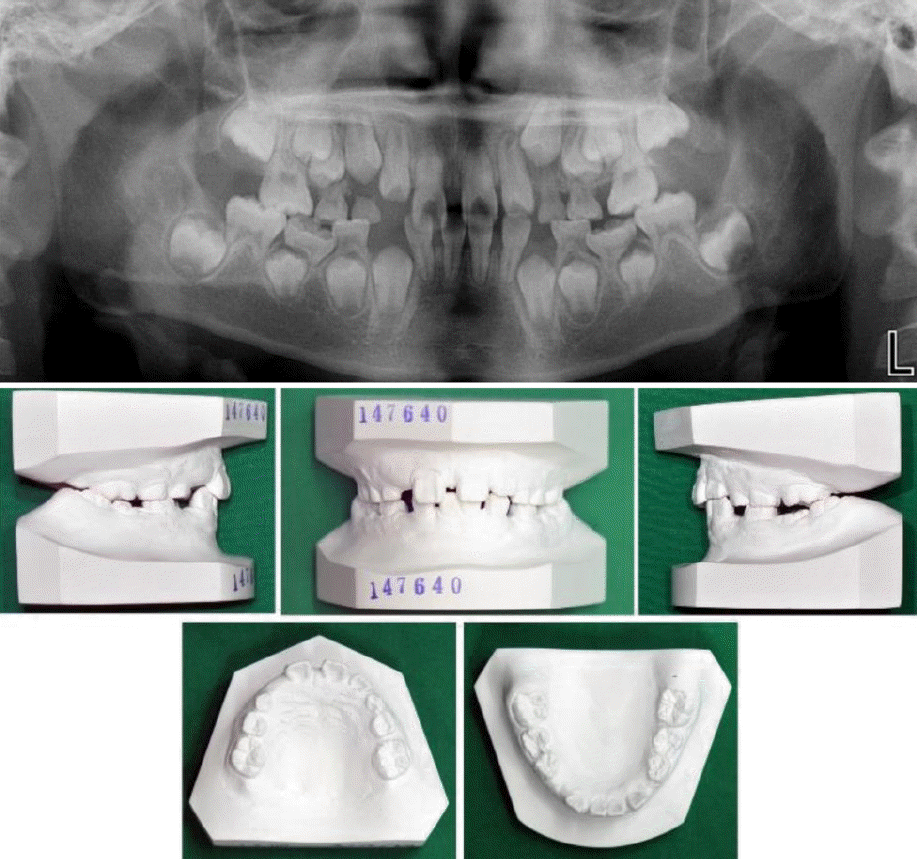
Pretreatment panoramic radiograph and cast models.
At first, the removable orthodontic appliance with bilateral screws was used to induce distalization of maxillary first molars. Especially, the screws were activated one by one unilaterally because of concern about weak anchorage. Nine months later, excessive overjet was observed. The use of the anterior teeth as an anchorage was stopped at this point, and fixed appliance with cervical headgear was adopted to allow distal traction using of extra-oral force. Initially, brackets were applied to maxillary four incisors and bilateral first molars. With the eruption of other teeth, brackets were attached sequentially. While a series of orthodontic wires universally applicable for Straight-Wire Appliance (SWA) system was used, every wire was kept for longer time than in usual cases. At this time, we could identify dens invaginatus clearly with periapical radiographs of maxillary lateral incisors (Fig. 3). The treatment with the headgear and fixed appliance continued for about 1 year, and then the second premolars were found to be locked in the mesial area of the first molars. Resorption of the first molars was suspected, which prompted surgical exposure and orthodontic traction of the second premolars (Fig. 4).
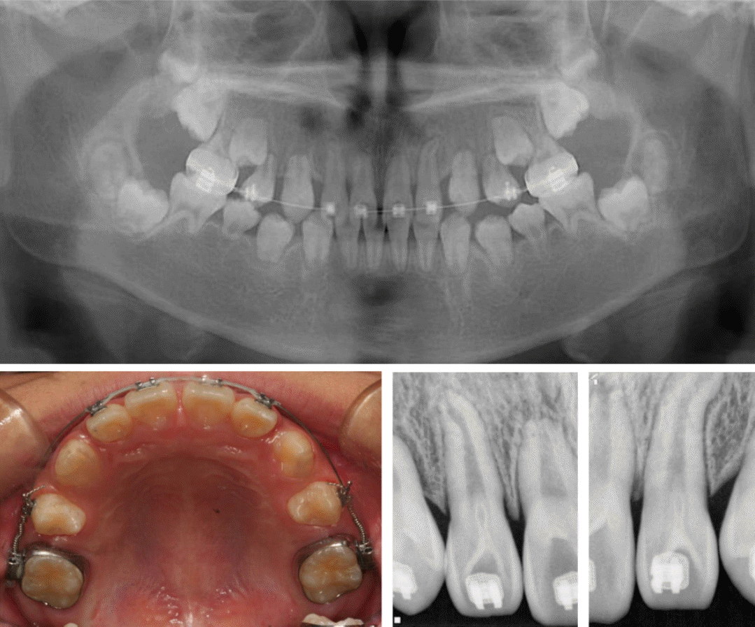
During treatment; intraoral appliance for headgear and dens invaginatus of maxillary lateral incisors.
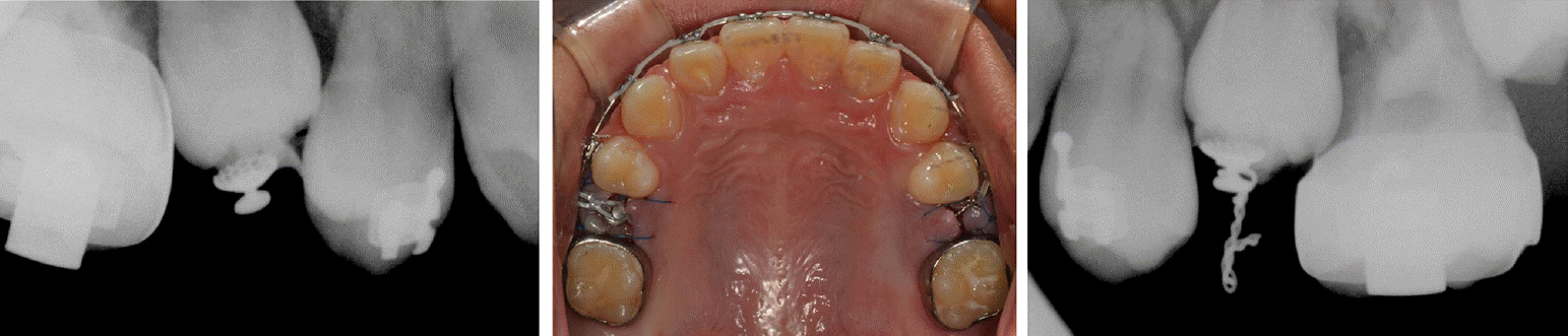
Surgical exposure and orthodontic traction of second premolars.
Toward the end of the teeth alignment, pinkish discoloration was observed on the maxillary left first molar, and radiographs indicated signs of internal resorption on its mesial side (Fig. 5). Based on consultation with the Department of Operative Dentistry at our institute, conservation therapy was infeasible due to signs of invasive resorption. Therefore, the tooth was extracted, and mesial eruption of the second molar was induced (Fig. 6).

Internal root resorption of maxillary left first molar.

After treatment; panoramic radiograph and intraoral photograph with a removable retainer.
The treatment lasted for approximately 3 years. Although the alignment of the maxillary dentition was completed, the left first molar was unfortunately lost. For nearly 2 years, the teeth has shown no significant mobility or root resorption since the treatment had been finished (Fig. 7).

Follow-up examination of 19 months.
Ⅲ. Discussion
This patient was unique in having idiopathic generalized SRA, which was not associated with any related systemic disease or family history. Idiopathic SRA is rare and difficult to predict. There have been several case reports on idiopathic generalized SRA accompanied by microdontia, taurodontia, dens invaginatus, agenesis, an ectopic canine, and obliteration of the pulp [10,11]. In this patient, the initial radiographs showed ectopic eruption and ankylosis, and indicated that the apical shape of the incisors and first molars differed from that of the normal immature permanent teeth. In addition, dens invaginatus of the maxillary lateral incisors were observed.
In a recent case report, tooth anomalies such as short root formation, abnormal shape of crown or root, and hypoplasia of enamel were assumed to be caused by epigenetic factors, such as damage, infection, medication exposure during the first 2 years of life, or damage at birth, especially damage affecting the central nervous system [12]. These events tended to match the time of development and damage to the teeth. However, our case involved a systemic anomaly, which appeared unconnected to such factors.
Several animal studies have identified various genes associated with root development, including Msx2 (msh homeobox 2), Sp6 (transcription factor), Shh (sonic hedgehog), and Nog(noggin) [13,14]. In particular, the Nfic (nuclear factor Ic) gene encodes a regulator that controls root dentin formation and is involved in odontoblast differentiation. The absence of this gene resulted in abnormal short roots in Nfic-mutant mice [6,14,15].
SRA has also been shown to be associated with other syndromes and systemic diseases [11,16]. If the related factors mentioned above cannot be identified, the prediction of SRA in mixed dentition showing immature permanent teeth is very challenging. The patient’s parents knew of no relevant family history and did not want to undergo further medical evaluation. Since there was no further evaluation, a potential relationship with a systemic disease or specific gene cannot be ruled out.
Teeth with short roots are at greater risk of root resorption, caused by orthodontic forces [7-9]. One study of the distribution of the orthodontic force using finite element methods showed that significant stress concentration occurs in the middle area of short roots, generating sufficient strength to trigger root resorption [17].
Some clinicians believe that orthodontic therapy is still possible for SRA patients, except in severe cases [4,5]. They insist that the risk of resorption can be controlled with clinical and radiological monitoring in most cases [4,18]. Precautions should be taken at all stages of orthodontic procedures. Lighter force needs to be applied because the center of resistance is closer to the crown than that of a normal tooth and the momentum/force ratio is easily affected [4]. Light, intermittent force may control resorption [19]. To allow sufficient time to repair the resorption, longer intervals between the activation of force are required [20]. Splints can be applied to ensure the stability of local SRA teeth, if needed [4,21]. Furthermore, in localized SRA, treatment methods that do not exert excessive force on the affected teeth should be considered. Orthopedic appliances, such as headgear or activators, can be used if the timing is appropriate. There is a referential report of a patient with class II malocclusion who underwent orthodontic treatment before the growth spurt and a desirable outcome was achieved using headgear without further damage to the maxillary central incisors in SRA [21].
Unfortunately, our patient had generalized, not localized, SRA so this strategy could not be used. A removable appliance was used with the anterior teeth as anchors during the early stages of treatment, and it was subsequently replaced by headgear to apply an extraoral force. In addition, light forces and a prolonged activation interval were applied as much as possible throughout the treatment. Without the use of the headgear, other anchor teeth may have been lost, leading to an unsuccessful result. Ultimately, this patient lost his left first molar, but coped with the associated complications by compensatory mesial eruption of the second molar. Furthermore, root development did not progress normally during the treatment period, and similar short roots were observed in the untreated mandibular dentition. This indicated a general state of arrested root formation, rather than iatrogenic root resorption caused by the orthodontic treatment.
Lind [1] stated that SRA has not received sufficient attention for several reasons. He mentioned that it could easily be misdiagnosed as root resorption because of a lower prevalence in Caucasians. SRA is more prevalent in Asians than Caucasians and ~10% of Japanese children with an average age of 9 years had central incisors with short roots [22]. However, the prevalence of SRA has been increasing in the USA recently, and Puranik et al. [23] suggested that this trend could be associated with the growing Latino population in the US. Moreover, teeth with SRA are generally asymptomatic, except in severe cases, with a normal crown shape and size, and related symptoms are usually absent. Unless excessive mobility or loss of teeth develops, SRA cannot be confirmed without radiographs. Valladares et al. [4] wrote that differential diagnosis should be carried out to distinguish SRA from other conditions such as incomplete root formation, external apical root resorption, dentin dysplasia type I, and post-trauma root hypoplasia.
Although the diagnosis of SRA can be missed easily for the reasons mentioned above, it should not be overlooked. The resistance against root resorption is lower, and the risk of tooth loss due to oral diseases is higher. The early detection of SRA is important and proper precautions must be taken during treatment. Further studies of SRA itself are required, as well as case reports on orthodontic, conservative, and periodontal treatment in SRA patients.
Ⅳ. Summary
This case confirms that orthodontic therapy is possible for SRA patients, but should be conducted with care. If SRA is confirmed during orthodontic treatment, a temporary cessation of treatment and appropriate modification of the treatment plan are required. Most importantly, the possibility of SRA in children should be evaluated with thorough assessments of the family history, systemic disease, and other dental anomalies before beginning orthodontic treatment.