


ŌģĀ. Introduction
Wolf-Hirschhorn syndrome (WHS), a congenital disorder associated with chromosome 4 microdeletion, is an uncommon but well-recognized disease. The syndrome was first described by Kurt Hirschhorn and his colleague Herbert Cooper [1] in 1961. They documented an individual with disturbed midline fusion resulting from deletion of a B-group chromosome, either 4 or 5, with the condition that became known as the Cri du Chat syndrome. Four years later, Ulrich Wolf [1] reported a similar deletion of the short arm of a B chromosome in a child that differed from the Cri du Chat syndrome; it turned out that the syndrome described by Hirschhorn and Cooper was different from Cri du Chat syndrome as well. The single eponym ŌĆ£WolfŌĆØwas employed, but the conjoined eponym ŌĆ£Wolf-HirschhornŌĆØhad become established by 1980. With increased interest in the molecular basis of the condition, ŌĆ£Deletion 4p SyndromeŌĆØwas used as an alternative to the eponymic version [1,2].
The incidence at birth is at least 1:50,000 births, and females are affected twice as often as males. There is no ethnic predilection. The amount of deleted material varies from a small break to 50% of the short arm of chromosome 4, and the more chromosomal material missing, the greater the extent to which symptoms of WHS are expressed [1,3].
The clinical signs of this syndrome are delayed growth, mental retardation, seizures, hypotonia, congenital heart defects, and atypical craniofacial features. The most characteristic feature is the ŌĆ£Greek warrior helmet appearanceŌĆØof the broad bridge of the nose continuing to the forehead. These characteristics are more pronounced during infancy [1,3]. Other characteristic craniofacial features are short philtrum, micrognathia, hypertelorism, high-arched eyebrows, ear anomalies, and bifid uvula. Oral manifestations include cleft lip with or without cleft palate, hypodontia, and taurodontism. The main factors that influence dental management are intellectual disability and epilepsy [2,3].
The aim of this study was to report the dental treatment of a 9-year-old girl with WHS and to present the clinical features and dental findings of this syndrome. This case report could help clinicians understand this syndrome and manage these patients.
ŌģĪ. Case report
A 9-year-old girl visited the department of pediatric dentistry for treatment of multiple caries. The patient was born after 39 weeksŌĆÖgestation with 1.8 kg weight by natural delivery. However, the patientŌĆÖs mother was not diagnosed with WHS during the prenatal ultrasound examination. When the patient came to the hospital, her height and weight were 128 cm and 13 kg, respectively, which were less than the third percentile. Both parents had no abnormal findings in their family history, and her brother also had no significant medical history. The patient presented motor and intellectual developmental delay, and verbal communication was not possible. The patient could not stand alone and could move only with the help of her parents. There were no prescribed drugs except phenobarbital for the management of seizures; the patient had not recently had any seizures. The patient was suffering from otitis media when she came to the hospital and had undergone surgery for the condition a month before the dental treatment at another hospital. The patient had feeding difficulties and was on a soft diet.
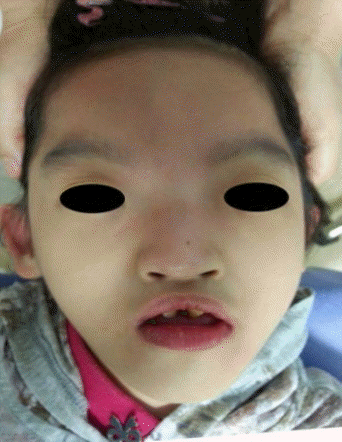
In extraoral examination, the patient had a high forehead with prominent glabella, broad nasal bridge, and hypertelorism. These characteristics were the typical facial features of WHS, the ŌĆ£Greek warrior helmet appearance.ŌĆØMicrognathia was present with down-turning of the corners of the mouth (Fig. 1).
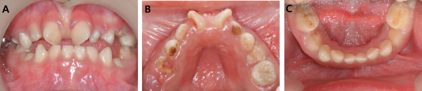
Dental examination was extremely difficult due to the mental handicap. Intraoral examination was performed under pedi-wrap with a mouth gag. The patientŌĆÖs mother brushed her teeth once or twice a day, and the patient had received a dental check-up when very young. (The patientŌĆÖs mother could not recall when.) There are no specific oral habits except occasional bruxism. The patient did not have cleft lip or palate, but did have poor oral hygiene with multiple caries and several residual roots. Of the erupted teeth, the only permanent teeth were the upper right central and lateral incisors and upper left central incisor; all of the lower teeth consisted of the primary dentition. Diastema between the upper central incisors was observed and heavy labial frenum extended the alveolar process (Fig. 2).
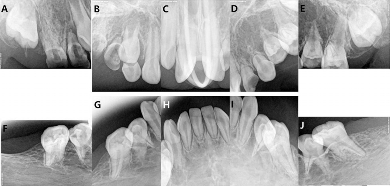
Intraoral radiography was taken in a wheelchair with a mouth gag, because the patient could not keep herself steady. In radiographic views, congenital missing teeth including the left upper lateral incisor and canine, upper permanent premolars, upper permanent second molars, and all lower permanent teeth were observed (Fig. 3).
General anesthesia (GA) was used during treatment of multiple caries. On physical examination, there were no other findings on the pre-operative work-up. Oral intubation was prepared due to the possibility of an anomaly of the airway tract; however, no abnormalities were found and nasal intubation was possible. Prior to the induction of anesthesia, oxygen was provided to a sufficient extent for more than three minutes, due to concern regarding the difficulty of endotracheal intubation.
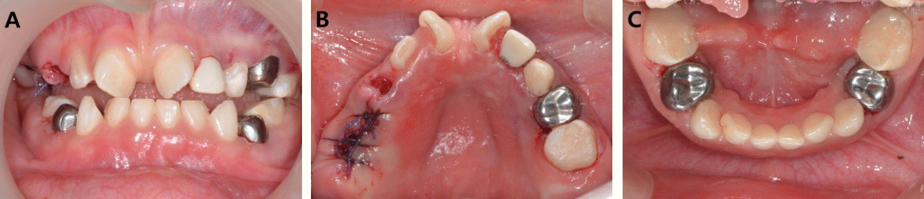
Under rubber dam isolation, stainless steel crowns (3M ESPE, St. Paul, MN, USA) were placed on the primary first molars, with the exception of the right primary maxillary first molar. Caries was not severe, but stainless steel crowns for space management were needed due to mesial tilting of the primary second molars caused by submerged primary first molars. On the mandibular teeth, maxillary contralateral stainless steel crowns were used due to deficient space caused by mesial tilting of the primary second molars. The primary second molars with the exception of the right primary maxillary second molar, the right primary mandibular canine, and the left primary maxillary canine were filled with composite resin (GC, Tokyo, Japan). Pulpectomy and a zirconia crown (NuSmile, Houston, TX, USA) were performed on the left primary maxillary lateral incisor. Finally, the residual roots of the right primary maxillary canine and first and second molars were surgically extracted and sutured. The patient showed stable vital signs without notable complications after GA (Fig. 4).
The patient had regular dental examinations every three months. Oral hygiene advice and fluoride application were performed to prevent dental caries and periodontal disease.
Ōģó. Discussion
WHS is a chromosomal abnormality caused by the deletion of the most distal or end portion of the short (p) arm of chromosome 4. The clinical symptoms are present from birth. Birth weight is low, and infants can have seizures and poor muscle tone from birth. About 1/3 of the patients die before reaching 2 years of age. The life expectancy ranges from 18 to 34 years depending on the extent of the genetic deletion, showing high mortality. As a larger deletion results in more severe congenital deformities, mortality is higher in individuals with larger deletions. Major causes of death are lower respiratory tract infection, multiple congenital anomalies, sudden unexplained death, and congenital heart disease [4,5].
Most cases of WHS result from a deletion in the 4p16.3 region, particularly in the WHS candidate genes, WHSC1 and WHSC2. These genes account for a number of the characteristic phenotypes of the syndrome; hence, a small deletion is easier to detect than a distal deletion. The deletions are caused by chromosomal structure deformities, such as ring chromosome, mosaicism, or translocation [1]. Most WHS patients have new mutations, and one of the parents of approximately 15% of the patients is known to have a balanced chromosomal translocation; therefore, genetic counselling plays an important role [2]. Recently, the WHSC1 gene was discovered to be present in sites responsible for DNA damage and replication stress and to be required to suppress DNA damage. WHSC1 is known to play a role in regulation of methylation of the K20 residue of histone H4 required for binding of p53-binding protein 1 (53BP1) on DNA damage sites. Moreover, WHSC1 is required for recruitment or retention of 53BP1 to DNA damage sites. DNA damage can cause developmental defects, immunodeficiency, neurological problems, and cancer. Therefore, WHSC1 plays an important role in DNA damage response and participates in human development and neuronal homeostasis [6]. Moreover, the MSX1 gene located about 3MB proximal to the critical region for WHS is known to be the most common site of mutation and is responsible for congenital absence of several permanent teeth and cleft deformities [1,7,8].
Characteristic features of WHS are hypospadias, congenital heart disease, renal and ophthalmic defects (such as iris coloboma, microphthalmia, and strabismus) and skeletal anomalies (concerning limbs and skeletal developmental retardation). A sacral dimple, hernia diaphragmatica, and omphalocele are also observed. The typical complications, such as epilepsy and mental retardation, are also among the characteristics. Children with WHS show growth and developmental retardation, as in this case [5]. Despite the age of 9 years, this patient was severely underweight at 13 kg, and the patientŌĆÖs growth parameter was below the third centile compared with a weight age of 9 years. The other deformities are muscle hypotonia and urinary tract malformations (such as renal agenesis, oligomeganephronia, bladder exstrophy, cystic renal dysplasia/hypoplasia, and obstructive uropathy) [1]. This patient also had severe muscle hypotonia, which made her incapable of holding up her head on her own. As ambulation was impossible, the parents accompanied her with a wheelchair. Also, the patient had a congenital defect on a kidney and had regular checkups at another hospital but had no problems involving the heart. As the patient was not toilet-trained despite the age of 9 years, diapers were worn in daily life. Moreover, the patient was suffering from otitis media upon arrival at the hospital, which could be attributed to the frequent occurrence of recurrent aspiration due to muscular hypotonia and gastroesophageal reflux dysfunction in WHS patients, leading to frequent occurrence of respiratory infections such as aspiration pneumonia, otitis media, sinusitis, or chronic cough. As hypotonia also causes swallowing difficulties and other gastrointestinal disorders [1], the patient could not chew solid foods and could only consume food in liquid form.
As WHS patients have very similar facial features, most patients have a distinctive craniofacial appearance such as a broad nasal bridge, high forehead, prominent glabella, hypertelorism, and highly arched eyebrows like the Greek warrior helmet appearance. Other characteristic features are protruding eyes, epicanthal folds, short philtrum, distinct mouth with downturned corners, micrognathia, and microcephalia. Such characteristics appear less in patients with smaller deletions9). In terms of oral characteristics, 30% of patients show uni- or bilateral cleft lip and/or palate and 50% show abnormal tooth development. Abnormal tooth development includes delayed tooth eruption with retention of deciduous teeth, peg-shaped teeth, and tooth agenesis [9]. Multiple tooth agenesis is caused by MSX1 gene anomalies and usually occurs in premolars and molars [2,7]. As the patients lack tooth buds, over-retained primary teeth are seen. Another characteristic is late dental development, leading to delayed tooth eruption and slower maturation [3]. A radiological study found taurodontism on primary molars of a WHS patient [4]. In a study by Dellavia et al. [10] that investigated oral features of 5 WHS patients, oral characteristics such as multiple coneshaped teeth, enamel hypoplasia, worn teeth, and discoloration of permanent dentition, as well as congenital taurodontism, spacing, and over-retained and misshapen primary molars were found. In this case, several permanent molars and premolars were congenitally missing, and over-retained primary molars were found as a consequence. Congenital absence of the anterior part of the mandible and some upper anterior permanent teeth was also present, and enamel hypoplasia and spacing of upper anterior permanent teeth were evident. Moreover, although the patient was 9 years old, the permanent first molars had not erupted and showed delayed development (Figs. 2 and 3).
As patients suffering from this disease require much medical care within the first 2 years after birth, the importance of oral health is easily overlooked. However, as hypodontia caused by anomalies in the MSX1 gene leads to absence of many permanent teeth, keeping the primary dentition as healthy as possible is important. Oral health directly influences the health status of patients, such that occlusal and dental alterations lead to nutritional abnormalities, and bacterial infection in the oral cavity leads to pathologies of the respiratory tract. Therefore, to maintain oral health, as soon as the primary dentition erupts, regular dental examinations and preventive care are needed [3,10].
The two factors that have the biggest influence on dental care of the patients are intellectual disability and epilepsy. Patients may experience difficulties in maintaining oral hygiene due to mental retardation, leading to occurrence of more severe dental caries and periodontal diseases. Due to lack of cooperation, dental treatment of WHS patients usually requires sedation or anesthesia. Therefore, maintaining regular oral hygiene prophylaxis is particularly important [2,10]. In this patient, considering the history of epilepsy and potential difficulties in securing the airway, caries treatment was performed under GA. Although the patient experienced no recent seizures, the same concentration of orally ingested phenobarbital was injected intravenously to control epilepsy, after consultation with a pediatrician. Also, as difficulties with endotracheal intubation are likely, consultation about the intubation method with an anesthesiologist is needed before GA. Although oral intubation was originally planned, nasal intubation was done with no great difficulty. As cases of malignant hyperthermia during GA in WHS patients have been reported, medications that may trigger malignant hyperthermia must not be used. Also, as delayed hyperthermia may occur even 11 h post-surgery, consistent monitoring of body temperature after recovery is required [11,12].
Further, as medications ingested for epilepsy may cause hypoplasia of the gingiva and ingestion of such medications may worsen periodontal diseases, consulting a medical doctor beforehand is recommended. Drooling and swallowing difficulties may affect dental procedures and, hence, more meticulous dental care compared with that in healthy individuals is required. Also, as there is the possibility of internal complications in the renal and cardiac systems, checking for them before dental treatment is important. Such issues need to be considered when deciding on the mode and type of anesthetic, and prophylactic antibiotic therapy may be considered before dental procedures when needed [2].
WHS patients have varying degrees of oro-dental abnormalities, and awareness of the oral, as well as systemic, features that may affect dental procedures is necessary for their dental care. Therefore, to maintain oral health, regular dental examination along with a multidisciplinary approach is important.
ŌģŻ. Summary
WHS, resulting from the deletion of the short arm of chromosome 4, is a malformation syndrome associated with growth and mental retardation and is characterized by a craniofacial appearance like a Greek helmet, accompanied by various oral manifestations, such as cleft lip with or without cleft palate, congenitally missing teeth, cone-shaped teeth, enamel hypoplasia, taurodontism, and delayed tooth eruption and slower maturation. The patient in this study showed multiple congenital missing teeth and mental retardation. Due to insufficient cooperation, caries treatment was performed under GA. The patient received regular dental check-ups and fluoride applications to manage oral hygiene.
 PDF Links
 PubReader
 ePub Link
 Full text via DOI
 Download Citation
 Print
Print


