


I. Introduction
Intrusion refers the condition that the traumatized tooth is driven into its socket and this can lead pulp and/or periodontal tissue damage and alveolar bone fracture, resulting in pulp necrosis, crown discoloration, pathologic root resorption, or ankylosis[1-3]. Primary tooth intrusion commonly occurs between the ages of 1 and 3 years which corresponds to the time of calcification of the tooth germ of permanent successor[4-6]. Therefore, primary tooth intrusion can affect a wide range of pathologic changes to permanent successors, including hypoplasia, crown/root dilacerations, cessation of root development, and eruption disturbance[1,2,7,8].
In primary dentition, intrusion usually does not occur solely but in the complicated form. Thus when anticipating the prognosis of intruded primary teeth, it is reasonable to examine whether the other adjacent tissue such as soft tissue is involved together.
Due to spatially and anatomically close relationship between apex of the primary tooth and its permanent successor, intrusive injuries in primary dentition can lead to sequelae in permanent dentition. Therefore, the treatment of primary tooth intrusion should aim to prevent the possible damage to the tooth germ of permanent successors[9]. Therefore, it is necessary to identify complications in the intruded primary tooth as well as effect on permanent successors.
The International Association for Dental Traumatology (IADT) recommends different treatment strategies according to the root orientation of affected primary tooth. If apex is labially displaced, the tooth is left for anticipating spontaneous repositioning. Otherwise, the tooth needs removed when apex is displaced palatally[9].
The purpose of this study was to investigate characteristics of primary tooth intrusion and to evaluate factors influencing prognosis of primary and permanent dentition during the long-time follow-up period.
II. Materials and Methods
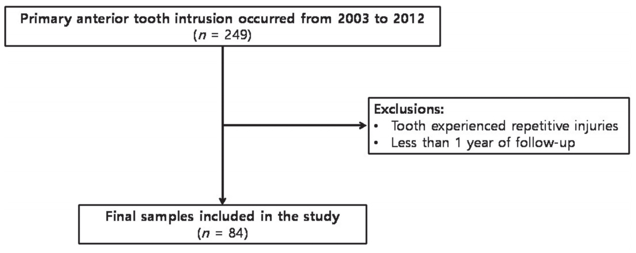
All patients who visited the Department of Pediatric Dentistry, School of Dentistry, Kyung Hee University, Seoul, Republic of Korea with chief complaints of traumatic dental injuries (TDIs) in the primary dentition between 2003 and 2012 were analyzed. Only patients who were diagnosed as primary anterior tooth intrusion were included. Patients with special medical history or repetitive TDIs during follow-up periods were excluded (Fig. 1). The study proposal was reviewed and approved by the ethics committee of Kyung Hee University Dental Hospital, Kyung Hee University, Seoul, Republic of Korea (KHDIRB-1408-1).
Based on trauma medical records, we collected data including clinical history, intraoral photographs and radiographic documentation of all patients. These data were organized according to age, gender, cause and site of trauma, severity of intrusion, other type of injuries accompanied at the time of injuries and complications during the follow-up periods in both primary and permanent dentition.
Severity of intrusion was classified into mild, moderate, or severe type by evaluation of clinical photographs and periapical radiographs at the time of injury. An adjacent primary incisor with no evidence of displacement was established as a baseline. Comparing with the baseline, it was defined as mild type when greater than half of clinical crown exposure was presented upon the gingival tissue, and moderate type as less than half of clinical crown exposure. Severe type was defined as a total of clinical crown was intruded.
Other types of injury accompanied at the time of TDI occurred were divided by classification of Andreasen et al .[2]; soft tissue injuries, hard dental tissue injuries, and periodontal tissue injuries.
1. Injuries to the soft tissue: abrasion, laceration and contusion;
2. Injuries to the hard tissue and the pulp: enamel crack, enamel fracture, enamel-dentin fracture without pulp exposure, enamel-dentin fracture with pulp exposure, enamel-dentin-cementum fracture without pulp exposure, enamel-dentin-cementum fracture with pulp exposure and root fracture;
3. Injuries to the periodontal tissue: concussion, subluxation, displacement, intrusion, extrusion and avulsion.
To evaluate clinical outcomes after primary tooth intrusion, only patients who were followed-up more than 1 year were included. Patients were scheduled for clinical and radiographic examination every 3 - 6 months until the physiologic exfoliation of intruded primary tooth and eruption of permanent successor. The follow-up period varied from 0.1 to 8.10 years (mean [SD], 1.8 [1.10] years).
Complication was grouped into 2 categories; (1) complication in primary tooth, and (2) complication in permanent successor. Crown discoloration, pulp canal obliteration, periapical abscess formation, inflammatory root resorption, and ankylosis were included as complication in primary tooth. To assess clinical outcome, clinical success was defined as discoloration only, pulp canal obliteration, and no complication and failure was defined when the traumatized tooth needed further treatment; periapical abscess formation, inflammatory root resorption, ankyloses. In permanent successor, enamel hypoplasia and crown shape malformation were recorded.
Data were analyzed using SPSS 22.0 software (SPSS Inc., Chicago, IL, USA). In order to determine the factors that affect prognosis, age, severity and type of accompanied injury were examined statistically. As the number of teeth which were followed-up until their successors erupt was small, influence of age and severity on prognosis of permanent tooth was statistically analyzed using FisherŌĆÖs exact test. For the same reason, difference in incidence of primary tooth complication according to a type of accompanied injury was analyzed using FisherŌĆÖ s exact test. On the other hand, effect of age and severity on primary tooth complication was statistically analyzed with Chisquare test.
III. Results
Of 176 children (249 teeth) who were diagnosed as primary tooth intrusion, 61 children (84 teeth) who met the inclusion criteria were evaluated. The mean age of these samples was 3.1 ┬▒ 1.3 years old and most prevalent age was 2 - 3 years in both boy and girl group. The final samples consisted of 40 (65.6%) boys and 21 (34.4%) girls. Intrusion occurred predominantly in males than in females, with a ratio of 1.90:1 (Table 1). Complication in primary and permanent tooth according to age is presented in Table 2. However, there was no statistical difference in incidence of complication in primary (Table 3, p= 0.197) and permanent tooth (Table 4, p= 0.237) according to age.
Causes and sites of intrusion were as follows (Table 5): Falls (51.2%) were the major sources of intrusion and most of intrusion occurred at home (56%) and the area around the home (31%).
Intrusion was commonly observed in primary maxillary central incisors (81%) with no difference between right and left teeth followed by primary maxillary lateral incisors (19%). 40 patients (65.6%) presented with a single affected tooth; 19 patients (31.1%) with 2 affected teeth; 2 patients (3.3%) with multiple affected teeth.
According to severity of intrusion, approximately half of tooth intrusion was mild type, followed by moderate type (32.1%) and severe type (14.3%). The severity of intrusion did not affect significantly the incidence of complication (Table 6, p= 0.082). However, it had an effect on the incidence of sequelae in permanent successors (Table 7, p= 0.014).
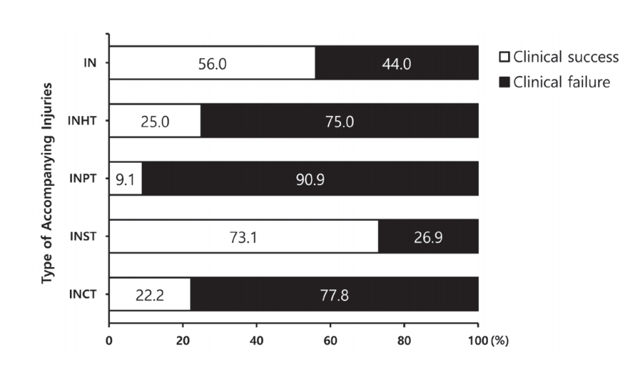
Of 84 teeth, 45 teeth (53.6%) exhibited clinical complications in affected primary teeth during the follow-up periods, and 39 teeth (46.4%) did not. Table 8 shows the clinical complications in primary tooth intrusion according to presence of other type of TDIs accompanied on the adjacent primary teeth. When primary tooth intrusion occurred with other periodontal tissue injuries simultaneously, the affected tooth was more likely to develop clinical complications. However, when intrusion was accompanied with soft tissue injuries, complications were significantly fewer (Fig. 2).
IV. Discussion
TDIs in the primary dentition can negatively affect the quality of life of children[10,11]. Especially in the children with developing permanent teeth, all kinds of TDIs can have a harmful effect on their quality of life for the whole their lives by causing developmental disorder on permanent teeth. Particularly the intrusive TDI to the primary teeth is likely to harm the successor of theirs due to spatial and anatomical position of primary and permanent teeth.
Most children suffer from primary tooth intrusion in the early stage of their lives. In the present study, higher incidence of intrusion was observed in 2 - 3 years. In this age group, children are likely to experience TDIs due to insufficient motor skills[4,12]. However, even in patients of the same age group, each patient may have all the different degrees of severity. And this can demonstrate why age was not a factor affecting the prognosis of primary and permanent tooth.
From this study, fall was predominant cause of intrusive dental injury accounting for 51.2% followed by collision and this result is in agreement with previous studies[13-15].
Moreover, approximately 84% of children in this age experienced intrusive luxation at home and around home in the present study. This is because children in this age spend most of their life time at home[16]. Based on these findings, educational program for caregivers regarding prevention and home care of TDIs can be beneficial.
Because of their anterior position in the dental arch, most intrusive luxation occurs in maxillary central incisors[5,7]. In this study, intrusive luxation usually involved a single primary tooth, which corresponded with previous studies[5,12].
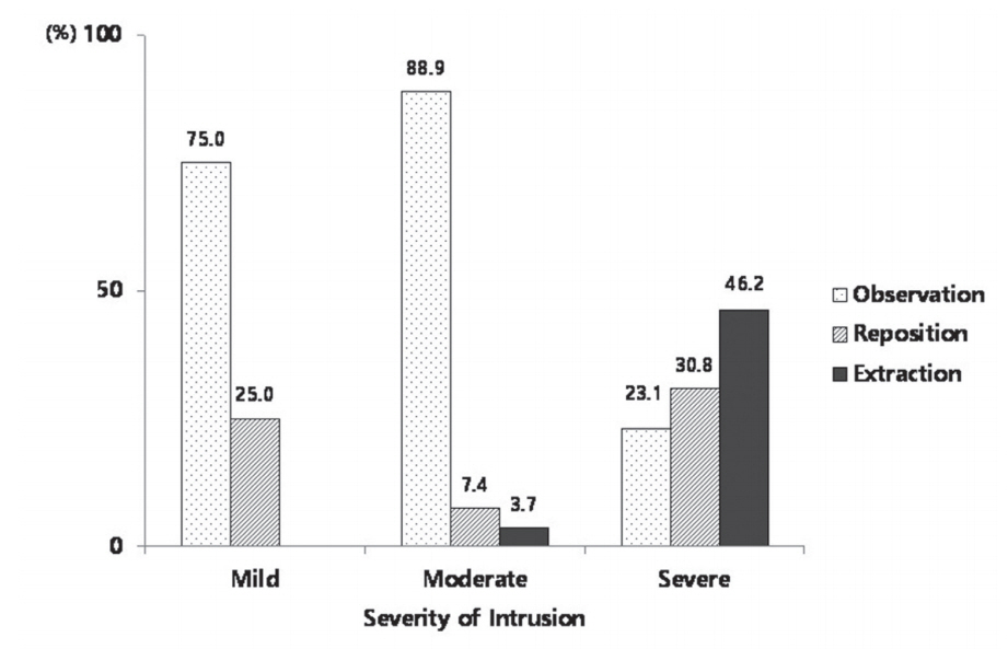
In terms of severity, mild type was most common followed by moderate type in this study. In severe intrusion cases, permanent successor presented unfavorable outcomes more frequently. Therefore, it is reasonable to rely not only on the path or direction of intrusion but also on the remaining portion of intruded tooth when selecting the treatment method and anticipating the prognosis of involved tooth. In the present study, 75% of mild type teeth, 88.9% of moderate type teeth and 23.1% of severe type teeth were observed without any intervention. And reposition was performed to 25% of mild type teeth, 7.4% of moderate type teeth and 30.8% of severe type tooth (Fig. 4).
In this study, 45 teeth (53.6%) of 84 teeth presented unfavorable outcomes in the primary teeth during the periodical check-up. Borum and Andreasen[17] reported inflammatory root resorption accounted for 14% of complications in the primary teeth following intrusion. Whereas in this study the inflammatory root resorption (71.1%) was the most common complication and followed by periapical abscess (35.6%). In the previous studies, the percentage of developmental disturbances of the permanent successors after intrusive dental injury ranges from 18% to 69%[6,18,19]. Likewise, 5 teeth (20.0%) of 25 successors presented complication in the permanent teeth in the study.
As most of intrusive dental injuries were consequences of fall accident, intrusive dental injuries usually occur in multiple teeth and are accompanied by multiple site injuries such as hard and soft tissues. In the present study, the intruded teeth accompanied with periodontal tissue injury showed unfavorable outcomes more frequently. While the intruded teeth accompanied with soft tissue injury were more likely to present favorable outcomes. This can be explained by the fact soft tissues like gingiva and buccal mucosa are softer than hard tissue and can absorb and favorably distribute impact from trauma[20-22].
However, there are several limitations in this study. First, since this study was performed retrospectively based on medical records, there was a possibility that majority of complicated type of injuries were included in ŌĆśintrusion onlyŌĆÖ group. For example concussion or subluxation of adjacent primary incisors could be ignored during the examination due to young patientŌĆÖs age. Second, as the number of intruded primary teeth which were followed-up until their permanent successors erupt was small, we could not statistically determine the effect of intrusive injuries on permanent successors development. Nonetheless, this study is significant in that a research on sequelae of permanent successors was carried out through long-term follow-up and in that this study determined factors influencing the outcome of intruded primary teeth and their successors. This retrospective study suggested that the severity of primary tooth intrusion can influence prognosis of permanent successor and when intrusion occurs with soft tissue injuries, traumatized primary tooth is likely to show favorable prognosis.
V. Conclusion
In this retrospective study, the intrusion occurred most frequently at home and fall accident was major cause of intrusion. Therefore caregivers such as parents should be trained about prevention of TDIs and should be encouraged to visit dental hospital in order to confirm whether the permanent successor is involved. Furthermore the clinician should provide periodic recall plan to the parents and exact periodic check with radiographs.
Within the limits of this study, age was not a factor which influence incidence of complication. However, severity of intrusion had an effect on incidence of sequelae in permanent successor and intrusion accompanied with soft tissue injury was likely to present better prognosis and show fewer complication.
On the basis of present study, when choosing a treatment method and predicting prognosis of intruded teeth, the clinician needs to depend not only on the intrusion path or direction, but also on the depth of intrusion or portion of crown above the gingiva and type of accompanied injuries.
Since the number of samples used in this study is limited, further studies with larger number of samples and longer follow-up period are needed.
 PDF Links
 PubReader
 ePub Link
 Full text via DOI
 Download Citation
 Print
Print


