


Ⅰ. Introduction
Dental caries is the most common chronic oral diseases in children[1]. Because enamel layer is thin in deciduous teeth, the dental pulp is easily involved as dental caries progress. An understanding of root canal morphology is essential in successful endodontic treatment[2,3].
It is difficult to obtain specific information about root canal configuration from periapical radiography since it only provides 2-dimensional view of 3-dimensional structure[2,4]. Several studies used some techniques such as canal staining[5] and micro computed tomography (CT) scanning of extracted teeth[6,7] to clarify the root canal morphology. These techniques have limitations such as the destruction of the tooth structure by the injection of staining material and small sample sizes of extracted teeth. In addition, there are not enough studies evaluating the root canal system in Korean pediatric patients.
The purpose of this study is to investigate the root canal morphology of the primary molars and to determine the symmetry in canal anatomy between left and right sides in the same patient.
Ⅱ. Materials and methods
This retrospective study was conducted at the Ewha Womans University Mokdong Hospital. The Institutional Review Board (IRB) of Hospital (IRB No. : 2019-02-001) approved the study prior to its implementation.
1. Study participants
A total of 114 children (81 boys, 33 girls) aged 3 to 7 years were included in this study. Participants did not exhibit any syndromic condition that could affect bone development. Over the period from January 1, 2007 to June 31, 2018, facial CT (SOMATOM perspective, SIEMENS, Germany) was carried out on the subject to assess various parameters such as trauma, facial swelling, and a presence of supernumerary teeth. All CT images were obtained with the following specifications : dose, 110 - 150 mAs; tube voltage, 80 - 120 kV; a matrix 512 × 512; pixel size, 0.3 mm; and slice thickness, 0.1 mm. To ensure the integrity of the morphology of the root canals, the following exclusion criteria were defined : molars with periapical lesions, a history from the patient of previous pulp treatment, and root resorption exceeding 1/5 of the root length. A total of 831 teeth (208 primary maxillary 1st molars, 195 primary maxillary 2nd molars, 205 primary mandibular 1st molars, 223 primary mandibular 2nd molars) were analyzed.
2. Evaluation of the image
Each CT image was evaluated twice by 2 pediatric dentists. Within 2 weeks, the assessments were conducted once again by the same pediatric dentists. All CT images were thoroughly screened at 1.0 mm intervals using 3 plane (sagittal, axial, and coronal) slices. The locations and numbers of roots and canals were determined for each tooth.
Cohen’s kappa coefficients were calculated to evaluate bilateral symmetry of the primary molars between left and right sides in the same patient. Statistical analysis was performed using SPSS 22.0 software (SPSS Inc., Chicago, IL, USA).
Ⅲ. Results
The kappa coefficients for the evaluation of intra-examiner and inter-examiner agreement were 0.910 and 0.969, respectively.
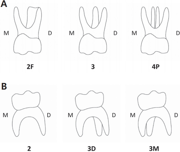
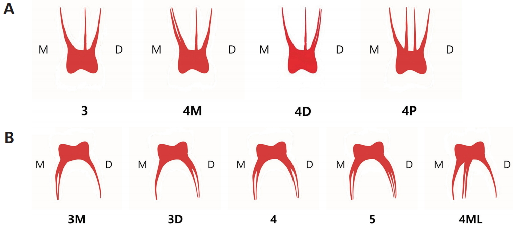
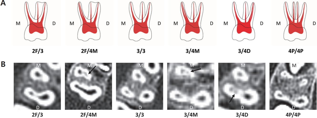
1. The types of root-canal, root, and canal in primary maxillary molars
Table 1 and 2 show the types of root and canal in primary maxillary molars. The most common type of root-canal in primary maxillary 1st molars found in this study was type 3/3, observed in 116 molars (55.8%), followed by type 2F/3 observed in 82 molars (39.4%). The types of canals were found to be 95.2% type 3 canal and 4.3% type 4M canal. Type 4D canal was found in only 1 case.
In primary maxillary 2nd molars, root-canal type 3/3 (30.3%) was the most commonly observed type, followed by type 2F/3 (29.7%). The types of canals were found to be 60% type 3 canal, 36.9% type 4M canal, and 3.1% type 4P canal.
2. The types of root-canal, root, and canal in primary mandibular molars
Table 3 and 4 show the results of the types of root and canal in primary mandibular molars. The root-canal type 2/4 (71.2%) was most frequently found in primary mandibular 1st molars, followed by type 2/3M (12.2%) and type 2/3D (12.2%). When the study focused on the types of canal, type 4 canal (73.7%) was the most common, followed by type 3D canal (14.1%) and type 3M (12.2%).
In primary mandibular 2nd molars, type 2/4 (63.2%) was the type observed most frequently, followed by type 3D/4 (25.6%). In terms of the types of canals, type 4 canal was the most common type and type 3M (9.9%) was the second most common type. Type 4ML canal was observed in only 1 case (0.4%), whereas type 5 canal was observed in only 2 cases (0.9%).
3. The symmetry of types of canal
Tables 5, 6, 7 and 8 show the types of canal of the primary molar on the left and right sides of the same patient. The kappa coefficient values, which test the relationships between the types of canal on both sides of the primary molars, are presented in Table 9. The kappa coefficient values in primary maxillary 1st and 2nd molars were 0.373 and 0.474 respectively, whereas the coefficients in primary mandibular 1st and 2nd molars were 0.809 and 0.601 respectively.
Ⅳ. Discussion
The purpose of this study was to investigate the root canal morphology of primary molars in pediatric patients. Dentist usually employ the periapical radiography for diagnosis and endodontic treatment. It can be a difficult task to find the exact location of the canal due to superimposition of structures in the images. It is not feasible to use CT for every pediatric patient in order to identify accurate locations of canals for endodontic treatment. Therefore, this study aimed to provide guidelines for clinical applications, by investigating the relationships between root canals of primary molars.
Each root of a primary maxillary molar tended to have 1 canal, similar the findings in previous studies[2,5,8]. 2 canals in mesio-buccal (MB) roots were observed in 4.3% of primary maxillary 1st molars and in 36.9% of 2nd molars. Joseph et al.[2] reported that 6.67% of primary maxillary 1st molars and 53.3% of 2nd molars had 2 canals in MB roots. A previous study on Korean pediatric patients[9] also reported that 22.8% of primary maxillary 1st molars and 66% of 2nd molars had 2 canals in MB root. These discrepancies in the results of different studies can be explained by the following reasons. First of all, the studies conducted by Joseph et al.[2] and Kim et al.[9] were carried out with extracted teeth and used small sample sizes. In addition, accessory canals were detected by Kim et al.[9] by observing canals directly in the cavities of extracted teeth. These different approaches seemed to be responsible for the different frequencies of 2 canals in MB roots among these studies.
The presence of 2 palatal roots was found only in maxillary 2nd molars. When there are 2 palatal roots, each root contains a canal. In other words, when 2 separated palatal roots are observed in a periapical X-ray, each root contains a canal. In 40% of primary maxillary molars, there was a fusion between the palatal and disto-buccal roots, although each root had a separate canal. This finding was consistent with those from previous studies by Wang et al.[7] and Bagherian et al.[5]. Clinicians should expect to find 2 separate canals in primary maxillary molars even when 1 fused root is observed in periapical radiographs.
In primary mandibular molars, each root seemed to have 1 or 2 canals. In the present study, 85.85% of mesial roots of primary mandibular 1st molars had 2 canals. This value was similar to the result from other previous studies, which were median values of 81.5%[5], 93.3%[2], and 100%[8]. Furthermore, each mesial root in a primary mandibular 2nd molar had 2 canals, which is consistent with the studies conducted by Joseph et al.[2], Bagherian et al.[5], Aminabadi et al.[8] and Gaurav et al .[3]. From this result, clinicians should expect a high probability of locating two canals in mesial roots of primary mandibular molars.
When only 1 distal root was observed in primary mandibular molars, 87.24% of the distal roots of the primary mandibular 1st molars contained 2 canals, and this percentage was higher than the results from studies carried out by Bagherian et al.[5] (22.2%), Wang et al.[7] (50%) and Joseph et al.[2] (60%). 2 canals were found in the distal roots in 84.94% of the primary mandibular 2nd molars, but this occurrence varies from 20%[7] to 100%[8] in previous studies. These varying results can be due to difference races and the different simple sizes in these studies.
2 separate distal roots were observed in 4.4% of primary mandibular 1st molars, whereas they were observed in 25.6% of primary mandibular 2nd molars. These results were consistent with those from a previous study performed in a Korean population where 2 separate distal roots were observed in 9.7% of primary mandibular 1st molars and 27.08% of primary mandibular 2nd molars[10]. According to the studies comparing the incidence rates of 2 separate distal roots among different races, 2 separate distal roots were found only in Chinese and Malassian populations, but not in European, Eurasian or Indian populations[11]. This finding indicates that 2 separate distal roots were found more often in Asian than in Caucasian populations[12,13]. This study also discovered that each separate distal root had its own canal. When 2 separated distal roots in primary mandibular molars are observed in periapical radiographs, clinicians should be able to predict that there are 2 canals in the distal roots.
With regard to the symmetry of the types of canal in primary molars on the left and right primary molars in the same patient, canal morphology was more symmetric in primary mandibular molars than in primary maxillary molars. Kappa coefficients of bilateral symmetry were 0.373 (confidence interval, CI = 0 - 0.765) and 0.474 (CI = 0.301 - 0.647) in primary 1st and 2nd maxillary molars respectively, whereas higher kappa coefficients were obtained for primary 1st and 2nd mandibular molars, 0.809 (CI = 0.686 - 0.932) and 0.601 (CI = 0.368 - 0.834) respectively. Based on Altman’s suggestion[14] on coincidence levels, 1st molars and 2nd molars can be interpreted as very high and high coincidence levels respectively. On the other hand, primary maxillary 1st molars showed fair coincidence between the left and right side of the molars, whereas the symmetry of primary maxillary 2nd molars showed moderate coincidence. A small number of reports have been published describing the symmetry of the root morphology of permanent molars[15,16], but only a few studies have been carried out on the symmetry of primary molars. Yang et al.[4] reported that 50.65% (39/77) of primary mandibular 2nd molars in Chinese children had symmetrical root morphology and canal morphology. In the present study on Korean children, the primary mandibular molars tended to show symmetry with regard to canal configuration. This was particularly evident in the primary mandibular 1st molars rather than in the primary mandibular 2nd molars. In accordance with the finding in this study, clinicians could readily predict the root canal morphology on the opposite side when the molars in both left and right sides needed endodontic treatment.
The limitation of this was that lower resolution facial CT was used rather than micro CT, which is used in more recent studies on root canal morphology[6,7,15]. Although it is difficult to locate small accessory canals with facial CT, the findings from our study are still valuable because of the large sample size used. Furthermore, our study observed canals that are clinically significant. More comprehensive investigations, taking root length, angulation, diameter and accessibility into account should be carried out in the future to overcome the limitations of the present study.
Ⅴ. Conclusions
Based on this research, the most commonly observed canal morphology in primary maxillary molars was mesio-buccal, disto-buccal and palatal canal, whereas it was were mesiobuccal, mesio-lingual, disto-buccal and disto-lingual canal in primary mandibular molars. All roots in primary maxillary molars, except for MB roots, tended to have 1 canal. The roots in primary mandibular molars tended to have 2 canals, especially the mesial root in primary mandibular 2nd molars. If either a palatal root in a primary maxillary molar or a distal root in a primary mandibular molar splits into 2 roots, there can be 1 canal for each root. Even in the fused types of disto-buccal and palatal roots of primary maxillary molars, there was 1 canal for each root as well. The symmetry of canals in primary molars was most common in primary mandibular 1st molars.

 PDF Links
 PubReader
 ePub Link
 Full text via DOI
 Download Citation
 Print
Print


