


ŌģĀ. ņä£ ļĪĀ
ņ£Āņ╣śņŚ┤ņØĆ ļ¦ÄņØĆ ĻĖ░ļŖźņØä ņłśĒ¢ēĒĢśļ®░ ņ¢┤ļ”░ņØ┤ņØś ņŗĀņ▓┤ ļ░£ļŗ¼ņŚÉ ņżæņÜöĒĢśļŗż. ņĀĆņ×æ, ļ░£ņØī, ņŗ¼ļ»ĖņĀüņØĖ ņŚŁĒĢĀņØä ĒĢśļ®░, ņØ┤ļź╝ ĒåĄĒĢ┤ ņĢģņĢłļ®┤ņØä ĒżĒĢ©ĒĢ£ ĻĄ¼Ļ░ĢņśüņŚŁņØś ļ░£ņ£ĪņŚÉ ņ¦ĆņåŹņĀüņØĖ ņ×ÉĻĘ╣ņØä ņżĆļŗż. ļśÉĒĢ£ ņśüĻĄ¼ņ╣śņŚ┤ņØś ņÖäņä▒ņØä ņ£äĒĢ£ Ļ│ĄĻ░ä ļ│┤ņĀäņØś ņŚŁĒĢĀņØä ļŗ┤ļŗ╣ĒĢ£ļŗż. ņ£Āņ╣śĻ░Ć ņś©ņĀäĒ׳ ļ│┤ņĀäļÉśņ¦Ć ļ¬╗ĒĢ£ ņ¢┤ļ”░ņØ┤ņŚÉņä£ ņśüĻĄ¼ņ╣śņŚ┤ņØś ĒśĢņä▒Ļ│╝ņĀĢņŚÉ ļ¦ÄņØĆ ļ│ĆņØ┤ņÖĆ ņןņĢĀĻ░Ć ļéśĒāĆļéśļŖö Ļ▓āņØä ĒØöĒ׳ ļ│╝ ņłś ņ׳ļŗż[1].
ņ£Āņ╣śļŖö ņ╣śĻĘ╝ ļ░Å ĻĘ╝Ļ┤ĆņØś Ēü¼ĻĖ░ņÖĆ ļé┤ļČĆ ļ░Å ņÖĖļČĆ ĒśĢĒā£ņŚÉņä£ ņśüĻĄ¼ņ╣śņÖĆ ĒśĢĒā£ĒĢÖņĀü ņ░©ņØ┤Ļ░Ć ņ׳ļŗż[2]. ĒŖ╣Ē׳, ņ£ĀĻĄ¼ņ╣śļŖö ņśüĻĄ¼ ļīĆĻĄ¼ņ╣śļ│┤ļŗż ņ╣śņłśĻ░ĢĻ│╝ ĻĘ╝Ļ┤ĆņØ┤ ņ×æĻ│Ā ĒĢśļČĆņØś ņśüĻĄ¼ņ╣ś ņ╣śļ░░ļź╝ ļ│┤ĒśĖĒĢśĻĖ░ ņ£äĒĢśņŚ¼ ņ╣śĻĘ╝ņØ┤ ņŗ¼ĒĢśĻ▓ī ņØ┤Ļ░£ ļÉśņ¢┤ņ׳ļŗż. ļśÉĒĢ£ ĻĘ╝Ļ┤ĆņØś ņłśĻ░Ć ļ¦ÄĻ│Ā ņ╣śņłśĻ░ĢņĀĆņŚÉ ļ¦ÄņØĆ ļČĆĻĘ╝Ļ┤ĆņØä Ļ░ĆņĀĖ ņ£Āņ╣śņØś ĻĘ╝Ļ┤Ć ņ╣śļŻīļŖö ļ¦żņÜ░ ļ│Ąņ×ĪĒĢ£ Ļ▓āņ£╝ļĪ£ Ļ░äņŻ╝ļÉ£ļŗż. ĻĘ╝Ļ┤ĆņØś ĒĢ┤ļČĆĒĢÖņĀü ļ│Ąņ×Īņä▒ņØĆ ņ×äņāü ņ╣śļŻīņØś ņ¢┤ļĀżņøĆņØä ļ│┤ņŚ¼ ņŻ╝ļ®░, ņ╣śĻ│╝ņØśņé¼ļŖö ĻĘ╝Ļ┤ĆņØś ĒśĢĒā£ĒĢÖņĀü ĒŖ╣ņä▒Ļ│╝ ļŗżņ¢æņä▒ņØä ņØĖņŗØĒĢśĻ│Ā ņ׳ņ¢┤ņĢ╝ ĒĢ£ļŗż[3]. ļŗżņłśņØś ĻĘ╝Ļ┤Ć ņ╣śļŻī Ļ│╝ņĀĢ ņżæņØ┤ļéś ņØ┤ĒøäņŚÉ ļ░£ņāØĒĢśļŖö ļ¼ĖņĀ£ļŖö ĻĘ╝Ļ┤ĆņŚÉ ļīĆĒĢ£ ļČĆņĀüņĀłĒĢ£ ņØ┤ĒĢ┤ ļĢīļ¼ĖņŚÉ ļ░£ņāØĒĢ£ļŗż. ņ£Āņ╣śņØś ĻĘ╝Ļ┤Ć ņ╣śļŻīņŚÉ ņ׳ņ¢┤ ņ£Āņ╣śņØś ĻĘ╝Ļ┤ĆņŚÉ ļīĆĒĢ£ ĒĢ┤ļČĆĒĢÖņĀü ņ¦ĆņŗØņØĆ ĻĘ╝Ļ┤Ć ņ╣śļŻīļź╝ ņ£äĒĢ£ ĒĢäņłś ņĀäņĀ£ ņĪ░Ļ▒┤ņØ┤ļ®░, ņ£Āņ╣śņØś ĻĘ╝Ļ┤Ć ĒśĢĒā£ņÖĆ ļ│ĆņØ┤ņŚÉ ļīĆĒĢ£ ņ¦ĆņŗØņØĆ ņ╣śņłśĻ░Ģ ļ░Å ĻĘ╝Ļ┤ĆņŚÉ ļīĆĒĢ£ ĒśĢĒā£ļź╝ ņŗ£Ļ░üĒÖöĒĢśņŚ¼ ņ╣śļŻīņŚÉ ļÅäņøĆņØ┤ ļÉ£ļŗż.
ĻĘ╝Ļ┤Ć ĒśĢĒā£ļź╝ ļČäņäØĒĢśļŖö ļŹ░ņŚÉ ņ׳ņ¢┤ ņØ╝ļ░śņĀüņ£╝ļĪ£ ņé¼ņÜ®ļÉśņŚłļŹś ļ░®ļ▓ĢņØĆ ļ░£Ļ▒░ ļÉ£ ņ╣śņĢä ņĪ░ņé¼, ņ╣śņłśĻ░Ģ ļé┤ ĻĘ╝Ļ┤Ć ņŚ╝ņāē ļ░Å ņŗżļ”¼ņĮś ņŻ╝ņ×ģ, ļ░®ņé¼ņäĀ ņé¼ņ¦ä ņ┤¼ņśü ļō▒ņØ┤ ņ׳ņ£╝ļéś ņ╣śĻĘ╝ ĒØĪņłś ņŚåņØ┤ ņåÉņāüļÉśņ¦Ć ņĢŖņØĆ ņ£Āņ╣ś Ēæ£ļ│ĖņØä ĻĄ¼ĒĢśĻĖ░ ņ¢┤ļĀĄĻ│Ā ņ╣©ņŖĄņĀüņØ┤ļØ╝ļŖö ĒĢ£Ļ│äĻ░Ć ņ׳ļŗż[3-5]. ļśÉĒĢ£ ņ╣śĻĘ╝ ņ×¼ĒØĪņłśĻ░Ć ņŚåļŖö ņåÉņāüļÉśņ¦Ć ņĢŖņØĆ ņ£Āņ╣śĻ░Ć ļČĆņĪ▒ĒĢśĻĖ░ ļĢīļ¼ĖņŚÉ ņ£Āņ╣śņØś ĻĘ╝Ļ┤Ć ĒśĢĒā£ņŚÉ ļīĆĒĢ£ ņŚ░ĻĄ¼ļŖö ļ¦Äņ¦Ć ņĢŖļŗż.
ņĄ£ĻĘ╝ Computed Tomography (CT)ļŖö ĻĘ╝Ļ┤Ć ĒśĢĒā£ļź╝ ņŚ░ĻĄ¼ĒĢśļŖöļŹ░ ņ׳ņ¢┤ ļäÉļ”¼ ņé¼ņÜ®ļÉśĻ│Ā ņ׳ļŗż. ņØ┤ ņżæ ĒĢśļéśņØĖ Cone-Beam CT (CBCT)ļŖö ĒśĢĒā£ĒĢÖņĀü ļČäņäØņŚÉ ņ׳ņ¢┤ ļ╣äņ╣©ņŖĄņĀüņØ┤ļ®░ ņŗ£ņāüļ®┤(sagittal), Ļ┤Ćņāüļ®┤(coronal), ņČĢļ®┤(axial)ņØś 3Ļ░Ćņ¦Ć ĒÅēļ®┤ņŚÉņä£ ļīĆņāüņØä Ļ┤Ćņ░░ĒĢĀ ņłś ņ׳ļŗż. ņØ┤ 3Ļ░Ćņ¦Ć ĒÅēļ®┤ņØś ņØ┤ļ»Ėņ¦Ć ņĪ░ĒĢ®ņØĆ ĒĢ┤ļČĆĒĢÖņĀü ĻĄ¼ņĪ░ļ¼╝ņØä 3ņ░©ņøÉņ£╝ļĪ£ ņŗ£Ļ░üĒÖöĒĢśņŚ¼ ņżæņ▓® ņŚåņØ┤ Ļ┤Ćņ░░ĒĢĀ ņłś ņ׳Ļ▓ī ĒĢśļ®░, ņÖ£Ļ│ĪņØ┤ ņĀüļŗż[5,6].
ņŚ¼ļ¤¼ ņŚ░ĻĄ¼ņŚÉņä£ ņ╣śĻĘ╝ ļ░Å ĻĘ╝Ļ┤ĆņØś ĒŖ╣ņ¦ĢņØĆ ņä▒ļ│ä, ņØĖņóģ, ņ¦ĆņŚŁņŚÉ ļö░ļØ╝ ļŗżņ¢æĒĢśļŗżĻ│Ā ļ░ØĒśĆņĪīļŗż[7-9]. Ēśäņ×¼ ĒĢ£ĻĄŁ ņ¢┤ļ”░ņØ┤ņØś ņśüĻĄ¼ņ╣ś ĻĘ╝Ļ┤Ć ĒśĢĒā£ņŚÉ ļīĆĒĢ£ ļ¦ÄņØĆ ņŚ░ĻĄ¼Ļ░Ć ņ׳ņ£╝ļéś[10,11] CBCTļź╝ ņØ┤ņÜ®ĒĢ£ ņ£ĀĻĄ¼ņ╣śņØś ņ╣śĻĘ╝ ļ░Å ĻĘ╝Ļ┤ĆņØś ĒśĢĒā£ņŚÉ ļīĆĒĢ£ ņŚ░ĻĄ¼ļŖö ļ¦Äņ¦Ć ņĢŖņØĆ ņŗżņĀĢņØ┤ļŗż.
ņØ┤ ņŚ░ĻĄ¼ļŖö ņĀäļé©ļīĆĒĢÖĻĄÉ ņ╣śĻ│╝ļ│æņøÉ ņåīņĢäņ╣śĻ│╝ņŚÉ ļé┤ņøÉĒĢ£ ĒÖśņĢä 68ļ¬ģņØś CBCT ņśüņāüņØä ņĪ░ņé¼ĒĢśņŚ¼ ņ£ĀĻĄ¼ņ╣śņØś ĻĘ╝Ļ┤Ć ņ╣śļŻī ņŗ£ ĒĢäņÜöĒĢ£ ņ╣śĻĘ╝ ļ░Å ĻĘ╝Ļ┤ĆņØś ĒśĢĒā£ĒĢÖņĀü ĒŖ╣ņ¦ĢņØä ĒÅēĻ░ĆĒĢśĻ│Ā ņ×äņāüņĀüņØĖ ĒÖ£ņÜ®ņØä ņ£äĒĢ£ ņĀĢļ│┤ļź╝ ņĀ£Ļ│ĄĒĢśĻ│Āņ×É ņłśĒ¢ēļÉśņŚłļŗż.
ŌģĪ. ņŚ░ĻĄ¼ ļīĆņāü ļ░Å ļ░®ļ▓Ģ
1. ņŚ░ĻĄ¼ ļīĆņāü
2017ļģä 5ņøöļČĆĒä░ 2018ļģä 8ņøöĻ╣īņ¦Ć Ļ│╝ņ×ēņ╣ś ļ░£Ļ▒░ļź╝ ņ£äĒĢ┤ ņĀäļé©ļīĆĒĢÖĻĄÉ ņ╣śĻ│╝ļ│æņøÉ ņåīņĢäņ╣śĻ│╝ņŚÉ ļé┤ņøÉĒĢśņŚ¼ CBCTļź╝ ņ┤¼ņśüĒĢ£ ļ¦ī 4 - 5ņäĖ ĒÖśņ×ÉņØś ņśüņāüņØä ļČäņäØĒĢśņśĆļŗż. ņĀäņŗĀņ¦łĒÖśņØ┤ ņŚåļŖö ĒÖśņ×ÉņØś ņ╣śĻĘ╝ļŗ© ļ│æļ│ĆĻ│╝ ņ╣śĻĘ╝ ĒØĪņłśĻ░Ć ņŚåļŖö ņ╣śņĢäļōżļĪ£ ņäĀļ│äļÉśņŚłĻ│Ā ĻĘ╝Ļ┤Ć ņČ®ņĀäļ¼╝ņØ┤ļéś ĻĖ░ņä▒ĻĖłņåŹĻ┤Ćņ£╝ļĪ£ ņłśļ│Ą ļÉ£ ņ╣śņĢäļŖö ņŚ░ĻĄ¼ņŚÉņä£ ņĀ£ņÖĖļÉśņŚłļŗż. ņāüņĢģ ņĀ£1ņ£ĀĻĄ¼ņ╣ś, ņāüņĢģ ņĀ£2ņ£ĀĻĄ¼ņ╣ś, ĒĢśņĢģ ņĀ£1ņ£ĀĻĄ¼ņ╣ś, ĒĢśņĢģ ņĀ£2ņ£ĀĻĄ¼ņ╣ś Ļ░üĻ░ü 40Ļ░£ņö®, ņ┤Ø 160Ļ░£ņØś ņ£ĀĻĄ¼ņ╣śĻ░Ć ļČäņäØļÉśņŚłĻ│Ā ņ┤Ø 68ļ¬ģņØś ĒÖśņĢäĻ░Ć ņŚ░ĻĄ¼ņŚÉ ĒżĒĢ©ļÉśņŚłņ£╝ļ®░, ĒÅēĻĘĀ ņŚ░ļĀ╣ņØĆ 4.9 ┬▒ 0.6ņäĖņśĆļŗż. ļ░®ņé¼ņäĀ ņśüņāüņØä ļ░öĒāĢņ£╝ļĪ£ ĒĢ£ ņØ┤ ņŚ░ĻĄ¼ņØś ĒöäļĪ£ĒåĀņĮ£ņØĆ ņĀäļé©ļīĆĒĢÖĻĄÉ ņ╣śĻ│╝ļ│æņøÉ ņāØļ¬ģņØśĒĢÖņŚ░ĻĄ¼ņ£żļ”¼ņŗ¼ņØśņ£äņøÉĒÜī(Institutional Review Board, IRB)ņØś ņŖ╣ņØĖņØä ļ░øĻ│Ā ņ¦äĒ¢ēļÉśņŚłļŗż(IRB NO.: CNUDH-2019-012).
2. ņŚ░ĻĄ¼ ļ░®ļ▓Ģ
CBCT ņśüņāüņØĆ CS9300(Carestream Dental, USA)ļĪ£ļČĆĒä░ ņ¢╗ņ¢┤ņĪīņ£╝ļ®░ ņØ┤ļŖö OnDemand3D software(Cybermed, Korea)ĒöäļĪ£ĻĘĖļשņØś 2D ļ░Å 3D ņØ┤ļ»Ėņ¦ĆļĪ£ ĻĄ¼ĒśäļÉśņŚłļŗż. Ļ░ü ņ╣śņĢäļōżņØś ņ╣śņłśĻ░Ģ ņĀĆņŚÉņä£ ņ╣śĻĘ╝ļŗ©Ļ╣īņ¦ĆņØś ņØ┤ļ»Ėņ¦ĆļŖö ņŗ£ņāüļ®┤, Ļ┤Ćņāüļ®┤, ņČĢļ®┤ņØś 3Ļ░Ćņ¦Ć ĒÅēļ®┤ņŚÉņä£ Ļ┤Ćņ░░ļÉśņŚłļŗż.
1) ņ╣śĻĘ╝ņØś ņłś
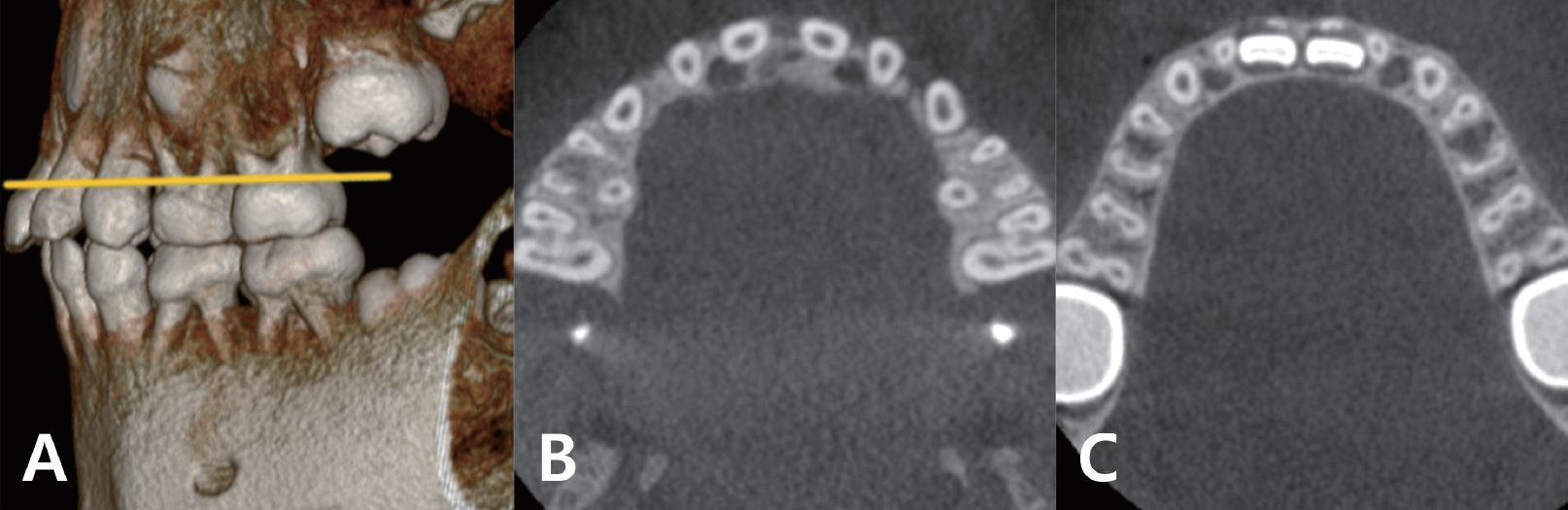
Ļ░üĻ░üņØś ņ╣śņĢäļ│äļĪ£ CBCTņØś ņČĢļ®┤ ņśüņāüņŚÉņä£ ĒĢ£ ņ╣śņĢä ļŗ╣ ņ╣śĻĘ╝ ņłśļź╝ ņĪ░ņé¼ĒĢśņśĆļŗż(Fig. 1A).
3) ņ╣śĻĘ╝ņØś ĻĖĖņØ┤
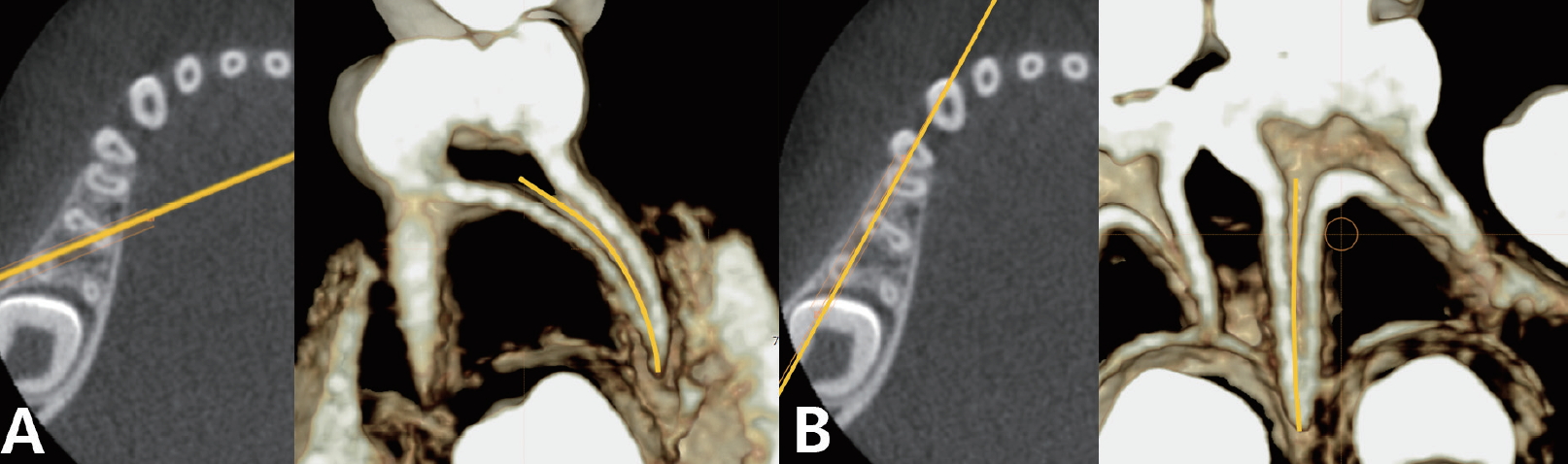
ņøÉĒśĢ ņ╣śĻĘ╝ņØś ĻĖĖņØ┤ļŖö ļ░▒ņĢģļ▓Ģļ×æĻ▓ĮĻ│äļź╝ ĻĘ╝ņøÉņŗ¼ņ£╝ļĪ£ ņŚ░Ļ▓░ĒĢ£ ņäĀņŚÉ ņ£äņ╣śĒĢ£ ĻĘ╝Ļ┤Ć ņ×ģĻĄ¼ņŚÉņä£ ĻĘ╝ņ▓©ļČĆĻ╣īņ¦Ć ļŗ©ļ®┤ĒÖö Ēøä ņżæņĢÖņØä ļö░ļØ╝ ĻĖĖņØ┤ļź╝ ņĖĪņĀĢĒĢśņśĆļŗż(Fig. 2A). ņøÉĒśĢņØ┤ ņĢäļŗī ĒśæņäżļĪ£ ĻĖĖĻ│Ā ļé®ņ×æĒĢ£ ĒśĢĒā£ņØś ņ╣śĻĘ╝ņØĆ Ēśæņäżļ®┤ņŚÉ ņłśņ¦üņ£╝ļĪ£ ļŗ©ļ®┤ĒÖöĒĢśņŚ¼ Ļ░Ćņן ĻĖ┤ ļČĆņ£äļź╝ ņĖĪņĀĢĒĢśņśĆļŗż(Fig. 2B).
4) ĻĘ╝Ļ┤ĆņØś ĻĖĖņØ┤
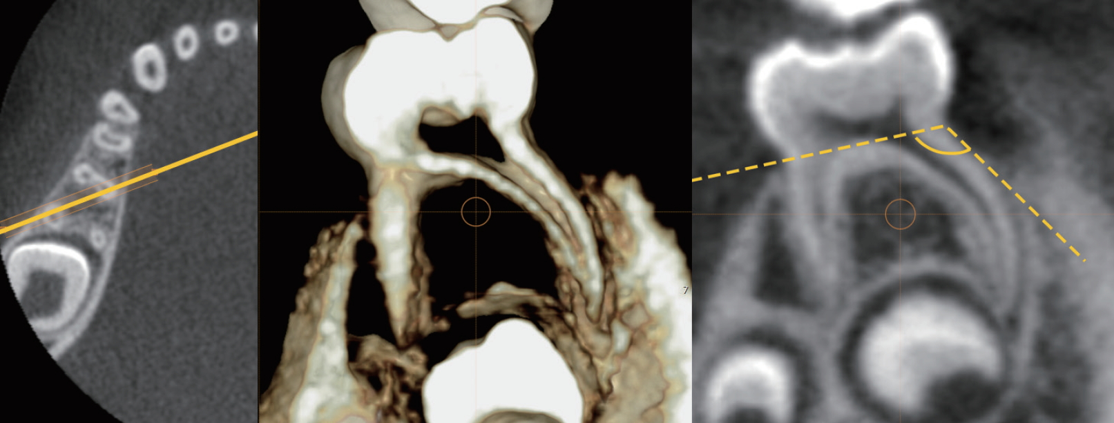
ĻĘ╝Ļ┤Ć ņ×ģĻĄ¼ņŚÉņä£ ĻĘ╝ņ▓©ļČĆĻ╣īņ¦Ć ļŗ©ļ®┤ĒÖöĒĢśņŚ¼ ļ░▒ņĢģļ▓Ģļ×æĻ▓ĮĻ│äņÖĆ ņ╣śņłśņĀĆļź╝ ņŚ░Ļ▓░ĒĢ£ ņäĀņŚÉ ņ£äņ╣śĒĢ£ ĻĘ╝Ļ┤Ć ņ×ģĻĄ¼ņŚÉņä£ ĻĘ╝Ļ┤Ć ļ¦Éļŗ©Ļ╣īņ¦Ć ņżæņĢÖņØä ļö░ļØ╝ ĻĖĖņØ┤ļź╝ ņĖĪņĀĢĒĢśņśĆļŗż(Fig. 3).
5) ņ╣śĻĘ╝ņØś ņØ┤Ļ░£ Ļ░üļÅä
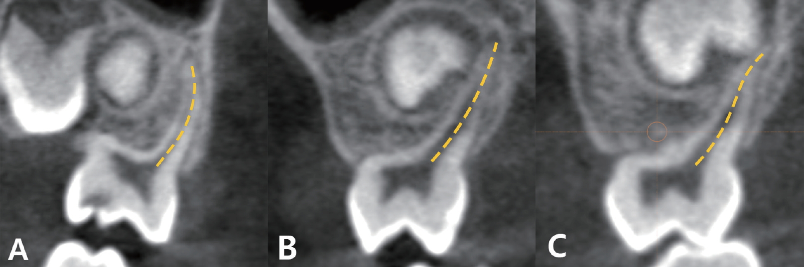
ņ╣śĻĘ╝ņØś ņØ┤Ļ░£ ņĀĢļÅäļź╝ ņĢīĻĖ░ ņ£äĒĢśņŚ¼ ļ░▒ņĢģļ▓Ģļ×æĻ▓ĮĻ│äņÖĆ ņ╣śņłśņĀĆļź╝ ņØ┤ņØĆ ņäĀņŚÉņä£ ņ╣śĻĘ╝ņØ┤ Ļ░Ćņן ļÅīņČ£ ļÉ£ ļČĆņ£äĻ╣īņ¦Ć Ļ░üļÅäļź╝ ņĖĪņĀĢĒĢśņśĆļŗż(Fig. 4).
3. Ēæ£ņżĆ ņØ╝Ļ┤Ćņä▒ ĒÅēĻ░Ć
40Ļ░£ņØś Ēæ£ļ│ĖņØä ļ¼┤ņ×æņ£äļĪ£ Ļ│©ļØ╝ ņĪ░ņé¼ņ×É ļé┤, ņĪ░ņé¼ņ×É Ļ░ä ņ×¼Ēśäņä▒ņØä ĒÅēĻ░ĆĒĢśņśĆļŗż. ņ╣śĻĘ╝ ļ░Å ĻĘ╝Ļ┤ĆņØś ņłś, ņ╣śĻĘ╝ ļ░Å ĻĘ╝Ļ┤ĆņØś ĻĖĖņØ┤, ņ╣śĻĘ╝ņØś ņØ┤Ļ░£ Ļ░üļÅäņŚÉ ļīĆĒĢśņŚ¼ 1ļ¬ģņØś ņ╣śĻ│╝ņØśņé¼Ļ░Ć Ļ░ÖņØĆ CBCT ņØ┤ļ»Ėņ¦Ćļź╝ 3ņŻ╝ Ļ░äĻ▓®ņ£╝ļĪ£ ĒīÉļÅģĒĢśņśĆĻ│Ā ņĪ░ņé¼ņ×É ļé┤ ņŗĀļó░ļÅäņØĖ Intraclass Correlation Coefficient (ICC) Ļ░ÆņØä ĻĄ¼ĒĢśņśĆņØä ļĢī ļ¬©ļōĀ ĒĢŁļ¬®ņŚÉņä£ 0.80 ņØ┤ņāüņ£╝ļĪ£ ļéśĒāĆļé¼ļŗż. ņ╣śĻĘ╝ ļ░Å ĻĘ╝Ļ┤ĆņØś ĒśĢĒā£ļŖö ņĪ░ņé¼ņ×É Ļ░ä Ēæ£ņżĆ ņØ╝Ļ┤Ćņä▒ Ļ▓Ćņé¼ļź╝ ņŗ£Ē¢ēĒĢśņśĆĻ│Ā 2ļ¬ģņØś ņ╣śĻ│╝ņØśņé¼Ļ░Ć Ļ░ÖņØĆ CBCT ņØ┤ļ»Ėņ¦Ćļź╝ ĒÅēĻ░ĆĒĢśņśĆļŗż. CohenņØś kappa Ļ│äņłśļŖö 0.74ļĪ£ ņŗĀļó░ņä▒ ņ׳ļŖö Ļ░Æņ£╝ļĪ£ ļéśĒāĆļé¼ļŗż.
4. ĒåĄĻ│ä ļČäņäØ
Ļ░ü ĒĢŁļ¬®ņŚÉ ļīĆĒĢ┤ ĒÅēĻĘĀ ļ░Å Ēæ£ņżĆĒÄĖņ░©ļź╝ ĻĄ¼ĒĢśņśĆļŗż. ņ╣śĻĘ╝ ļ░Å ĻĘ╝Ļ┤ĆņØś ĻĖĖņØ┤ņÖĆ ņ╣śĻĘ╝ņØś ņØ┤Ļ░£ Ļ░üļÅä ĒĢŁļ¬®ņŚÉ Ļ┤ĆĒĢśņŚ¼ ņä▒ļ│äņŚÉ ļö░ļźĖ ņ░©ņØ┤ ņ£Āļ¼┤ļź╝ ĒÖĢņØĖĒĢśĻĖ░ ņ£äĒĢśņŚ¼ ļÅģļ”Į Ēæ£ļ│Ė t-Ļ▓ĆņĀĢ(Independent t-test)ņØä ņé¼ņÜ®ĒĢśņŚ¼ ļČäņäØĒĢśņśĆļŗż. ļśÉĒĢ£ Ļ░ÖņØĆ ņ╣śņĢä ļé┤ņŚÉņä£ ņ╣śĻĘ╝ ļ░Å ĻĘ╝Ļ┤Ć ĻĖĖņØ┤ņÖĆ ņ╣śĻĘ╝ ņØ┤Ļ░£ Ļ░üļÅä ĒĢŁļ¬®ņŚÉ Ļ┤ĆĒĢśņŚ¼ ņ╣śĻĘ╝ļü╝ļ”¼ņØś ņ£ĀņØśĒĢ£ ĒśĢĒā£ ņ░©ņØ┤Ļ░Ć ņ׳ļŖö ņ¦Ć ĒåĄĻ│äņĀüņ£╝ļĪ£ Ļ▓Ćņ”ØĒĢśĻĖ░ ņ£äĒĢ┤ ņØ╝ņøÉ ļ│Ćļ¤ē ļČäņé░ļČäņäØ(One-way repeated ANOVA)Ļ│╝ ļÅģļ”Į Ēæ£ļ│Ė t-Ļ▓ĆņĀĢ(Independent t-test)ņØä ņé¼ņÜ®ĒĢśņŚ¼ ļČäņäØĒĢśņśĆļŗż. 1Ļ░£ņØś ņ╣śĻĘ╝ņŚÉ 2Ļ░£ņØś ĻĘ╝Ļ┤ĆņØ┤ ņ׳ļŖö Ļ▓ĮņÜ░, ĻĖĖņØ┤Ļ░Ć ĻĖ┤ ņŻ╝ĻĘ╝Ļ┤ĆņØä ĒåĄĻ│ä ļČäņäØņŚÉ ņØ┤ņÜ®ĒĢśņśĆļŗż.
ĒåĄĻ│ä ļČäņäØņØĆ SPSS 22.0 (Statistical Package for Social Sciences, IBM Corp., USA)ņØä ņØ┤ņÜ®ĒĢśņŚ¼ ĒÅēĻ░ĆĒĢśņśĆļŗż.
Ōģó. ņŚ░ĻĄ¼ ņä▒ņĀü
ļ¬©ļōĀ ņ╣śņĢäĻĄ░ņŚÉņä£ Ļ░üĻ░üņØś ņ£ĀĻĄ¼ņ╣śņØś ņ╣śĻĘ╝Ļ│╝ ĻĘ╝Ļ┤Ć ņłśļŖö ļŗżņ¢æĒĢśĻ▓ī ļČäĒżĒ¢łļŗż(Table 1). ņāüņĢģ ņ£ĀĻĄ¼ņ╣śņŚÉņä£ ļ¬©ļōĀ ņ╣śņĢäĻ░Ć ĻĘ╝ņŗ¼ ĒśæņĖĪ, ņøÉņŗ¼ ĒśæņĖĪ, ĻĄ¼Ļ░£ņĖĪ ņ╣śĻĘ╝ņØä Ļ░ĆņĪīļŗż. ņāüņĢģ ņĀ£2ņ£ĀĻĄ¼ņ╣śņØś ĻĘ╝ņŗ¼ ĒśæņĖĪ ņ╣śĻĘ╝ņØĆ ņ£ĀņØ╝ĒĢśĻ▓ī 1Ļ░£ņØś ņ╣śĻĘ╝ņŚÉ 2Ļ░£ņØś ĻĘ╝Ļ┤ĆņØ┤ Ļ┤Ćņ░░ļÉśņŚłĻ│Ā(17.5%) ĻĘĖ ņÖĖ ņāüņĢģ ņ£ĀĻĄ¼ņ╣śņØś ņ╣śĻĘ╝ņØĆ ļ¬©ļæÉ ļŗ©ņØ╝ ĻĘ╝Ļ┤ĆņØä Ļ░ĆņĪīļŗż. ĒĢśņĢģ ņ£ĀĻĄ¼ņ╣śņŚÉņä£ļŖö 1Ļ░£ņØś ĻĘ╝ņŗ¼ ņ╣śĻĘ╝Ļ│╝ 1Ļ░£ņØś ņøÉņŗ¼ ņ╣śĻĘ╝ņ£╝ļĪ£ ņ┤Ø 2Ļ░£ņØś ņ╣śĻĘ╝ņØä Ļ░¢ļŖö Ļ▓āņØ┤ Ļ░Ćņן ĒØöĒĢ£ ĒśĢĒā£ņśĆņ£╝ļéś ĒĢśņĢģ ņĀ£2ņ£ĀĻĄ¼ņ╣śņŚÉņä£ļ¦ī 2Ļ░£ļĪ£ ļČäļ”¼ļÉ£ ņøÉņŗ¼ ņ╣śĻĘ╝ Ēæ£ļ│Ė 9Ļ░£Ļ░Ć ļ░£Ļ▓¼ļÉśņŚłļŗż(22.5%). ĒĢśņĢģ ņ£ĀĻĄ¼ņ╣śņØś ĻĘ╝ņŗ¼ ņ╣śĻĘ╝ņØĆ 2Ļ░£ņØś ĻĘ╝Ļ┤ĆņØä Ļ░¢ļŖö Ļ▓āņØ┤ ļīĆļČĆļČäņØ┤ņŚłĻ│Ā ĻĘĖ ļ╣äņ£©ņØĆ ĒĢśņĢģ ņĀ£1ņ£ĀĻĄ¼ņ╣śņŚÉņä£ 90%, ņĀ£2ņ£ĀĻĄ¼ņ╣śņŚÉņä£ 100% ņśĆļŗż. ņøÉņŗ¼ ņ╣śĻĘ╝ņØ┤ 1Ļ░£ņØĖ Ļ▓ĮņÜ░, 1Ļ░£ņØś ĻĘ╝Ļ┤ĆņØä Ļ░¢ļŖö Ļ▓ĮņÜ░ņÖĆ 2Ļ░£ņØś ĻĘ╝Ļ┤ĆņØä Ļ░¢ļŖö Ļ▓ĮņÜ░ļĪ£ ļéśļēśņŚłĻ│Ā 1Ļ░£ņØś ĻĘ╝Ļ┤ĆņØä Ļ░¢ļŖö Ļ▓āņØ┤ ņØ╝ļ░śņĀüņØ┤ņŚłļŗż. 2Ļ░£ņØś ĻĘ╝Ļ┤Ćņ£╝ļĪ£ ļéśļłäņ¢┤ņ¦ä Ļ▓ĮņÜ░, ĻĘ╝Ļ┤ĆņØĆ ņÖäņĀäĒ׳ ļČäļ”¼ļÉśņ¢┤ ņ׳ņŚłĻ│Ā ĒśæņäżļĪ£ ļéśļēśņ¢┤ņĪīļŗż(Table 2).
ĻĖĖņØ┤ņÖĆ Ļ┤ĆļĀ©ĒĢśņŚ¼ ņāüņĢģ, ĒĢśņĢģ Ļ┤ĆĻ│äņŚåņØ┤ ņĀ£2ņ£ĀĻĄ¼ņ╣śĻ░Ć ņĀ£1ņ£ĀĻĄ¼ņ╣śļ│┤ļŗż ĻĖĖņŚłļŗż(Table 3). ņāüņĢģ ņ£ĀĻĄ¼ņ╣śņØś ĒÅēĻĘĀ ņ╣śĻĘ╝ ĻĖĖņØ┤ļŖö ĻĄ¼Ļ░£ ņ╣śĻĘ╝ņØ┤ ņ£ĀņØśĒĢśĻ▓ī Ļ░Ćņן ĻĖĖĻ│Ā ņøÉņŗ¼ ĒśæņĖĪ ņ╣śĻĘ╝ņØ┤ Ļ░Ćņן ņ¦¦ņØĆ Ļ▓āņ£╝ļĪ£ ļéśĒāĆļé¼ļŗż. ĒĢśņĢģņŚÉņä£ļŖö ņ£ĀĻĄ¼ņ╣ś ĻĘ╝ņŗ¼ ņ╣śĻĘ╝ņØ┤ ņøÉņŗ¼ ņ╣śĻĘ╝ļ│┤ļŗż ņ£ĀņØśĒĢśĻ▓ī ĻĖĖņŚłļŗż. Ļ░ü ņ╣śņĢäņØś ņ╣śĻĘ╝ ĻĖĖņØ┤ļŖö ņä▒ļ│äņŚÉ ļīĆĒĢ┤ņä£ ņ£ĀņØśĒĢ£ ņ░©ņØ┤ļź╝ ļ│┤ņØ┤ņ¦Ć ņĢŖņĢśļŗż(Table 4).
ņ╣śĻĘ╝Ļ│╝ ĻĘ╝Ļ┤Ć ĻĖĖņØ┤ ņé¼ņØ┤ņŚÉļŖö ņĢĮ 1.16 mmņØś ņ░©ņØ┤Ļ░Ć ņ׳ņŚłļŗż. 1Ļ░£ņØś ņ╣śĻĘ╝ņŚÉ ĻĘ╝Ļ┤ĆņØ┤ 2Ļ░£Ļ░Ć ņĪ┤ņ×¼ĒĢĀ Ļ▓ĮņÜ░, ĒśæņĖĪ ĻĘ╝Ļ┤ĆņØś ĻĖĖņØ┤Ļ░Ć ņäżņĖĪ ĻĘ╝Ļ┤Ćļ│┤ļŗż ĻĖĖņŚłļŗż(Table 3). ņä▒ļ│äņŚÉ ļö░ļźĖ ĻĘ╝Ļ┤Ć ĻĖĖņØ┤ļŖö ĒĢśņĢģ ņĀ£1ņ£ĀĻĄ¼ņ╣śņØś ĻĘ╝ņŗ¼ ĒśæņĖĪ ĻĘ╝Ļ┤ĆĻ│╝ ĻĘ╝ņŗ¼ ņäżņĖĪ ĻĘ╝Ļ┤ĆņŚÉņä£ļ¦ī ņ£ĀņØśĒĢ£ ņ░©ņØ┤ļź╝ ļ│┤ņśĆļŗż(Table 4, p = 0.002, 0.030).
ņ╣śĻĘ╝ņØś ņØ┤Ļ░£ņĀĢļÅäļź╝ ņĪ░ņé¼ĒĢśņśĆņØä ļĢī, ņāüņĢģ ņ£ĀĻĄ¼ņ╣ś ļ¬©ļæÉņŚÉņä£ ĻĄ¼Ļ░£ ņ╣śĻĘ╝ņØ┤ Ļ░Ćņן Ēü░ ņØ┤Ļ░£ Ļ░üļÅäļź╝ ļ│┤ņśĆĻ│Ā, ĒĢśņĢģ ņ£ĀĻĄ¼ņ╣śņŚÉņä£ļŖö ņøÉņŗ¼ ņ╣śĻĘ╝ņØ┤ Ļ░Ćņן Ēü░ Ļ░üļÅäļź╝ ļ│┤ņśĆļŗż(Table 5). ņä▒ļ│äņŚÉ ļö░ļźĖ ņØ┤Ļ░£ ņĀĢļÅäļŖö ņāüņĢģ ņĀ£2ņ£ĀĻĄ¼ņ╣śņØś ņøÉņŗ¼ ĒśæņĖĪ ņ╣śĻĘ╝ņŚÉņä£ļ¦ī ņ£ĀņØśĒĢ£ Ļ▓āņ£╝ļĪ£ ļ░£Ļ▓¼ļÉśņŚłļŗż(Table 4, p = 0.043).
ņāüņĢģ ņĀ£1ņ£ĀĻĄ¼ņ╣śņØś ņ╣śĻĘ╝ņØĆ ņĀäļ░śņĀüņ£╝ļĪ£ ņ¦üņäĀĒśĢņØ┤ņŚłņ£╝ļ®░, ņāüņĢģ ņĀ£2ņ£ĀĻĄ¼ņ╣śļŖö ņ¦üņäĀĒśĢļ│┤ļŗż Ļ│ĪņäĀĒśĢ ņ╣śĻĘ╝ņØ┤ ļŹö ļ¦ÄņĢśļŗż. Sņ×ÉĒśĢ ņ╣śĻĘ╝ņØĆ ņāüņĢģ ņĀ£2ņ£ĀĻĄ¼ņ╣śņŚÉņä£ļ¦ī ļ│╝ ņłś ņ׳ņŚłļŗż. ĒĢśņĢģ ņ£ĀĻĄ¼ņ╣ś ņ╣śĻĘ╝ņØĆ ņĀäļ░śņĀüņ£╝ļĪ£ ņ¦üņäĀĒśĢņØ┤ņŚłņ£╝ļéś ĒĢśņĢģ ņĀ£2ņ£ĀĻĄ¼ņ╣śņØś ņøÉņŗ¼ ņ╣śĻĘ╝ņØĆ Ļ│ĪņäĀĒśĢ ĒśĢĒā£ļź╝ ļ│┤ņśĆļŗż(Table 6).
ŌģŻ. ņ┤ØĻ┤ä ļ░Å Ļ│Āņ░░
ņ£Āņ╣śņØś Ēü¼ĻĖ░, ĒśĢĒā£, ĻĘ╝Ļ┤ĆņŚÉ ļīĆĒĢ£ ņ¦ĆņŗØņØĆ ņ╣śņłśĻ░Ģ ļ░Å ĻĘ╝Ļ┤ĆņŚÉ ļīĆĒĢ£ ĒśĢĒā£ļź╝ ņØ┤ĒĢ┤ĒĢśņŚ¼ ņ╣śļŻīņŚÉ ļÅäņøĆņØ┤ ļÉ£ļŗż.
ņØ┤ ņŚ░ĻĄ¼ļŖö ņĀäļé©ļīĆĒĢÖĻĄÉ ņ╣śĻ│╝ļ│æņøÉ ņåīņĢäņ╣śĻ│╝ņŚÉ ļé┤ņøÉĒĢ£ ņ¢┤ļ”░ņØ┤ņØś ņāüņĢģĻ│╝ ĒĢśņĢģ ņ£ĀĻĄ¼ņ╣śņØś ņ╣śĻĘ╝ ļ░Å ĻĘ╝Ļ┤Ć ĒśĢĒā£ņŚÉ ļīĆĒĢ£ ņĀĢļ│┤ļź╝ ļČäņäØĒĢśĻ│Ā ĻĘ╝Ļ┤Ć ņ╣śļŻīņŚÉ ļÅäņøĆņØ┤ ļÉśļŖö ĒśĢĒā£ĒĢÖņĀüņØĖ ņĀĢļ│┤ļź╝ ņĀ£Ļ│ĄĒĢśĻ│Āņ×É ĒĢ£ļŗż. CBCTļŖö ļ╣äņ╣©ņŖĄņĀüņ£╝ļĪ£ ĻĘ╝Ļ┤ĆņØś ĒĢ┤ļČĆĒĢÖņØä ņŚ░ĻĄ¼ĒĢĀ ņłś ņ׳ļŖö ņ£ĀņÜ®ĒĢ£ ļÅäĻĄ¼ņØ┤ļ®░[12,13] ņ╣śĻĘ╝Ļ│╝ ĻĘ╝Ļ┤ĆņØĆ ņØ┤ļź╝ ĒåĄĒĢ┤ ļ¬ģĒÖĢĒĢśĻ▓ī ņŗ£Ļ░üĒÖöļÉĀ ņłś ņ׳ļŗż.
ņāüņĢģ ņĀ£1ņ£ĀĻĄ¼ņ╣śņØś ņ╣śĻĘ╝ ļ░Å ĻĘ╝Ļ┤Ć ĒśĢĒā£ļŖö ņāüņĢģ ņĀ£2ņ£ĀĻĄ¼ņ╣śņÖĆ ļ¦żņÜ░ ņ£Āņé¼ĒĢśļ®░, ĒĢśņĢģ ņĀ£1ņ£ĀĻĄ¼ņ╣ś ļśÉĒĢ£ ĒĢśņĢģ ņĀ£2ņ£ĀĻĄ¼ņ╣śņÖĆ ĒśĢĒā£Ļ░Ć ļ¦żņÜ░ ņ£Āņé¼ĒĢśļŗż. ņāüņĢģ ņ£ĀĻĄ¼ņ╣śļŖö ļ¬©ļæÉ 3Ļ░£ņØś ņ╣śĻĘ╝ņØä Ļ░ĆņĪīĻ│Ā ņØ┤ļŖö ĻĄ¼Ļ░£, ĻĘ╝ņŗ¼ ĒśæņĖĪ, ņøÉņŗ¼ ĒśæņĖĪ ņ╣śĻĘ╝ņ£╝ļĪ£ ņØ┤ļŻ©ņ¢┤ņĪīļŗż.
ĒĢśņĢģ ņ£ĀĻĄ¼ņ╣śļŖö ĻĘ╝ņŗ¼Ļ│╝ ņøÉņŗ¼ņ£╝ļĪ£ 2Ļ░£ņØś ņ╣śĻĘ╝ņØä Ļ░¢ļŖö ĒśĢĒā£Ļ░Ć ņØ╝ļ░śņĀüņØ┤ņŚłļŗż. ņØ┤ ņŚ░ĻĄ¼ņŚÉņä£ ĒĢśņĢģ ņĀ£1ņ£ĀĻĄ¼ņ╣śņŚÉņä£ 2Ļ░£ņØś ņøÉņŗ¼ ņ╣śĻĘ╝ņØä Ļ░¢ļŖö Ļ▓ĮņÜ░ļŖö ņŚåņŚłņ¦Ćļ¦ī ĒĢśņĢģ ņĀ£2ņ£ĀĻĄ¼ņ╣śņŚÉņä£ 22.5%ņØś ļ╣äņ£©ļĪ£ ņøÉņŗ¼ ĒśæņĖĪ ņ╣śĻĘ╝Ļ│╝ ņøÉņŗ¼ ņäżņĖĪ ņ╣śĻĘ╝ņ£╝ļĪ£ ļČäļ”¼ ļÉ£ 2Ļ░£ņØś ņøÉņŗ¼ ņ╣śĻĘ╝ņØä Ļ░¢ļŖö Ļ▓ĮņÜ░ļź╝ ļ░£Ļ▓¼ĒĢĀ ņłś ņ׳ņŚłļŗż. ĻĖ░ņĪ┤ ņŚ░ĻĄ¼ņŚÉņä£ļŖö ĒĢśņĢģ ņĀ£1ņ£ĀĻĄ¼ņ╣śņŚÉņä£ 5%, 9.7%, ĒĢśņĢģ ņĀ£2ņ£ĀĻĄ¼ņ╣śņŚÉņä£ 6.2%, 27.8%ļĪ£ ļČäļ”¼ļÉ£ ņøÉņŗ¼ ņ╣śĻĘ╝ņØś ņČ£Ēśäņ£©ņØ┤ ļ│┤Ļ│Ā ļÉśņŚłļŗż[14-16]. Song ļō▒[16]ņØś ņŚ░ĻĄ¼ņŚÉņä£ ĒĢ£ĻĄŁ ņ¢┤ļ”░ņØ┤ņØś 9.7%Ļ░Ć ĒĢśņĢģ ņĀ£1ņ£ĀĻĄ¼ņ╣śņŚÉņä£ ĻĘ╝ņŗ¼, ņøÉņŗ¼ ĒśæņĖĪ, ņøÉņŗ¼ ņäżņĖĪņ£╝ļĪ£ ļÉ£ 3Ļ░£ņØś ņ╣śĻĘ╝ņØä Ļ░Ćņ¦äļŗżĻ│Ā ļ│┤Ļ│ĀļÉśņŚłĻ│Ā 27.8%ņØś ļ░£ņāØļźĀļĪ£ 3Ļ░£ņØś ņ╣śĻĘ╝ņØä Ļ░Ćņ¦ä ĒĢśņĢģ ņĀ£2ņ£ĀĻĄ¼ņ╣śļź╝ Ļ░Ćņ¦äļŗżĻ│Ā ļ│┤Ļ│ĀļÉśņŚłļŗż. ņØ┤ļŖö ĒĢśņĢģ ņĀ£1ņ£ĀĻĄ¼ņ╣śļ│┤ļŗż ņĢĮ 3ļ░░ Ēü░ Ļ░ÆņØ┤ņŚłļŗż. Tu ļō▒ [15]ņØĆ 121ļ¬ģņØś ļīĆļ¦ī ņ¢┤ļ”░ņØ┤ņŚÉņä£ 5%ļĪ£ 3Ļ░£ņØś ņ╣śĻĘ╝ņØä Ļ░Ćņ¦ä ĒĢśņĢģ ņ£ĀĻĄ¼ņ╣śĻ░Ć ļ░£ņāØĒĢ£ļŗżĻ│Ā ļ│┤Ļ│ĀĒĢśņśĆļŗż. ĒĢśņĢģ ņ£ĀĻĄ¼ņ╣śņŚÉņä£ 3Ļ░£ņØś ņ╣śĻĘ╝ņØä Ļ░Ćņ¦ĆļŖö Ļ▓āņØĆ ļ¬ĮĻ│© ĻĖ░ņøÉ ņØĖņóģņØś ĒŖ╣ņ¦Ģņ£╝ļĪ£ ņé¼ļŻīļÉ£ļŗż[8].
ļīĆļČĆļČäņØś ņāüņĢģ ņ£ĀĻĄ¼ņ╣śļŖö ņ╣śĻĘ╝ ļŗ╣ ļŗ©ņØ╝ĻĘ╝Ļ┤Ćņ£╝ļĪ£ ņ┤Ø 3Ļ░£ņØś ĻĘ╝Ļ┤ĆņØä Ļ░ĆņĪīņ£╝ļéś ņāüņĢģ ņĀ£2ņ£ĀĻĄ¼ņ╣śņŚÉņä£ļ¦ī 17.5%ņØś ļ╣äņ£©ļĪ£ 4Ļ░£ņØś ĻĘ╝Ļ┤ĆņØä Ļ░Ćņ¦ä ņ╣śņĢäĻ░Ć ļ░£Ļ▓¼ļÉśņŚłĻ│Ā ņØ┤ļŖö ĻĘ╝ņŗ¼ ĒśæņĖĪ ņ╣śĻĘ╝ņŚÉņä£ ļČäļ”¼ļÉ£ ĻĘ╝Ļ┤ĆņØä Ļ░Ćņ¦ä Ļ▓ĮņÜ░ņśĆļŗż. ĻĖ░ņĪ┤ņØś ņŚ░ĻĄ¼ņŚÉņä£ 1Ļ░£ņØś ņ╣śĻĘ╝ņŚÉ 2Ļ░£ņØś ĻĘ╝Ļ┤ĆņØ┤ ļ░£ņāØĒĢśļŖö ĒÖĢļźĀņØĆ ņāüņĢģ ņĀ£1ņ£ĀĻĄ¼ņ╣śņŚÉņä£ 6.7%, 7.4%, 24.3%ļĪ£ ļŗżņ¢æĒ¢łĻ│Ā[17-19] ņāüņĢģ ņĀ£2ņ£ĀĻĄ¼ņ╣śņŚÉņä£ļŖö 53.3%, 77.7%[18,20]ļĪ£ ņāüļīĆņĀüņ£╝ļĪ£ ļåÆņØĆ ļ╣äņ£©ņØä ļ│┤ņśĆņ£╝ļ®░ ļīĆļČĆļČäņØ┤ ĻĘ╝ņŗ¼ ĒśæņĖĪ ņ╣śĻĘ╝ņŚÉņä£ ļéśĒāĆļé£ Ļ▓āņ£╝ļĪ£ ļ│┤Ļ│ĀļÉśņŚłļŗż.
ņØ┤ ņŚ░ĻĄ¼ņŚÉņä£ ĒĢśņĢģ ņĀ£1ņ£ĀĻĄ¼ņ╣śļŖö ļīĆļČĆļČäņØś ĻĘ╝ņŗ¼ ņ╣śĻĘ╝ņØ┤ ĒśæņäżļĪ£ ļČäļ”¼ ļÉ£ 2Ļ░£ņØś ĻĘ╝Ļ┤ĆņØä Ļ░¢ļŖö Ļ▓āņ£╝ļĪ£ ĻĖ░ņĪ┤ņØś ņŚ░ĻĄ¼ņÖĆ ļ╣äņŖĘĒĢśņśĆļŗż. ņØ┤ ņŚ░ĻĄ¼ņŚÉņä£ ĒĢśņĢģ ņĀ£2ņ£ĀĻĄ¼ņ╣śņØś ĻĘ╝ņŗ¼ ņ╣śĻĘ╝ņØĆ 100%ņØś ļ░£ņāØļźĀļĪ£ 2Ļ░£ņØś ĻĘ╝Ļ┤ĆņØä Ļ░ĆņĀĖ Aminabadi ļō▒[21], Gaurav ļō▒[22]Ļ│╝ Bagherain ļō▒[19]ņØś ņŚ░ĻĄ¼ņÖĆ ņØ╝ņ╣śĒĢśļŖö Ļ▓░Ļ│╝ļź╝ ļ│┤ņśĆļŗż. ĒĢśņĢģ ņĀ£1ņ£ĀĻĄ¼ņ╣śņØś ņøÉņŗ¼ ņ╣śĻĘ╝ņØĆ 42.5%ņØś ļ╣äņ£©ļĪ£ 2Ļ░£ņØś ĻĘ╝Ļ┤ĆņØä ļ│┤ņśĆņ£╝ļ®░ ĻĖ░ņĪ┤ņØś ņŚ░ĻĄ¼ļŖö 19.6%, 22.2%, 60%ņØś ļ╣äņ£©ņØä ļ│┤ņśĆļŗż[18,19,21]. ĒĢśņĢģ ņĀ£2ņ£ĀĻĄ¼ņ╣śņØś ņøÉņŗ¼ ņ╣śĻĘ╝ņØĆ 45.1%ņØś ļ╣äņ£©ļĪ£ 2Ļ░£ņØś ĻĘ╝Ļ┤ĆņØä Ļ░ĆņĪīĻ│Ā 36.4%, 53.3%ņØś Ļ░ÆņØä ļ│┤ņØ┤ļŖö ĻĖ░ņĪ┤ņØś ņŚ░ĻĄ¼ņÖĆ ņ£Āņé¼ĒĢśņśĆļŗż[18,19,23]. ĻĖ░ņĪ┤ ņŚ░ĻĄ¼ņŚÉņä£ ņøÉņŗ¼ ņ╣śĻĘ╝ņŚÉ 2Ļ░£ņØś ĻĘ╝Ļ┤ĆņØä Ļ░Ćņ¦ĆļŖö Ļ▓ĮņÜ░ļŖö 19.6%ņŚÉņä£ 60%ņŚÉ ņØ┤ļź┤ĻĖ░Ļ╣īņ¦Ć ļŗżņ¢æĒ¢łļŗż[18,19,21].
ņØ┤ ņŚ░ĻĄ¼ņŚÉņä£ ņāüņĢģ ņĀ£1ņ£ĀĻĄ¼ņ╣śņØś ĻĘ╝ņŗ¼ ĒśæņĖĪ, ņøÉņŗ¼ ĒśæņĖĪ ļ░Å ĻĄ¼Ļ░£ ņ╣śĻĘ╝ņØś ĒÅēĻĘĀ ĻĖĖņØ┤ļŖö 7.7 mm, 6.7 mm, 7.9 mmļĪ£ ĻĄ¼Ļ░£ ņ╣śĻĘ╝ņØś ĻĖĖņØ┤Ļ░Ć Ļ░Ćņן ĻĖĖņŚłĻ│Ā ņøÉņŗ¼ ĒśæņĖĪ ņ╣śĻĘ╝ņØ┤ ņ£ĀņØśĒĢśĻ▓ī Ļ░Ćņן ņ¦¦ņĢśļŗż(p < 0.001). Ozcan ļō▒[17]ņŚÉ ņØśĒĢ┤ ļ│┤Ļ│Ā ļÉ£ ņŚ░ĻĄ¼ņŚÉ ļö░ļź┤ļ®┤ ņāüņĢģ ņĀ£1ņ£ĀĻĄ¼ņ╣śņŚÉņä£ ĻĘ╝ņŗ¼ ĒśæņĖĪ, ņøÉņŗ¼ ĒśæņĖĪ ļ░Å ĻĄ¼Ļ░£ ņ╣śĻĘ╝ņØś ĒÅēĻĘĀ ĻĖĖņØ┤ļŖö Ļ░üĻ░ü 6.9 mm, 6.1 mm, 7.7 mmņśĆĻ│Ā Fumes ļō▒[24]ņØś ņŚ░ĻĄ¼ņŚÉņä£ Ļ░üĻ░ü 7.9 mm, 6.7 mm, 5.9 mmņśĆļŗż. ņØ┤ ņŚ░ĻĄ¼ņŚÉņä£ ņāüņĢģ ņĀ£2ņ£ĀĻĄ¼ņ╣śņØś ĒÅēĻĘĀ ĻĖĖņØ┤ļŖö Ļ░üĻ░ü 7.2 mm, 6.9 mm, 8.3 mmņśĆļŗż. Fumes ļō▒ [24]ņØś ņŚ░ĻĄ¼ņŚÉņä£ ņāüņĢģ ņĀ£2ņ£ĀĻĄ¼ņ╣śņØś ĻĘ╝ņŗ¼ ĒśæņĖĪ, ņøÉņŗ¼ ĒśæņĖĪ ļ░Å ĻĄ¼Ļ░£ ņ╣śĻĘ╝ņØś ĒÅēĻĘĀ ĻĖĖņØ┤ļŖö Ļ░üĻ░ü 8.5 mm, 6.5 mm, 7.4 mmļĪ£ ņØ┤ ņŚ░ĻĄ¼ļ│┤ļŗż ņĀäļ░śņĀüņ£╝ļĪ£ ĻĖ┤ ĻĖĖņØ┤ļź╝ ļ│┤ņśĆļŗż.
ĒĢśņĢģ ņ£ĀĻĄ¼ņ╣śņŚÉņä£ ĻĘ╝ņŗ¼ ņ╣śĻĘ╝ņØś ĒÅēĻĘĀ ĻĖĖņØ┤ļŖö ņøÉņŗ¼ ņ╣śĻĘ╝ļ│┤ļŗż ļŹö ĻĖĖņŚłļŗż. ĒĢśņĢģ ņĀ£1 ņ£ĀĻĄ¼ņ╣ś ĻĘ╝ņŗ¼ ņ╣śĻĘ╝ņŚÉņä£ 7.9 mm, ņøÉņŗ¼ ņ╣śĻĘ╝ņŚÉņä£ 6.4 mmņØś Ļ░ÆņØä Ļ░ĆņĪīņ£╝ļ®░, ĒĢśņĢģ ņĀ£2ņ£ĀĻĄ¼ņ╣śņŚÉņä£ļŖö 8.4 mm, 8.1 mmņØś ĻĖĖņØ┤ļź╝ ļ│┤ņŚ¼ ĒĢśņĢģ ņĀ£2ņ£ĀĻĄ¼ņ╣śņØś ņ╣śĻĘ╝ ĻĖĖņØ┤Ļ░Ć ņĀäļ░śņĀüņ£╝ļĪ£ ĻĖĖņŚłļŗż.
ĻĘ╝Ļ┤ĆņØś ĻĖĖņØ┤ļŖö ņāüņĢģ ņĀ£1ņ£ĀĻĄ¼ņ╣śņŚÉņä£ ĻĘ╝ņŗ¼ ĒśæņĖĪ, ņøÉņŗ¼ ĒśæņĖĪ ļ░Å ĻĄ¼Ļ░£ ņ╣śĻĘ╝ņØ┤ Ļ░üĻ░ü 6.6 mm, 5.7 mm, 6.9 mm, ņāüņĢģ ņĀ£2ņ£ĀĻĄ¼ņ╣śņŚÉņä£ 7.3 mm, 6.5 mm, 7.6 mm ņśĆļŗż. ņāüņĢģ ņ£ĀĻĄ¼ņ╣śņŚÉņä£ļŖö ļ¬©ļæÉ ĻĄ¼Ļ░£ ĻĘ╝Ļ┤ĆņØś ĻĖĖņØ┤Ļ░Ć Ļ░Ćņן ĻĖĖņŚłĻ│Ā ņØ┤ņ¢┤ņä£ ĻĘ╝ņŗ¼ ĒśæņĖĪ, ņøÉņŗ¼ ĒśæņĖĪĻĘ╝Ļ┤Ć ņł£ņØ┤ņŚłĻ│Ā ņ£ĀņØśņä▒ņØĆ ņøÉņŗ¼ ĒśæņĖĪ ĻĘ╝Ļ┤ĆņŚÉņä£ļ¦ī ļéśĒāĆļé¼ļŗż(p < 0.001). ĻĘĖļ¤¼ļéś Reddy ļō▒[25]ņØś ņŚ░ĻĄ¼ņŚÉņä£ļŖö ņāüņĢģ ņĀ£1ņ£ĀĻĄ¼ņ╣śņŚÉņä£ ĻĘ╝ņŗ¼ ĒśæņĖĪ ĻĘ╝Ļ┤ĆņØś ĻĖĖņØ┤Ļ░Ć Ļ░Ćņן ĻĖ┤ļŹ░, ņØ┤ ņ░©ņØ┤ļŖö ļŗżļźĖ Ēæ£ļ│ĖņØś ļŗżņ¢æņä▒ņŚÉ ĻĖ░ņØĖ ĒĢ£ Ļ▓ā ņØ╝ ņłś ņ׳ņ£╝ļ®░ ļŗżļźĖ ņŚ░ĻĄ¼ļōż ļśÉĒĢ£ Reddy ļō▒[25]ņØś ņŚ░ĻĄ¼ņÖĆ ņ£Āņé¼ĒĢ£ Ļ▓ĮĒ¢źņØä ļ│┤ņśĆļŗż[8,22].
ĒĢśņĢģ ņĀ£1ņ£ĀĻĄ¼ņ╣śņŚÉņä£ ĻĘ╝ņŗ¼ ĒśæņĖĪ ĻĘ╝Ļ┤ĆņØĆ 6.6 mmļĪ£ Ļ░Ćņן ĻĖĖņŚłņ£╝ļ®░, ņøÉņŗ¼ ņäżņĖĪ ĻĘ╝Ļ┤ĆņØś ĻĖĖņØ┤ļŖö 4.6 mmļĪ£ Ļ░Ćņן ņ¦¦ņĢśļŗż. ĒĢśņĢģ ņĀ£2ņ£ĀĻĄ¼ņ╣śņŚÉņä£ļŖö ĻĘ╝ņŗ¼ ĒśæņĖĪ, ņøÉņŗ¼ ĒśæņĖĪ ĻĘ╝Ļ┤ĆņØĆ Ļ░üĻ░ü 7.2 mm, 6.7 mmņśĆĻ│Ā Fumes ļō▒[24]ņØś ņŚ░ĻĄ¼ņÖĆ ļ╣äĻĄÉĒ¢łņØä ļĢī 0.2 mmņĀĢļÅä ĻĖĖņŚłļŗż. ĒĢśņĢģ ņ£ĀĻĄ¼ņ╣ś ĻĘ╝Ļ┤ĆņŚÉņä£ 2Ļ░£ņØś ĻĘ╝Ļ┤ĆņØä Ļ░Ćņ¦ĆļŖö Ļ▓ĮņÜ░, ĒśæņĖĪ ĻĘ╝Ļ┤ĆņØś ĻĖĖņØ┤Ļ░Ć ņäżņĖĪ ĻĘ╝Ļ┤Ćļ│┤ļŗż ĻĖĖņŚłļŗż. ĻĘ╝Ļ┤Ć ĻĖĖņØ┤ņŚÉ ļīĆĒĢśņŚ¼ ņä▒ļ│ä Ļ░äņØś ņ£ĀņØśĒĢ£ ņ░©ņØ┤ļŖö ĒĢśņĢģ ņĀ£1ņ£ĀĻĄ¼ņ╣śņØś ĻĘ╝ņŗ¼ ĒśæņĖĪ ĻĘ╝Ļ┤ĆĻ│╝ ĻĘ╝ņŗ¼ ņäżņĖĪ ĻĘ╝Ļ┤ĆņŚÉņä£ ļ░£Ļ▓¼ļÉśņŚłļŗż.
ņØ┤ ņŚ░ĻĄ¼ņŚÉņä£ļŖö ņāüņĢģ ņ£ĀĻĄ¼ņ╣śņØś ĻĄ¼Ļ░£ ņ╣śĻĘ╝ņŚÉņä£ ņāüņĢģ ņĀ£1ņ£ĀĻĄ¼ņ╣ś 114.2┬░, ņāüņĢģ ņĀ£2ņ£ĀĻĄ¼ņ╣ś 118.4┬░ļĪ£ ņ╣śĻĘ╝ ņżæ Ļ░Ćņן Ēü░ Ļ░üļÅäļź╝ ļ│┤ņśĆĻ│Ā, ĒĢśņĢģ ņ£ĀĻĄ¼ņ╣śļŖö ĒĢśņĢģ ņĀ£1ņ£ĀĻĄ¼ņ╣ś 107.4┬░, ĒĢśņĢģ ņĀ£2ņ£ĀĻĄ¼ņ╣ś 111.7┬░ļĪ£ ņøÉņŗ¼ ņ╣śĻĘ╝ņŚÉņä£ Ļ░üļÅäĻ░Ć Ļ░Ćņן ņ╗Ėļŗż. ņØ┤ Ļ▓░Ļ│╝ļŖö Reddy ļō▒ [25]Ļ│╝ Gaurav ļō▒[22]ņØś ņŚ░ĻĄ¼ņÖĆ ļ╣äĻĄÉĒĢĀ ļĢī ņĢĮĻ░ä ņ░©ņØ┤Ļ░Ć ņ׳ļŖöļŹ░, ļæÉ ņŚ░ĻĄ¼ņŚÉņä£ļŖö ņøÉņŗ¼ ĒśæņĖĪ ņ╣śĻĘ╝ņØ┤ Ēü░ Ļ░üļÅäļź╝ ļ│┤ņŚ¼ Ļ░Ćņן ņØ┤Ļ░£ ļÉśņ¢┤ņ׳ņŚłļŗż. ņä▒ļ│äņŚÉ ļö░ļźĖ ņ╣śĻĘ╝ ņØ┤Ļ░£ ņĀĢļÅäņØś ņ░©ņØ┤ļŖö ņāüņĢģ ņĀ£2ņ£ĀĻĄ¼ņ╣śņØś ņøÉņŗ¼ ĒśæņĖĪ ĻĘ╝Ļ┤ĆņŚÉņä£ļ¦ī ņ£ĀņØśĒĢ£ Ļ▓āņ£╝ļĪ£ ļéśĒāĆļé¼ļŗż. ĻĘĖļ¤¼ļéś ņØ┤ļ¤¼ĒĢ£ Ļ▓░Ļ│╝ļŖö Ēæ£ļ│ĖņØ┤ ņĀüņ¢┤ Ēæ£ļ│Ė ļé┤ ņä▒ļ│ä ļ╣äņ£©ņØ┤ ņŚ░ĻĄ¼ Ļ▓░Ļ│╝ļź╝ ĒÄĖĒ¢źņŗ£ņ╝░ņØä Ļ░ĆļŖźņä▒ņØ┤ ņ׳ņ£╝ļ®░, ņä▒ļ│äņŚÉ ļö░ļźĖ ĻĖ░ņĪ┤ņØś ņŚ░ĻĄ¼ ņ×ÉļŻīĻ░Ć ļ¦Äņ¦Ć ņĢŖņĢä Ē¢źĒøä ņČöĻ░ĆņĀüņØĖ ņŚ░ĻĄ¼Ļ░Ć ĒĢäņÜöĒĢĀ Ļ▓āņ£╝ļĪ£ ņé¼ļŻīļÉ£ļŗż.
ņØ┤ ņŚ░ĻĄ¼ņŚÉņä£ ņ╣śĻĘ╝ ļ░Å ĻĘ╝Ļ┤ĆņØś ĒśĢĒā£ļŖö ņāüņĢģĻ│╝ ĒĢśņĢģ ņ£ĀĻĄ¼ņ╣ś ļ¬©ļæÉņŚÉņä£ Ļ│ĪņäĀĒśĢņØ┤Ļ▒░ļéś ņ¦üņäĀĒśĢņ£╝ļĪ£, Sņ×ÉĒśĢņØĆ ņāüņĢģ ņĀ£2ņ£ĀĻĄ¼ņ╣śņŚÉņä£ļ¦ī ļ░£Ļ▓¼ļÉśņŚłļŗż. Bagherian ļō▒[19]ņØś ņŚ░ĻĄ¼ņÖĆ Reddy ļō▒[25]ņØś ņŚ░ĻĄ¼ņŚÉņä£ Sņ×ÉĒśĢ ņ╣śĻĘ╝ņØĆ ņāüņĢģ ņ£ĀĻĄ¼ņ╣śņŚÉņä£ļ¦ī ļ│╝ ņłś ņ׳ņŚłĻ│Ā ņØ┤ ņŚ░ĻĄ¼ņÖĆ ļ¦łņ░¼Ļ░Ćņ¦ĆļĪ£ ņøÉņŗ¼ ĒśæņĖĪ ņ╣śĻĘ╝ņŚÉņä£ļŖö ļ░£Ļ▓¼ļÉśņ¦Ć ņĢŖņĢśļŗż.
ĒĢśņĢģ ņ£ĀĻĄ¼ņ╣śņŚÉņä£ļŖö ņĀ£1ņ£ĀĻĄ¼ņ╣ś, ņĀ£2ņ£ĀĻĄ¼ņ╣ś ņāüĻ┤ĆņŚåņØ┤ ļīĆļČĆļČäņØś ņ╣śĻĘ╝ņØ┤ ņ¦üņäĀĒśĢņØ┤ņŚłļŗż. ĻĘĖļ¤¼ļéś ĒĢśņĢģ ņĀ£2ņ£ĀĻĄ¼ņ╣śņŚÉņä£ ļČäļ”¼ļÉ£ ņøÉņŗ¼ ņ╣śĻĘ╝ņØä Ļ░¢ļŖö Ļ▓ĮņÜ░ ņøÉņŗ¼ ĒśæņĖĪņØś ņ╣śĻĘ╝ņØĆ Ļ│ĪņäĀĒśĢņØ┤ ļŹö ļ¦ÄņĢśĻ│Ā ĻĘ╝ņŗ¼ņŚÉņä£ļŖö ņ¦üņäĀĒśĢņØ┤ ņØ╝ļ░śņĀüņØ┤ņŚłļŗż. ņØ┤ļŖö Bagherian ļō▒[19]ņØś ņŚ░ĻĄ¼ņÖĆ Reddy ļō▒[25]ņØś ņŚ░ĻĄ¼ņŚÉņä£ ĒĢśņĢģ ņ£ĀĻĄ¼ņ╣śņØś ĻĘ╝ņŗ¼ ņ╣śĻĘ╝ņØ┤ ņøÉņŗ¼ ņ╣śĻĘ╝ļ│┤ļŗż ļŹö Ļ│ĪņäĀĒśĢņØ┤ļØ╝Ļ│Ā ĒĢ£ Ļ▓āĻ│╝ ļ╣äĻĄÉļÉśļŖö Ļ▓░Ļ│╝ļź╝ ļ│┤ņØĖļŗż.
ņ£Āņ╣śņØś Ēü¼ĻĖ░, ĒśĢĒā£, ĻĘ╝Ļ┤ĆņØś ļŗżņ¢æĒĢ£ ļ│ĆņØ┤ņŚÉ ļīĆĒĢ£ ņ¦ĆņŗØņØĆ ĻĘ╝Ļ┤ĆņØä ņ╣śļŻī ĒĢśļŖöļŹ░ ņżæņÜöĒĢśļŗż. ĻĘ╝Ļ┤Ć ņ╣śļŻī ņżæņØś ļ¦ÄņØĆ ņśżļźśļŖö ņ╣śņłśĻ░ĢņŚÉ ļīĆĒĢ£ ņÖĆļÅÖ ĒśĢņä▒ ņżæ ļśÉļŖö ĻĘ╝Ļ┤Ćņ×ģĻĄ¼ļź╝ ņ░ŠļŖö ņżæņŚÉ ņØ╝ņ¢┤ļéśĻ│Ā ņØ┤ļŖö ņ╣śĻ│╝ņØśņé¼ņØś ņ┤ēĻ░üņĀü ņØĖņ¦Ć, Ļ▓ĮĒŚśņĀü ņ░©ņØ┤ ļ░Å ņ╣śĻ│╝ņĀü ĒĢ┤ļČĆĒĢÖ ņ¦ĆņŗØņŚÉ ļ¦ÄņØĆ ļČĆļČäņØä ņØśņĪ┤ĒĢ£ļŗż[26]. ĻĘĖļ¤¼ļ»ĆļĪ£ ņä▒Ļ│ĄņĀüņØĖ ĻĘ╝Ļ┤Ć ņ╣śļŻīļŖö ņłĀņ×ÉĻ░Ć ļ│Ąņ×ĪĒĢ£ ĻĘ╝Ļ┤Ć ņ▓┤Ļ│äļź╝ ņØ┤ĒĢ┤ĒĢśļŖö ļŹ░ņŚÉ ļŗ¼ļĀż ņ׳ļŗżĻ│Ā ļ│╝ ņłś ņ׳ļŗż. ņ£ĀĻĄ¼ņ╣śņØś ĒśĢĒā£ĒĢÖņĀü ĒŖ╣ņä▒ ņāü ļČĆĻĘ╝Ļ┤ĆņØś ņĪ┤ņ×¼ļĪ£ ņØĖĒĢśņŚ¼ ņÖäņĀäĒĢ£ ĻĘ╝Ļ┤Ć ņ╣śļŻīņŚÉļŖö ĒĢ£Ļ│äĻ░Ć ņ׳ņØä ņłś ņ׳ļŗż. ĻĘĖļ¤¼ļ»ĆļĪ£ ņ╣śĻ│╝ņØśņé¼ļŖö ĻĘ╝Ļ┤Ć ĒśĢĒā£ņŚÉ ļīĆĒĢ£ ņĀĢĒÖĢĒĢ£ ņØ┤ĒĢ┤ļź╝ ļ░öĒāĢņ£╝ļĪ£ Ļ░ÉņŚ╝ļÉ£ ĻĘ╝Ļ┤ĆņØś ņĪ░ņ¦üņØä ņĄ£ļīĆĒĢ£ ņĀ£Ļ▒░ĒĢĀ ņłś ņ׳ņ¢┤ņĢ╝ ĒĢ£ļŗż. ņ╣śĻĘ╝ ļ░Å ĻĘ╝Ļ┤ĆņØś ĻĖĖņØ┤ņŚÉ ļīĆĒĢ£ ļŹö ļéśņØĆ ņ¦ĆņŗØņØĆ ĻĘ╝Ļ┤Ć ņ×æņŚģņןņØä ņś¼ļ░öļź┤Ļ▓ī Ļ▓░ņĀĢĒĢśņŚ¼ ĻĘ╝Ļ┤Ć ĒśĢņä▒ņØ┤ Ļ│╝ĒĢśĻ▓ī ļÉśĻ▒░ļéś ņĀüĻ▓ī ļÉśņ¢┤ ĻĘ╝Ļ┤Ć ļé┤ ņ×öņé¼ ņĀ£Ļ▒░Ļ░Ć ņלļ¬╗ļÉśĻ▒░ļéś ĻĖ░ĻĄ¼ ĒīīņĀł, ņ▓£Ļ│ĄņØ┤ļéś ļĀøņ¦ĆĻ░Ć ĒśĢņä▒ ļÉśļŖö ņØ╝ņØä ņśłļ░®ĒĢśļŖö ļŹ░ ļÅäņøĆņØ┤ ļÉĀ Ļ▓āņØ┤ļŗż. ņ╣śĻĘ╝ņØ┤Ļ░£ ņĀĢļÅäļéś ņ╣śĻĘ╝ ĒśĢĒā£ņŚÉ ļīĆĒĢ£ ņĀĢļ│┤ļŖö ĻĘ╝Ļ┤Ć ņ╣śļŻī ņŗ£ ĻĘ╝Ļ┤ĆņŚÉ ļīĆĒĢ£ ņĀæĻĘ╝ņØä ņēĮĻ▓ī ĒĢśĻĖ░ ņ£äĒĢ£ ĒīīņØ╝ņØś Ēöäļ”¼ņ╗żļ╣ÖņŚÉ ļÅäņøĆņØ┤ ļÉśņ¢┤ ņ▓£Ļ│ĄņØ┤ļéś ļĀøņ¦Ć ĒśĢņä▒ ļō▒ņØä Ēö╝ĒĢśļŖö ļŹ░ņŚÉ ņ£ĀņÜ®ĒĢśļŗż.
ņ£ĀĻĄ¼ņ╣śļŖö ņ×äņāüņĀü ļéśņØ┤ņØĖ 10 - 12ņäĖĻ╣īņ¦Ć Ļ░Ćņן ņśżļל ņé¼ņÜ®ĒĢśļŖö ņ£Āņ╣śņØ┤ļŗż. ņ£ĀĻĄ¼ņ╣śļź╝ ĒżĒĢ©ĒĢ£ ņĖĪļ░®ņ╣śņŚ┤ĻĄ░ņØĆ Ļ│ĄĻ░ä Ļ░Éļ”¼ņŚÉ ņ׳ņ¢┤ņä£ ļ¦żņÜ░ ņżæņÜöĒĢ£ Leeway spaceļź╝ ņĀ£Ļ│ĄĒĢśļ®░ ņśüĻĄ¼ņ╣śņŚ┤ņØś ļ░░ņŚ┤Ļ│╝ ļīĆĻĄ¼ņ╣śņØś ĻĄÉĒĢ®Ļ┤ĆĻ│ä ĒśĢņä▒ņŚÉ ņżæņÜöĒĢ£ ņŚŁĒĢĀņØä ĒĢ£ļŗż[27]. ņĀ£2ņ£ĀĻĄ¼ņ╣śĻ░Ć ņØ╝ņ░Ź ĒāłļØĮĒĢ£ Ļ▓ĮņÜ░, ņĀ£1ļīĆĻĄ¼ņ╣śĻ░Ć ļ╣Āļź┤Ļ▓ī ĻĘ╝ņŗ¼ ņØ┤ļÅÖļÉśņ¢┤ ļ¦╣ņČ£ Ļ│ĄĻ░äņØ┤ ņāüņŗżļÉśĻ│Ā ņ╣śņŚ┤ĻČüņØś ļæśļĀłĻĖĖņØ┤ļź╝ Ļ░Éņåīņŗ£ĒéżļŖö ļō▒ņØś ļČĆņĀĢĻĄÉĒĢ®ņØä ņ£Āļ░£ĒĢĀ ņłś ņ׳ļŗż. ļśÉĒĢ£ ņĀ£3ļīĆĻĄ¼ņ╣śļź╝ ņĀ£ņÖĖĒĢśĻ│Ā ņĀ£2ņåīĻĄ¼ņ╣śļŖö ĒØöĒĢ£ Ļ▓░ņåÉņ╣śņĢäļĪ£[28,29], ņĀ£2ņ£ĀĻĄ¼ņ╣śļŖö ņ╣śņĢä ļ░░ņŚ┤ ļō▒ņØś ņ╣śļŻīĻ│äĒÜŹ ņŗ£ ļ¦żņÜ░ ņżæņÜöĒĢĀ ņłś ņ׳ļŗż. ņØ┤ļ¤¼ĒĢ£ ņĖĪļ®┤ņŚÉņä£ ņ£ĀĻĄ¼ņ╣śņØś ņ£Āņ¦ĆļŖö ļŗ©ņł£ĒĢ£ ņĀĆņ×æ ņ×æņÜ® ņØ┤ņāüņØś ņżæņÜöĒĢ£ ņŚŁĒĢĀņØä ĒĢ£ļŗż.
ĻĖ░ņĪ┤ņØś ļ░®ņé¼ņäĀ ņé¼ņ¦äņØĆ ĻĄ¼ņĪ░ļ¼╝ņØś ņżæņ▓®ņ£╝ļĪ£ ņØĖĒĢśņŚ¼ ļČäņ¦Ć ļÉ£ ĻĘ╝Ļ┤ĆņØ┤ļéś ļČĆĻĘ╝Ļ┤Ć ļō▒ņØś ĒŖ╣ņ¦ĢņØä ņĀĢĒÖĢĒ׳ ļ░£Ļ▓¼ĒĢĀ ņłś ņŚåņ¢┤[30], ļ░®ņé¼ņäĀ ņé¼ņ¦äļ¦īņ£╝ļĪ£ ņ╣śĻĘ╝ ļ░Å ĻĘ╝Ļ┤ĆņØä ņØĖņ¦ĆĒĢśĻĖ░ņŚÉļŖö ĒĢ£Ļ│äĻ░Ć ņ׳ļŗż[31]. CBCTļŖö 3ņ░©ņøÉņĀü ņĀĢļ│┤ļź╝ ņĀ£Ļ│ĄĒĢśļŖö ĒÜ©Ļ│╝ņĀüņØ┤Ļ│Ā ņĀĢĒÖĢĒĢ£ ņ¦äļŗ© ļÅäĻĄ¼ņØ┤ļ®░ ĻĘ╝Ļ┤ĆņØś ļ¼śņé¼ļéś ņ£äņ╣śņØś ĒŖ╣ņĀĢņØ┤ ņ¢┤ļĀżņÜ┤ ņāüĒÖ®ņŚÉņä£ ņĀäĒåĄņĀüņØĖ ļ░®ņé¼ņäĀ ņé¼ņ¦äņØä ļ│┤ņÖäĒĢĀ ņłś ņ׳ļŗż. ļö░ļØ╝ņä£ ĻĘ╝Ļ┤Ć ĒśĢĒā£ņØś ĒŖ╣ņĀĢ ļ®┤ņØä ņé┤ĒÄ┤ļ│╝ ļĢī 3D ņØ┤ļ»Ėņ¦ĆļŖö ĻĘ╝Ļ┤Ć ĒśĢĒā£ņØś ļ│ĆĒÖöņŚÉ ļīĆĒĢ£ ļŹö ļéśņØĆ ļŗ©ņä£ņÖĆ ņ¦ĆņŗØņØä ņĀ£Ļ│Ą ĒĢĀ ņłś ņ׳ņ£╝ļ®░ ņØ┤ļŖö ņś¼ļ░öļźĖ ņ╣śĻ│╝ ņØśņé¼ņØś ņ¦äļŗ©ĒĢ┤ņäØ ļŖźļĀźņŚÉ ļÅäņøĆņØä ņżä ņłś ņ׳ļŗż. ĻĘĖļ¤¼ļéś ņ£ĀĻĄ¼ņ╣ś ņ╣śĻĘ╝Ļ│╝ ĻĘ╝Ļ┤ĆņØś ĒśĢĒā£ņØś ņŚ░ĻĄ¼ņŚÉ Ļ┤ĆĒĢ£ ļ¼ĖĒŚīņØ┤ ļ¦Äņ¦Ć ņĢŖņĢä Ļ│ĀĒĢ┤ņāüļÅä CBCTņÖĆ ╬╝CTļź╝ ĒåĄĒĢ£ ļīĆĻĘ£ļ¬© ņāśĒöīņØä ļīĆņāüņ£╝ļĪ£ ĒĢ£ ņŚ░ĻĄ¼Ļ░Ć ņłśĒ¢ēļÉĀ ĒĢäņÜöĻ░Ć ņ׳ļŗż. ļ┐Éļ¦ī ņĢäļŗłļØ╝, ņś©ņĀäĒ׳ ļ░£Ļ▒░ ļÉ£ ņ╣śņĢäņŚÉ ļīĆĒĢ£ ņČöĻ░ĆņĀüņØĖ ņŚ░ĻĄ¼Ļ░Ć ĒĢäņÜöĒĢĀ Ļ▓āņ£╝ļĪ£ ņé¼ļŻīļÉ£ļŗż.
ņØ┤ ņŚ░ĻĄ¼ņØś ĒĢ£Ļ│äļŖö ļīĆņāü ņ╣śņĢäņØś Ļ░£ņłśĻ░Ć ņĀüĻ│Ā ņĀüņØĆ ņĪ░ņé¼ņ×ÉņŚÉ ņØśĒĢ┤ ņĖĪņĀĢļÉśņŚłļŗżļŖö Ļ▓āņØ┤ļŗż. ļśÉĒĢ£ ņ£ĀĻĄ¼ņ╣śņØś ĒśĢĒā£ĒĢÖņĀü ĒŖ╣ņ¦ĢņŚÉ ļīĆĒĢśņŚ¼ ņä▒ļ│äņŚÉ ļö░ļźĖ ĒåĄĻ│äņĀüņØĖ ņ£ĀņØśņä▒ņØä ņĀ£Ļ│ĄĒĢśļŖö ņäĀĒ¢ē ņŚ░ĻĄ¼Ļ░Ć ļ¦Äņ¦Ć ņĢŖĻ│Ā ĻĘĖņŚÉ ļīĆĒĢ£ ņĀĢļ│┤ļź╝ ņĀ£Ļ│ĄĒĢśĻĖ░ņŚÉļŖö Ēæ£ļ│ĖņØ┤ ņĀüļŗżļŖö ĒĢ£Ļ│äĻ░Ć ņ׳ļŗż. ļö░ļØ╝ņä£ Ē¢źĒøä ņŚ░ĻĄ¼ņŚÉņä£ļŖö ņČöĻ░ĆņĀüņØĖ ļīĆļ¤ē Ēæ£ļ│ĖņŚÉ ļīĆĒĢ£ ņŚ░ĻĄ¼Ļ░Ć ĒĢäņÜöĒĢśļŗż.
Ōģż. Ļ▓░ ļĪĀ
ņØ┤ ņŚ░ĻĄ¼ļŖö ņ£ĀĻĄ¼ņ╣śņØś ĒśĢĒā£ĒĢÖņĀü ĒŖ╣ņ¦ĢņØä ņĢīņĢäļ│┤ĻĖ░ ņ£äĒĢśņŚ¼, 68ļ¬ģņØś ĒÖśņĢäņŚÉņä£ ņ¢╗ņ¢┤ņ¦ä 160Ļ░£ņØś CBCT ņśüņāüņØä ļīĆņāüņ£╝ļĪ£ ļČäņäØĒĢśņŚ¼ ļŗżņØīĻ│╝ Ļ░ÖņØĆ Ļ▓░ļĪĀņØä ņ¢╗ņŚłļŗż.
ņāüņĢģ ņ£ĀĻĄ¼ņ╣śņŚÉņä£ ļŗ©ņØ╝ ņ╣śĻĘ╝ņŚÉ ļŗ©ņØ╝ ĻĘ╝Ļ┤ĆņØ┤ ļīĆļČĆļČäņØś ņ╣śņĢä Ēæ£ļ│ĖņŚÉņä£ Ļ┤Ćņ░░ļÉśņŚłļŗż. 2Ļ░£ņØś ĻĘ╝Ļ┤ĆņØĆ ņāüņĢģ ņĀ£2ņ£ĀĻĄ¼ņ╣śņØś ĻĘ╝ņŗ¼ ĒśæņĖĪ ņ╣śĻĘ╝ņŚÉņä£ļ¦ī ļ░£Ļ▓¼ļÉśņŚłļŗż. ĒĢśņĢģ ņ£ĀĻĄ¼ņ╣śņŚÉņä£ļŖö 2Ļ░£ņØś ņ╣śĻĘ╝ņØä Ļ░¢ļŖö Ļ▓āņØ┤ ņØ╝ļ░śņĀüņØ┤ņŚłĻ│Ā ĻĘ╝ņŗ¼ ņ╣śĻĘ╝ņŚÉņä£ 2Ļ░£ņØś ĻĘ╝Ļ┤ĆņØä Ļ░¢ļŖö Ļ▓āņØ┤ ļīĆļČĆļČäņØś ļ╣äņ£©ņØä ņ░©ņ¦ĆĒĢśņśĆļŗż. ĒŖ╣ņ¦ĢņĀüņ£╝ļĪ£ ĒĢśņĢģ ņĀ£2ņ£ĀĻĄ¼ņ╣śņŚÉņä£ ļČäļ”¼ļÉ£ ņøÉņŗ¼ ņ╣śĻĘ╝ņØä Ļ░ĆņĀĖ ņ┤Ø 3Ļ░£ņØś ņ╣śĻĘ╝ņØä Ļ░¢ļŖö Ļ▓ĮņÜ░Ļ░Ć ļ░£Ļ▓¼ļÉśņŚłļŗż.
ņāüņĢģ ņ£ĀĻĄ¼ņ╣śņŚÉņä£ļŖö ĻĄ¼Ļ░£ņĖĪ ņ╣śĻĘ╝Ļ│╝ ĻĘ╝Ļ┤ĆņØ┤ Ļ░Ćņן ĻĖĖņŚłņ£╝ļ®░, ĒĢśņĢģņŚÉņä£ļŖö ĻĘ╝ņŗ¼ ņ╣śĻĘ╝Ļ│╝ ĻĘ╝Ļ┤ĆņØś ĻĖĖņØ┤Ļ░Ć Ļ░Ćņן ĻĖĖņŚłļŗż. ņä▒ļ│äņŚÉ ļö░ļźĖ ņ╣śĻĘ╝ ļ░Å ĻĘ╝Ļ┤ĆņØś ĻĖĖņØ┤ļŖö ĒåĄĻ│äņĀüņ£╝ļĪ£ ņ£ĀņØśĒĢśņ¦Ć ņĢŖņĢśļŗż. ņāüņĢģ ņ£ĀĻĄ¼ņ╣śļŖö ĻĄ¼Ļ░£ņĖĪ ņ╣śĻĘ╝ņØ┤ Ļ░Ćņן Ēü░ Ļ░üļÅäļź╝ ļ│┤ņŚ¼ Ļ░Ćņן ņØ┤Ļ░£ ļÉśņ¢┤ ņ׳ņŚłĻ│Ā ĒĢśņĢģ ņ£ĀĻĄ¼ņ╣śļŖö ņøÉņŗ¼ ņ╣śĻĘ╝ņØ┤ Ļ░Ćņן Ēü░ Ļ░üņØä ļ│┤ņśĆļŗż.
ņāüņĢģ ņĀ£2ņ£ĀĻĄ¼ņ╣śņŚÉņä£ Ļ│ĪņäĀĒśĢņØś ņ╣śĻĘ╝ņØ┤ ļ¦ÄņĢśņ£╝ļ®░ Sņ×ÉĒśĢ ĻĘ╝Ļ┤ĆņØĆ ņāüņĢģ ņĀ£2ņ£ĀĻĄ¼ņ╣śņØś ĻĄ¼Ļ░£ ņ╣śĻĘ╝Ļ│╝ ĻĘ╝ņŗ¼ ĒśæņĖĪ ņ╣śĻ┤ĆņŚÉņä£ļ¦ī ļ░£Ļ▓¼ļÉśņŚłļŗż. ĒĢśņĢģ ņ£ĀĻĄ¼ņ╣śļŖö ļīĆļČĆļČäņØ┤ ņ¦üņäĀĒśĢņØ┤ņŚłņ£╝ļéś ĻĘ╝ņŗ¼ ņ╣śĻĘ╝ņŚÉņä£ Ļ│ĪņäĀĒśĢņØ┤ ļŹö ļ¦ÄņĢśļŗż.
ņØ┤ļ¤¼ĒĢ£ Ļ▓░Ļ│╝ļź╝ ļ░öĒāĢņ£╝ļĪ£, ņāüņĢģ ņ£ĀĻĄ¼ņ╣śņØś ĻĘ╝Ļ┤Ć ņ╣śļŻī ņŗ£ ļŗżļźĖ ņ╣śĻĘ╝ļōżņŚÉ ļ╣äĒĢ┤ ņøÉņŗ¼ ĒśæņĖĪ ņ╣śĻĘ╝ ļ░Å ĻĘ╝Ļ┤ĆņŚÉņä£ ĻĖĖņØ┤Ļ░Ć ņ£ĀņØśĒĢśĻ▓ī ņ¦¦Ļ│Ā ņØ┤Ļ░£ ļÉśņ¢┤ ņ׳ņ¢┤ ĻĘ╝Ļ┤Ćņן ņĖĪņĀĢĻ│╝ Ēöäļ”¼ņ╗żļ╣Ö ņŗ£ ļŹö ņŻ╝ņØś Ļ╣ŖĻ▓ī ĒÖĢņØĖĒĢ┤ņĢ╝ ĒĢĀ Ļ▓āņ£╝ļĪ£ ņé¼ļŻīļÉśļ®░ ĒĢśņĢģ ņ£ĀĻĄ¼ņ╣śņØś ĻĘ╝Ļ┤Ć ņ╣śļŻī ņŗ£ņŚÉļŖö ņøÉņŗ¼ ņ╣śĻĘ╝ ļ░Å ĻĘ╝Ļ┤ĆņØś ĻĖĖņØ┤Ļ░Ć ņ¦¦Ļ│Ā ņØ┤Ļ░£ ļÉśņ¢┤ ņ׳ņ£╝ļéś ļīĆļČĆļČä ņ¦üņäĀĒśĢņØś ņ╣śĻĘ╝ņØä Ļ░Ćņ¦ĆĻ│Ā ņ׳ņ£╝ļ»ĆļĪ£ ĻĘ╝Ļ┤ĆņןņØś ņĖĪņĀĢĻ│╝ ĒīīņØ╝ņØś ĻĘ╝Ļ┤Ć ņ×ģĻĄ¼ņØś ņĀæĻĘ╝ Ļ░üļÅäņŚÉ ņŻ╝ņØśļź╝ ĻĖ░ņÜĖņŚ¼ņĢ╝ ļÉĀ Ļ▓āņ£╝ļĪ£ ņé¼ļŻīļÉ£ļŗż.
ņØ┤ņāüņØś ņŚ░ĻĄ¼ļź╝ ĒåĄĒĢśņŚ¼ ņ£ĀĻĄ¼ņ╣śņØś ņ╣śĻĘ╝ ļ░Å ĻĘ╝Ļ┤ĆņØś ĒśĢĒā£ņŚÉ ļīĆĒĢ£ ņĀĢļ│┤ņÖĆ ņČöĻ░Ć ļČäņäØņØś ĻĖ░ņ┤łĻ░Ć ļÉĀ ņłś ņ׳ļŖö ņ×ÉļŻīļź╝ ņĀ£Ļ│ĄĒĢśņŚ¼ ņ×äņāüņØśĻ░Ć ĻĘ╝Ļ┤Ćņ╣śļŻīļź╝ ņłśĒ¢ēĒĢśļŖö ļŹ░ņŚÉ ļÅäņøĆņØ┤ ļÉśĻ│Āņ×É ĒĢ£ļŗż.

 PDF Links
 PubReader
 ePub Link
 Full text via DOI
 Download Citation
 Print
Print


