


Introduction
A tooth avulsion is the complete dislocation of a tooth from its alveolar socket due to a traumatic dental injury[1]. Immediate replantation is recommended to ensure better healing of the avulsed tooth and to prevent complications[2]. In a previous study, 73% of teeth immediately replanted within 5 minutes showed favorable healing of periodontal ligament (PDL) cells[3]. However, immediate replantation at the site of injury is seldom performed due to a lack of knowledge and training about the replantation procedure, complex injuries to the periodontal tissues, or life-threatening injuries[4]. If immediate replantation is impossible, it is important to store the avulsed tooth in an appropriate storage medium to maintain the viability of the PDL cells and ensure a good prognosis for the tooth.
Several studies have investigated suitable materials as storage media for avulsed teeth. The conditions for an ideal storage medium include osmolality, a pH similar to that of blood plasma, nutrients for cell metabolism, availability, and accessibility[5]. Hank’s balanced salt solution (HBSS) is considered as the gold standard due to its several favorable characteristics[6,7]. In addition, pasteurized whole milk is often recommended at the scene of an accident, due to its advantages such as high availability, accessibility, and physiological compatibility[8].
In particular, various natural products are currently being investigated for their suitability as storage media due to their cost-effectiveness and easy availability[9,10]. Some studies have shown that natural products positively affect the viability of PDL cells and may be suitable for use as storage media. One study demonstrated that green tea extract maintains the viability of PDL cells better than milk[11]. Another study evaluated the suitability of 10% propolis solution as a transport medium for avulsed teeth[12]. Delphinidin, a type of anthocyanidin found in blueberries, has also been reported to preserve the viability of PDL cells and exhibit anti-inflammatory effects[13].
Polydeoxyribonucleotide (PDRN) is a registered drug with various beneficial activities, including wound healing, therapeutic angiogenesis, and anti-inflammatory and anti-ischemic effects. It has been used for therapeutic purposes in clinical medicine[14-17]. PDRN is derived through the processing of sperm DNA obtained from chum salmon or salmon trout. It is composed of a mixture of deoxyribonucleotides with molecular weights between 50 and 1500 KDa[18]. Recently, PDRN was obtained from plants, especially seaweed, and found to exhibit wound-healing effects[19,20].
PDRN has been shown to be effective in reducing the inflammation response in human cells. It exerted anti-inflammatory effects on macrophage cells pre-treated with lipopolysaccharide (LPS)[21]. Furthermore, PDRN suppressed the production of inflammatory cytokines such as interleukin (IL) -6 and tumor necrosis factor α (TNF-α) in human chondrocytes stimulated by IL-1β[22].
The efficacy of PDRN has been explored and applied in clinical practice in the field of dentistry. A study assessing the impact of PDRN during sinus elevation surgery found that PDRN stimulated early bone formation[23]. In addition, PDRN, combined with demineralized dentin matrix, induced bone formation[24]. In a retrospective clinical study, the injection of PDRN around the wound after surgical debridement in medication-related osteonecrosis of the jaw patients enhanced wound healing and reduced the recurrence of osteonecrosis[25]. To the best of our knowledge, no study has investigated the impact of PDRN on replanted teeth to date. We expected that PDRN could exert anti-inflammatory and wound-healing effects on human PDL tissues in replanted teeth. Therefore, this study aimed to evaluate the effects of PDRN on PDL cells and determine its potential and suitability as a storage medium for avulsed teeth.
Materials and Methods
1. Research ethics
This study was conducted with the approval of the Institutional Review Board of Kyung Hee University Dental Hospital (KH-DT23004).
2. Cell culture of human PDL cells
Human PDL fibroblasts (donor’s age: 19 weeks gestation, passage: 1) were obtained from ScienCell Research Laboratories (Young Science Inc., Seoul, Korea). The cells were cultured in culture media composed of Dulbecco’s modified Eagle’s medium (Gibco BRL, Life Technologies, Grand Island, NY, USA) with 10% fetal bovine serum, 100 U/mL penicillin, and 100 μg/mL streptomycin at 37°C in a 5% CO2 atmosphere. The passage 4 cells were used to perform all experiments.
3. Preparation of storage media for PDL cells
PDRN stock at a concentration of 2 mg/mL was provided by GENOSS Company Limited (Suwon, Korea). The stock solution was diluted in HBSS (Sigma-Aldrich, Seoul, Korea) at concentrations 0, 10, 25, 50, and 100 μ g/mL to prepare PDRN media. Tap water was used as a negative control.
4. Cell viability test
1) Cell Counting Kit-8 assay
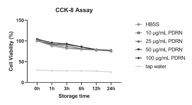
The viability of human PDL cells stored in each medium was evaluated using Cell Counting Kit-8 (CCK-8; Dojindo Molecular Technologies, Kumamoto, Japan). First, PDL cells were seeded at 5 × 10³ cells/well with 200 μL of culture media into a 96-well plate. After incubation for 24 hours, the culture media were removed and the following prepared solutions were treated: (a) HBSS; (b) 10 μg/mL PDRN; (c) 25 μg/mL PDRN; (d) 50 μg/mL PDRN; (e) 100 μg/mL PDRN; and (f) tap water. The cells were stored in each solution for 0, 1, 3, 6, 12, and 24 hours and exposed to room temperature and a normal atmospheric environment to imitate the extraoral condition. Then, 20 μg of CCK-8 solution was added to each plate. After incubation for 2 hours at 37°C, the optical density was measured at 450 nm (A450) using a Benchmark Plus Multiplate Spectrophotometer (Bio-Rad, Hercules, CA, USA) to determine the cell viability. The relative cell viability (%) was calculated as follows: (A450 [treated] - A450 [blank]) ÷ (A450 [control] - A450 [blank]) × 100%. The assays were performed in triplicate, and each experiment involved three samples.
2) Live/Dead assay
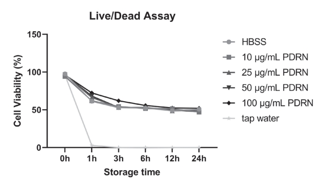
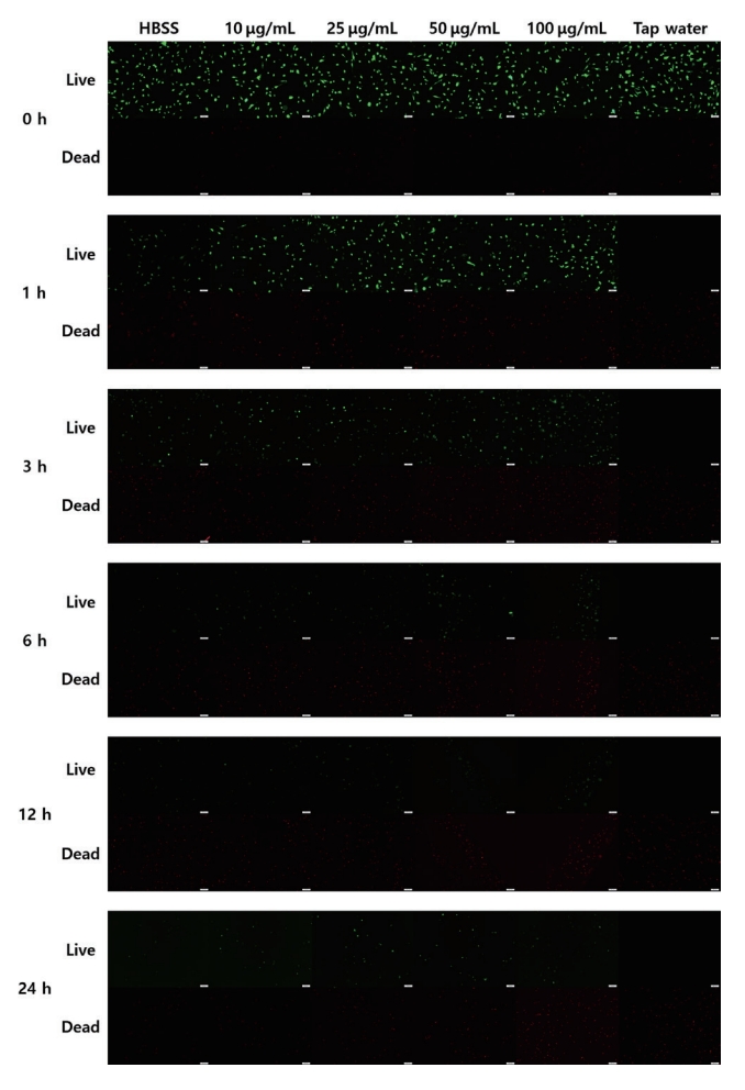
The viability of human PDL cells stored in each medium was evaluated by using LIVE/DEAD Viability/Cytotoxicity Kit for mammalian cells (Invitrogen, Paisley, UK). PDL cells were seeded at 2 × 10⁴ cells/well with 2 mL of culture media into a 12-well plate. After incubation for 24 hours, the culture media were removed, and the prepared solutions (HBSS, 10 μg/mL PDRN, 25 μg/mL PDRN, 50 μg/mL PDRN, 100 μg/mL PDRN, and tap water) were treated for 0, 1, 3, 6, 12, and 24 hours. The dye reagent was prepared: 5 μL of 2 μM calcein AM and 20 μL of 2 mM ethidium homodimer-1 were added to 10 mL of Dulbecco’s phosphate-buffered saline (Gibco BRL, Life Technologies, Grand Island, NY, USA). The resulting solution was vortexed to ensure thorough mixing. The storage media were removed from each plate, and the staining reagent was treated for 10 minutes at room temperature. A fluorescence microphotograph of live and dead PDL cells in each storage medium was obtained three times at different sites using fluorescence microscopy (IX71; Olympus, Tokyo, Japan). The number of PDL cells was counted using the ImageJ software (National Institutes of Health, Bethesda, MD, USA). The relative cell viability (%) was calculated as follows: the number of live cells ÷ (the number of live cells + the number of dead cells) × 100. All assays were done in triplicate.
5. Nitric oxide (NO) assay
First, 2 × 10⁵ PDL cells were seeded into a 6-well plate and incubated at 37°C in a 5% CO2 atmosphere for 24 hours. The culture media were removed from the plates, and each well was treated with 2 mL of HBSS and 50 and 100 μg/mL PDRN for 0, 1, 3, and 6 hours at room temperature and normal atmospheric conditions. After storage each time, the cell supernatants were collected from each well, and the NO assay was conducted using the NO Plus Detection Kit (iNtRON Biotechnology, Inc., Seoul, Korea). The supernatant samples (100 μL each) were added to a 96-well plate, and a pre-reaction was induced by mixing 50 μL of N1 buffer with each sample. After incubation for 10 minutes at room temperature, 50 μL of N2 buffer was treated to each well to conduct the final reaction. The absorbance of each sample was measured at 540 nm using Epoch (Agilent Technologies, Santa Clara, CA, USA). The nitrite concentration was determined using the standard curve of the formula in accordance with the manufacturer’s instructions. All assays were performed five times, and each experiment involved two samples.
6. RNA isolation
First, 2 × 10⁵ PDL cells were seeded into a 6-well plate and incubated at 37°C in a 5% CO2 atmosphere for 24 hours. The culture media were removed from the plates, and wells were treated with 2 mL of HBSS and 50 and 100 μg/mL PDRN for 3 hours at room temperature and normal atmospheric conditions. RNA was extracted from three groups of PDL cells using the AccuPrep Universal RNA Extraction Kit (Bioneer, Daejeon, Korea), according to the manufacturer’s instructions.
7. Quantitative real-time polymerase chain reaction (qRT-PCR)
qRT-PCR was performed to investigate the expression levels of inflammation-related cytokines. Total RNA (5 μg) was converted into complementary DNA (cDNA) using SuperScript II Reverse Transcriptase (Invitrogen, Carlsbad, CA, USA). The Power SYBR Green Master Mix (Thermo Fisher Scientific, Carlsbad, CA, USA) was used to assess SYBR green fluorescence using the QuantStudio 5 Real-Time PCR Ultimate Simplicity System. The cycling conditions were as follows: denaturation for 10 minutes at 95°C, followed by 40 amplification cycles of denaturation for 15 seconds at 95°C, and annealing for 1 minute at 60°C. The cDNA levels were normalized to those of glyceraldehyde 3-phosphate dehydrogenase (GAPDH) using the 2-ΔΔCt method. The primer sequences used for PCR are listed in Table 1. All experiments were performed in triplicate.
8. Statistical analysis
SPSS software (Version 21.0; IBM Corp., Armonk, NY, USA) was used to perform the statistical analysis. Oneway analysis of variance was used to validate statistical significance (p < 0.05), followed by Tukey’ honestly significant difference post hoc test was performed to determine the statistical significance of multiple comparisons. The significance level was adjusted according to the number of groups. Data are expressed as the mean ± the standard deviation value.
Results
1. Cell-preserving effect of PDRN on human PDL cells
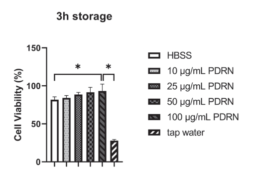
The results of the CCK-8 assay are shown in Fig. 1 and Table 2. At 3 hours of storage, the viability of PDL cells stored in 100 μg/mL PDRN was significantly higher than that of cells stored in HBSS and tap water (p < 0.01, Fig. 2).
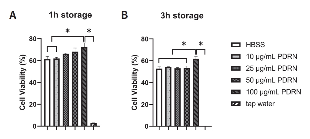
The results of the Live/Dead assay are shown in Fig. 3 and Table 3. At 1 hour of storage, the viability of PDL cells stored in 100 μg/mL PDRN solution was significantly higher than that of cells stored in HBSS, 10 μg/mL PDRN solution, and tap water (p < 0.01, Fig. 4A). At 3 hours of storage, the viability of PDL cells stored in 100 μg/mL PDRN solution was significantly higher than that of cells stored in all other storage media (p < 0.01, Fig. 4B).
Fluorescence microscopic images obtained from the Live/Dead assay are shown in Fig. 5. Although the number of surviving PDL cells stored in HBSS and 10, 25, 50, and 100 μg/mL PDRN solutions decreased with the increase in storage time, the cell viability was maintained for up to 24 hours of storage. However, PDL cells stored in tap water were not viable after 1 hour of storage. The number of viable cells in the 50 and 100 μg/mL PDRN solutions was higher than that in the other solutions until 6 hours.
2. Effect of PDRN on NO production
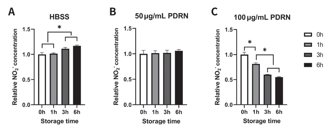
The production of NO in PDL cells stored in HBSS after more than 3 hours of storage was significantly higher than that observed at 0 and 1 hour (p < 0.0001, Fig. 6A). A similar increase was observed in cells stored in 50 μg/mL PDRN solution, statistical significance notwithstanding (Fig. 6B). Alternatively, the NO production in cells stored in 100 μg/mL PDRN solution significantly decreased with the increase in storage time until 3 hours (p < 0.0001, Fig. 6C).
3. Effect of PDRN on the expression of inflammation-related cytokines
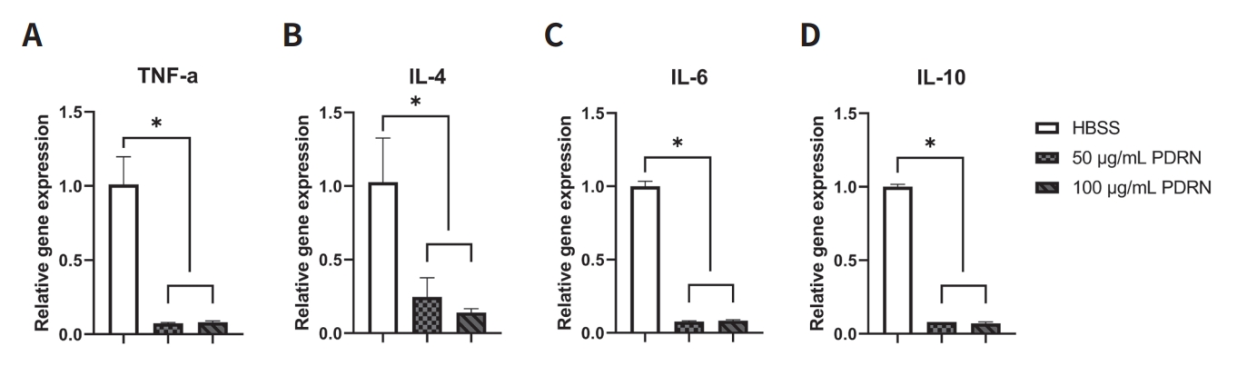
qRT-PCR analysis showed that PDRN significantly reduced the expression levels of inflammation-related cytokines. The expression levels of TNF-α and IL-6 in cells stored in the 50 and 100 μg/mL PDRN solution groups were significantly lower than those in cells stored in the HBSS group (p < 0.01, Fig. 7A, C). No significant differences in the expression levels of TNF-α and IL-6 were seen between cells in the 50 and 100 μg/mL PDRN solutions. The expression levels of IL-4 and IL-10 were decreased in a concentration-dependent manner. IL-4 and IL-10 levels in cells stored in the 50 and 100 μg/mL PDRN solution groups were significantly downregulated compared to those in cells stored in the HBSS (p < 0.01, Fig. 7B, D).
Discussion
An inflammatory reaction is a common complication in the PDL tissues of an avulsed tooth. The inflammatory response of a replanted tooth after delayed replantation is greater than that after immediate replantation. A previous study on inflammatory cytokines and matrix metalloproteinases (MMPs) in replanted teeth found that the expression levels of IL-1β, IL-6, and MMP-1, MMP-2, MMP-7, MMP-8, and MMP-9 were significantly upregulated with the increase in the dry time[4]. Furthermore, another study reported a significant difference in the healing process via TNF-α signaling between teeth that were immediately replanted and those that underwent a delay in replantation[26]. In a retrospective clinical study that evaluated the survival and complications of avulsed and replanted teeth, the probability of tooth loss and inflammatory tooth resorption increased with the increase in the extra-alveolar storage time[27]. It is important to preserve the vital and intact PDL tissues and reduce the inflammatory response in order to prevent inflammatory root resorption[28,29]. Therefore, a storage medium that can ensure the viability of the PDL cells and suppress the inflammatory reaction is required to improve the prognosis of replanted teeth.
PDRN has been reported to enhance tissue regeneration and wound healing through the salvage pathway and via the activation of the adenosine A2A receptor[30,31]. Therefore, it has been widely investigated in the fields of regenerative medicine and biomedical engineering[32]. Additionally, the anti-inflammatory properties and therapeutic effects of PDRN on inflammatory diseases have been studied[33]. Due to these characteristics of PDRN, we thought that it would have a positive effect on the healing of replanted teeth. Thus, PDRN was selected as the subject of subsequent experiments to develop a storage medium for avulsed teeth.
In both the CCK-8 and Live/Dead assays, cells stored in the 100 μg/mL PDRN solution showed the highest cell viability. Especially, in both experiments with 3 hours of storage, 100 μg/mL PDRN showed a significantly higher cell-preserving effect than HBSS. This finding is consistent with those of previous studies. In a study targeting human corneal endothelial cells, PDRN (100 μg/mL) significantly increased the survival rate of cells under oxidative stress[34]. In another in vitro study, PDRN significantly enhanced the proliferation of human skin fibroblasts in a concentration-dependent manner, and the effect was highest at a concentration of 100 μg/mL[35]. Furthermore, 100 μg/mL PDRN in serum-free medium significantly increased the cell viability of RAW 264.7 cells treated with zoledronic acid and LPS[36].
To ascertain whether PDRN has anti-inflammatory effects on PDL cells, a NO detection assay was performed. NO production was significantly increased in PDL cells stored in HBSS after 3 hours of storage. However, the production was significantly decreased in PDL cells stored in 100 μg/mL PDRN solution. These results suggest that PDRN exerts an anti-inflammatory effect on PDL cells by suppressing nitric oxide synthase (NOS). PDRN was reported to inhibit NO production in other human cells[21,31]. An excessive amount of NO can contribute to the destruction of periodontal tissues. Inducible NOS (iNOS) expression and NO production were found to be increased in inflamed periodontal tissues[37-39]. Therefore, PDRN could protect PDL cells from the oxidative damage of NO and reduce the inflammation of periodontal tissues surrounding the replanted tooth.
Additionally, we conducted the qRT-PCR analysis to evaluate the anti-inflammatory effect of PDRN on PDL cells by identifying the expression levels of inflammatory cytokines. PDRN significantly suppressed the expression levels of TNF-α, IL-4, IL-6, and IL-10. TNF-α is a critical pro-inflammatory mediator in the destruction of periodontal tissues. Additionally, it exerts a significant impact by impeding the migration of PDL cells and inhibiting PDL regeneration[40,41]. IL-6 plays crucial roles in various aspects, including the initiation and progression of inflammation and the resorption of alveolar bone[42]. The findings of the current study suggest that PDRN inhibits the inflammatory response by reducing the expression levels of several inflammatory cytokines. In previous studies, PDRN has been reported to have antiinflammatory effects by reducing the inflammatory cytokines such as TNF-α, IL-1β, IL-6, and high mobility group box 1, via adenosine A2A receptor signaling[14,33-35,43]. Adenosine is an important modulator of inflammatory function in periodontal tissues[44]. In particular, activation of the adenosine A2A receptor was reported to inhibit the expression of pro-inflammatory cytokines[45]. PDRN, an adenosine A2A receptor agonist, significantly inhibited the inflammatory cascade and the apoptotic reaction in PDL tissues via adenosine A2A receptor stimulation[46]. Thus, storing the avulsed teeth in storage media containing PDRN could enhance the prognosis of replanted teeth by suppressing the inflammatory response of the PDL cells and periodontal tissues.
Furthermore, PDRN can promote angiogenesis by increasing the expression levels of vascular endothelial growth factor (VEGF)[47-51]. The application of VEGF during the replantation of avulsed teeth increased angiogenesis. Therefore, it reduced external root resorption and ankylosis and enhanced the reattachment of the teeth to the surrounding tissues[52]. In addition, topical treatment with VEGF improved neovascularization in the human dental pulp and could be beneficial for the replantation of avulsed immature teeth[53]. Therefore, the PDRN storage medium may be useful in regenerating the pulp tissues and repairing the periodontal tissues in replanted immature teeth.
However, our study has some limitations. Although PDL cells were stored in an environment to imitate the extraoral condition, the dry conditions before transferring the avulsed tooth to the storage medium were not considered in this study. To comprehensively understand the biological effect of PDRN on replanted teeth, it is necessary to investigate the impacts of PDRN on cell cycle regulation, cell proliferation, and angiogenesis. However, our research only focused on evaluating the cell-preserving and anti-inflammatory properties of PDRN. Furthermore, since the scope of this study is solely focused on the effects at the cellular level, it may be challenging to apply the findings directly to clinical situations.
Additional in vitro experiments are needed to evaluate the wound healing and angiogenesis effects of PDRN on PDL cells. Subsequent investigations involving human and animal studies are warranted to validate the findings of the current study.
 PDF Links
 PubReader
 ePub Link
 Full text via DOI
 Download Citation
 Print
Print


