DIAGNOcam의 임상적 적용
Clinical Application of DIAGNOcam
Article information
Abstract
치아 우식증은 어린이와 청소년에서 일생에 거쳐 지속되는 질병이며 가장 중요한 치과질환이다. 유치열기의 치아우식증은 영구치에도 영향을 미치기 때문에 유치의 치아우식증을 조기에 진단하는 것은 매우 중요하다. 또한 예방을 하는 것이 매우 중요한데 치아 우식증을 조기에 발견하여 우식 위험율이 높은 아이들에서 적극적으로 불소 도포, 치면 열구 전색 등을 미리 해주는 것이 중요하다.
치아우식증을 조기에 발견하기 위한 방법들이 많이 개발되어 왔지만 더 효율적인 방법을 찾기 위해 여전히 많은 연구가 진행 중이다. 일반적으로 시진과 방사선 사진을 많이 사용해왔지만 시진의 부정확성과 방사선의 조사량 등 많은 한계점을 보이고 있다.
이번 연구에서는 치아 우식증에 있어서 새로 개발된 DIAGNOcam의 임상적 적용에 대해 알아보고 그 활용 가능성을 알아보고자 하였다. 소아치과에 내원한 환자들을 대상으로 구치부에 DIAGNOcam을 이용하여 진단한 결과 인접면 우식, 수복물의 변연 확인 및 우식의 범위 확인에 유용하게 사용될 수 있다는 점을 확인하였다.
Trans Abstract
Dental caries is an important dental disease among children and adolescents that can continue for a lifetime. Early detection of dental caries in deciduous dentition is significant because it can influence the permanent teeth. It is also critical to prevent dental caries by performing fluoride treatment and pit-and-fissure sealant for high-risk children.
Various methods have been developed for the early detection of dental caries; however, many studies are still seeking to discover more effective methods. In general, visual examination and radiographic images are used, but these techniques have several limitations such as errors and radiation exposure. In this study, clinical application of the newly developed DIAGNOcam caries identification device and its possible applications were examined. DIAGNOcam was applied to diagnose dental caries in the posterior teeth of patients in the Department of Pediatric Dentistry, and it was confirmed that it could be used to detect proximal caries, the margin of restoration, and the extent of dental caries lesions.
Ⅰ. Introduction
Dental caries is a common dental disease among children and adolescents, and can cause pain and eating disorders [1]. In treating caries lesions, the most important factor is an effective method for early detection. The major advantages of early detection are that treatment plans can be less invasive, treatment can be delayed by managing the incipient caries lesions, and dental tissues can be preserved.
To diagnose dental caries, visual examination, probing, and radiographic images have been used; however, these methods lack specificity and sensitivity, leading to low accuracy in diagnosing dental caries [2]. Thus, there is a continuing need for better methods to detect dental caries. The new methods of caries diagnosis include fiber-optic trans-illumination (FOTI) and digital imaging FOTI (DIFOTI), which use differences in light absorption among carious lesions; quantitative light-induced fluorescence (QLF), which uses laser and visible light rays; and electrical conductance measurements (ECM), which use electric conduction rates of carious lesions [3].
Recently, DIAGNOcam (Kavo, Biberach, Germany), which uses near-infrared rays to confirm dental caries in real time, was developed. This device is similar to DIFOTI, but uses different wavelengths of light to detect initial dental caries. This case report demonstrates various clinical situations in which DIAGNOcam can be used.
Ⅱ. Case Report
Cases where DIAGNOcam can be used to detect dental caries can be summarized into identifying restoration margins, confirming the removal of carious lesions, and characterizing proximal early caries in deciduous teeth.
The instructions for using this device and the program are shown in Figures 1 and 2.
The directions for use of DIAGNOcam.

The program of DIAGNOcam.
1. Identifying restoration margins
To identify the restoration margins, 23 amalgam restoration surfaces were observed with intraoral cameras and the DIAGNOcam, of which 7 restorations had poor marginal adaptation. Restorations that were visually considered to have no microleakage were identified by DIAGNOcam as having microleakage (Fig. 3). At the buccal margins of amalgam restorations, no microleakage was identified, but at the lingual margins of amalgam restorations and buccal margins of resin restorations, obvious microleakage was identified.
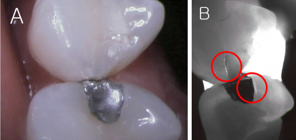
(A) Intraoral camera and (B) DIAGNOcam images of resin and amalgam restoration margins.
2. Confirming the removal of carious lesions
Dental caries was detected with the DIAGNOcam, using the difference in light absorption between sound regions and carious lesions. After observing 16 carious lesions, their boundaries were confirmed by differences in light absorption, which were quite practical in establishing the extent of caries lesions.
Before treatment, DIAGNOcam was used (Fig. 4D). After removing the carious lesions, DIAGNOcam was used again to check the residual carious lesions (Fig. 4E). The last picture shows resin-filled teeth (Fig. 4F).
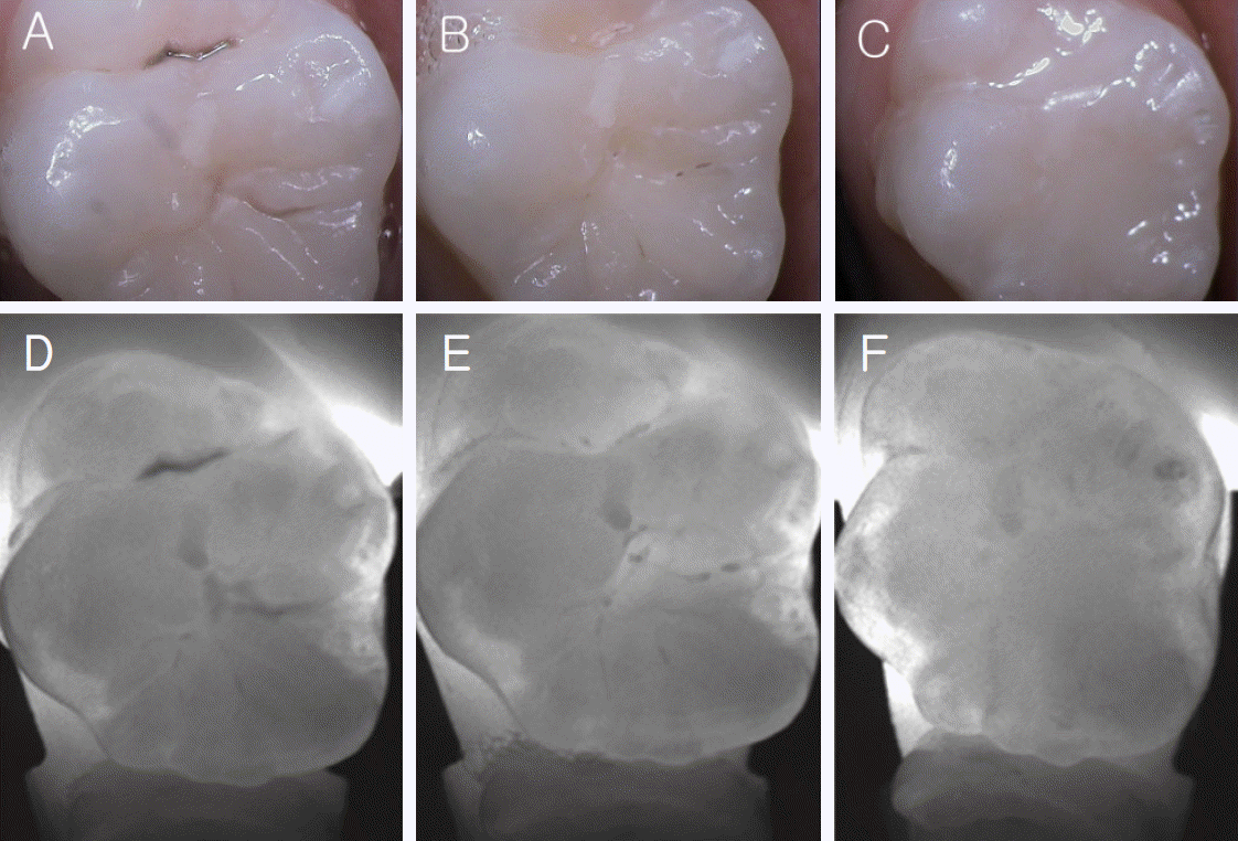
Intraoral camera images of (A) before, (B) during, and (C) after the dental caries treatment of maxillary molars. DIAGNOcam images of (D) before, (E) during, and (F) after the dental caries treatment of maxillary molars.
3. Proximal caries of deciduous teeth
DIAGNOcam confirmed proximal caries that could not be identified by eye. Of the 70 deciduous teeth observed, 12 were confirmed to have proximal caries by DIAGNOcam images (Fig. 5A) that could not be identified by visual inspection (Fig. 5B). This highlights a major advantage of DIAGNOcam, which is that it can be applied to the initial diagnosis of dental caries.

(A) Intraoral camera, (B) DIAGNOcam and (C) radiographic images of proximal dental caries.
Proximal caries that could not be identified by visual inspection could also be confirmed by radiographic imaging (Fig. 5C).
Ⅲ. Conclusion and Discussion
Various methods for early detection of dental caries have been developed, but they have limitations and are not sufficient to accurately diagnose caries lesions [2]. Visual examination and probing are frequently used to observe dental caries, but they lack sensitivity and reproducibility [4]. In addition, probing can cause initial carious lesions to progress, and allow cariogenic microorganisms to migrate to sound regions [5]. Furthermore, the force applied to the probe can cause a larger cavity to form [6].
To avoid these problems, radiographic imaging has been recommended. Nevertheless, there are concerns about radiation exposure, and caries progression of at least half the thickness of the enamel is required for caries detection [7].
Various studies for early detection have been performed on new methods. FOTI uses the difference in light absorption between carious lesions and sound regions, making it practical, easy, and affordable to use [8]. DIFOTI combines FOTI and a digital charge-coupled device camera to reduce the drawbacks of FOTI [1]. Although DIFOTI has lower specificity than radiographic imaging, its sensitivity is higher and it is more capable of detecting dental caries [9].
QLF diagnoses initial caries lesions by laser and visible light rays, and is particularly useful for diagnosing smooth surface caries and the progression of caries lesions [10]. Shi et al. [11] studied QLF and DIAGNOdent and concluded that QLF was more effective for diagnosing the decalcification and remineralization of smooth surfaces. ECM uses the phenomenon that sound enamel has no electrical conductivity, but caries lesions show higher electrical conductivity. With two tips, one is placed on the skin or gingiva, and the other is placed on the teeth to diagnose the extent of dental caries via electrical conductivity. Although this method has high sensitivity, it has low specificity, and thus a low capability of diagnosing initial dental caries. Therefore, ECM is no longer currently used [12].
In this case report, initial dental caries could be observed with DIAGOcam, and a similar accuracy to radiographic imaging was obtained in detecting initial proximal caries [13]. FOTI and DIFOTI use visible light wavelengths, but the new DIAGNOcam uses near-infrared wave-lengths, 0.7-2.0 μm, with less absorption by stains and extensive infiltration into teeth, making it quite practical in diagnosing dental caries [14]. In addition, real-time videos are effective for educating patients and observing the progression of this condition.
When the DIAGNOcam was applied to patients, 7 of 23 restorations showed microleakage, which were impossible to observe by visual inspection, even with clinical pictures (Fig. 3). If timely restoration after applying DIAGNOcam is possible, secondary caries beneath existing restorations could be prevented, and more successful restorations with regular checks after restoration would be possible.
Moreover, DIAGNOcam could be applied to evaluate the extent of caries lesions when removing dental tissues. It is possible to use DIAGNOcam to set the extent of removal by the difference in light absorption between a sound region and caries lesions. By observing 16 caries lesions, complete removal was performed by relying on the dark color of the caries lesions. Before treatment, appropriate caries lesions were observed with DIAGNOcam, which was used again after removal. Through this procedure, excessive removal of teeth was prevented by accurately diagnosing the extent of dental caries, and secondary caries was prevented by not leaving residual caries lesions. However, in the case of existing residual carious lesions, continuous DIAGNOcam taking was required and the process became more complex.
Finally, the DIAGNOcam could be applied to detect proximal dental caries that could not be seen by visual inspection when examining patients who did not have a chief complaint of dental caries. During dental examination of infants and children, DIAGNOcam detected proximal dental caries with no radiation exposure. Upon observing 70 deciduous teeth, 12 had proximal dental caries, which was confirmed by radiographic imaging. With early detection possible, progression of dental caries and excessive removal of teeth can be prevented.
DIAGNOcam does have some limitations. First, it cannot be applied to the anterior teeth, because only tips for posterior teeth exist. Second, it is impossible to use with rubber dam clamps, because DIAGNOcam covers the whole tooth. In addition, it may be less sensitive than DIAGNOdent, which shows fairly different scores according to the condition of the tooth surface, but DIAGNOcam also needs a clean tooth surface to accurately diagnose dental caries [15].
Ⅳ. Summary
DIAGNOcam can be used to confirm proximal dental caries, margins of restorations, and the extent of dental caries. Although it is not capable of completely replacing radiographic imaging, it is practical as a supplement.
Moreover, compared to other methods, DIAGNOcam is effective, convenient, and can be used in real time. However, more studies are needed before DIAGNOcam can replace other methods.