구개확장장치와 facemask를 이용한 교정치료 환자의 구개주름 평가
Evaluation of Palatal Rugae Following Orthopedic Treatment Using Rapid Maxillary Expander and Facemask
Article information

Abstract
이 연구는 혼합치열기 3급 부정교합 환자에서 RME와 facemask치료 전후에 구개 및 구개주름의 횡적 및 전후방적 변화를 평가하고, 구개주름이 교정 전후의 모델 중첩을 위한 적절한 기준 영역으로 사용될 수 있는지를 확인하는 것을 목적으로 수행되었다.
RME와 facemask를 이용한 교정치료를 받은 52명의 혼합치열기 3급 부정교합 환자의 교정치료 전, 후 상악 진단모형을 이용하여 치료 전(T1), 후(T2)의 구개 및 구개주름을 계측하였다.
RME와 facemask 치료로 인해 상악골과 구개에 나타나는 골성 변화에도 불구하고 구개와 구개주름의 전후방적 변화는 유의하지 않았다. 구개와 구개주름의 형태학적 변화는 주로 횡적 변화로 나타났다. 구개주름은 하방의 경조직의 해부학적 변화에 적응하여 늘어나는 것으로 보인다. 계측된 구개주름의 영역 중 3번째 구개주름의 내측점이 가장 적은 변화를 나타내었다.
이 연구에서 관찰된 구개주름의 변화는 3번째 구개주름의 내측점이 RME와 facemask 치료를 받은 환자의 치아 이동을 연구하기 위한 목적으로 교정 전후 모델을 중첩할 때 안정적인 기준 영역으로 사용될 수 있는 가능성을 보여준다.
Trans Abstract
The purpose of this study was to determine whether the palatal rugae could be used as an appropriate reference area for serial model superimposition following Rapid maxillary expansion(RME) and facemask treatment.
A total of 52 pediatric patients who had undergone RME and facemask treatment were selected. Palate and palatal rugae in the pre- and post- treatment casts from the patients were measured.
In spite of dentoalveolar changes occurred by RME and facemask, anteroposterior changes in palate and palatal rugae were not significant. Anatomical changes of palate and palatal rugae were mostly shown in the transverse dimension. The soft tissue of the palatal rugae stretches in adaptation to hard tissue movement. Among the evaluated landmarks, the medial point of the third palatal rugae seemed to be the most stable.
The observed alterations in the palatal rugae demonstrated the potential of medial points of third palatal rugae as a reference point in model superimpositions to evaluate dental movement within the maxillary arch following RME and facemask treatment.
Ⅰ. 서 론
교정 치료의 결과를 평가하기 위해 사용되는 측모 두부 방사선 계측 중첩에는 몇 가지 단점과 한계점이 존재한다. 환자가 방사선에 노출되어야 하며, 술자가 방사선 사진 상에서 해부학적 랜드마크를 정확하게 판단하기 어렵고, 환자의 자세에 따라 방사선 사진의 왜곡이 존재할 수 있다. 반면에 모델 중첩 방법은 환자가 방사선에 노출되는 것을 최소화 할 수 있으며 그 과정이 더 단순하다[1]. 그러나 모델 중첩을 통해 치아의 정확한 움직임을 비교하기 위해서는 기준으로 사용할 수 있는 안정적인 연조직 구조물이 필요하다[2].
구개주름은 구개 점막 전방부에 있으며 절치공 후방에 위치하는 불규칙한 굴곡들이다[3]. 구개주름은 개인마다 그 형태가 다르며 일생동안 변화가 아주 적으므로 법치의학 분야에서 지문처럼 개인 식별을 위한 도구로 사용되어왔다[4]. 이러한 특성에 기반하여 구개주름이 교정치료 전 후 모델 중첩 시 기준 영역으로 사용하기에 적당한지를 알아보기 위한 연구들이 진행되어왔다[5,6].
급속구개확장장치(RME)와 facemask를 이용한 교정 치료가 상악골 복합체와 치열에 미치는 영향은 잘 연구되어 있다. RME와 facemask를 이용한 교정치료는 상악골 복합체의 전방 성장을 일으키므로 3급 부정교합의 교정치료에 널리 사용된다[7-9]. 급속구개확장을 통한 구개봉합의 개방과 분리는 대부분 횡적 측면에서의 상악과 구개의 형태학적 리모델링을 초래한다[10-14].
일부 연구자들은 치열궁에서 RME와 facemask를 이용한 치료 효과를 확인하기 위해 모델 중첩을 사용하였으며 이때 구개주름을 기준영역으로 사용하였다[15,16]. 그러나 RME와 facemask 치료 전후 구개주름의 안정성에 대한 기존 연구는 거의 없다. 따라서 RME와 facemask를 이용한 교정치료에 따른 치열궁의 변화에도 불구하고 안정적인 구개주름 영역이 있는지를 조사하는 것은 임상적으로 의미가 있다.
이 연구는 소아 환자에서 RME와 facemask 치료 전후에 구개 및 구개주름의 횡적 및 전후방적 변화를 평가하고, 구개주름 부위 중 안정적인 영역을 확인하기 위해 수행되었다.
Ⅱ. 연구 대상 및 방법
1. 연구 대상
RME와 facemask를 이용한 교정치료를 받은 혼합치열기 3급 부정교합 환자52명을 연구대상으로 하였다(남성 21명; 여성 31명, 나이 8.11 ± 1.02세). 이 환자들의 교정치료 전과 후에 채득한 진단모형이 연구에 사용되었다. RME 확장 기간은 7일에서 14일 이었으며 평균 확장량은 2.44 ± 0.66 mm 였다. RME와 facemask를 이용한 교정 기간은 평균 1.01 ± 0.05년이었다. 전신질환이 있는 환자, RME와 facemask 이외에 다른 교정 장치로 치료받은 환자, 진단모형 상에서 구개주름을 명확하게 구별하기 어려운 환자는 연구 대상에서 제외되었다(Table 1). 이 연구는 전남대학교 치과병원 임상 연구 윤리 위원회 (Institutional Review Board, IRB)의 승인을 받아 시행되었다(IRB NO.: CNUDH-2019-011).

Inclusion criteria for the subjects
2. 연구 방법
1) 구개의 횡적 및 전후방적 계측
구개의 횡적 변화를 측정하기 위해 양측 유견치, 제1유구치, 제1대구치 간의 거리를 치아 치은 경계부의 중간점에서 각각 측정하였다(PW-C; PW-P; PW-M). 구개의 전후방적 변화를 측정하기 위해 절치 유두의 최전방점에서부터 제1대구치 최후방을 연결한 점까지의 측정하였다(PL)(Table 2, Fig. 1).

Definition of measurements

Palatal measurements. PW-C = Distances between right and left primary canines, PW-P = Distances between right and left first primary molars, PW-M = Distances between right and left first permanent molars, PL= Palatal length.
2) 구개주름의 횡적 및 전후방적 계측
구개주름을 Lysell[3]의 정의를 이용하여 5 mm 이상의 일차 주름을 평가 대상으로 선정하여 연필로 표시하였다. 구개주름은 전방에서부터 후방으로 갈수록 각각 1, 2, 3으로 번호를 매겼고 오른쪽과 왼쪽은 R, L로 표시하였다. 좌우측 구개주름 각각의 길이를 측정하였다(R1, R2, R3, L1, L2, L3). 구개주름의 가장 내측점(m)과 가장 외측점(l)을 표시하였다(Table 2, Fig. 2). 구개주름의 내측점 간의 횡적 거리(Tm1, Tm2, Tm3) 및 외측점 간의 횡적 거리(Tl1, Tl3, Tl3)를 측정하였다(Table 2, Fig. 3).

Rugae landmarks. m = medial, l = lateral, R = right, L= left.

Transverse palatal rugae measurements. R1, R2, R3, L1, L2, L3 = Lengths of 1st, 2nd and 3rd rugae on the right(R) and left(L) from medial to lateral points. Tm1, Tm2, Tm3 = Transverse distances between opposing medial rugae points. Tl1, Tl2, Tl3 = Transverse distances between opposing lateral rugae points.
구개주름의 전후방적 변화를 평가하기 위해 오른쪽 구개주름의 내측점 사이의 전후방 거리(APmR-12, APmR-23), 왼쪽 구개주름의 내측점 사이의 전후방 거리(APmL-12, APmL-23), 오른쪽 구개주름의 외측점 사이의 전후방 거리(APlR-12, APlR-23), 왼쪽 구개주름의 외측점 사이의 전후방 거리(APlL-12, APlL-23)를 측정하였다(Table 2, Fig. 4).
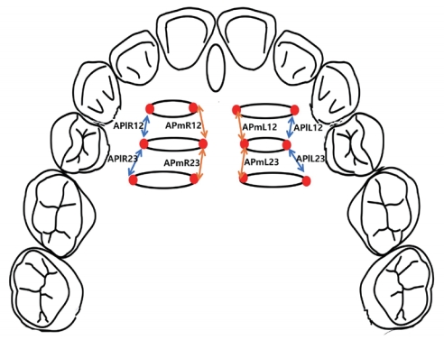
Anteroposterior palatal rugae measurements. APmR12; APmR-23 = Anteroposterior distances between medial points of (1st and 2nd) and (2nd and 3rd) right-side rugae, APmL-12; APmL-23 = Anteroposterior distances between medial points of (1st and 2nd) and (2nd and 3rd) leftside rugae, APlR-12; APlR-23 = Anteroposterior distances between lateral points of (1st and 2nd) and (2nd and 3rd) right-side rugae, APlL-12; APlL-23 = Anteroposterior distances between lateral points of (1st and 2nd) and (2nd and 3rd) left-side rugae.
3. 표준 일관성 평가
조사자 내 신뢰도를 평가하기 위해 초기 평가 3주 후에 무작위로 선택된 치료 전 및 치료 후 모형 10개에서 모든 측정을 반복하여 실시하였다. 조사자 내 신뢰도인 Intraclass Correlation Coefficient(ICC) 값을 구하였을 때 모든 항목에서 0.80 이상으로 나타났다.
4. 통계학적 분석
RME와 facemask를 이용한 교정치료 전후의 구개 및 구개주름의 변화를 비교하기 위해 대응 표본 t-검정(paired t-test)을 사용하여 분석하였다. 또한 구개의 변화와 구개주름의 변화 간의 상관관계를 파악하기 위해 피어슨 상관계수(Pearson's productmoment correlation)를 사용하여 분석하였다. 프로그램은 SPSS 22.0(Statistical Package for Social Sciences, IBM Corp., USA)을 이용하였다.
Ⅲ. 연구 성적
1. 구개의 횡적 및 전후방적 계측치
2.44 ± 0.66 mm의 구개 확장과 1.01 ± 0.05 년의 확장 유지 및 facemask 치료 후 구개 폭경은 유견치, 제1유구치, 제1대구치 각각의 위치에서 각각 평균 1.68 ± 2.64 mm (p < 0.001), 1.93 ± 2.51 mm (p < 0.001), 1.20 ± 3.75 mm (p= 0.025) 증가하였으며, 이러한 증가는 모두 통계적으로 유의하였다(Table 3). 그러나 구개 길이의 증가(0.78 ± 4.03 mm)는 유의하지 않았다(Table 3, p= 0.171).

Changes in palate following rapid maxillary expansion and facemask (n = 52)
2. 구개주름의 횡적 및 전후방적 계측치
구개주름의 횡적 길이는 모든 구개주름에서 증가하는 양상이 나타났으며 이 중 R1(0.37 ± 1.32 mm, p= 0.026), R2(0.49 ± 1.59 mm, p= 0.029), L2(0.55 ± 1.77 mm, p= 0.029)의 길이 증가는 통계적 유의성이 있는 것으로 나타났다. 구개주름의 내측점 간 거리는 1번째 구개주름(Tm1: 0.34 ± 1.32 mm, p= 0.021)과 2번째 구개주름(Tm2: 0.96 ± 2.09 mm, p= 0.002)에서 유의한 증가를 나타내었다. 그러나 3번째 구개주름의 내측점 간 거리(Tm3: 0.008 ± 2.87 mm, p= 0.849)에서는 유의한 횡적 변화가 나타나지 않았다. 구개주름의 외측점 간 거리는 Tl1(0.76 ± 2.15 mm, p= 0.013), Tl2(1.23 ± 2.18 mm, p < 0.001), Tl3(1.28 ± 2.56 mm, p= 0.001) 모두에서 유의한 증가를 나타냈다(Table 4). 구개주름의 전후방적 길이 변화는 모두 유의하지 않은 것으로 나타났다(Table 5).

Changes in transverse palatal rugae measurements following rapid maxillary expansion and facemask (n = 52)
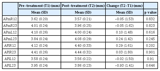
Changes in anteroposterior palatal rugae measurements following rapid maxillary expansion and facemask (n = 52)
3. 구개의 변화와 구개주름의 변화간의 상관관계
구개 폭경의 변화와 구개주름의 횡적 변화 간의 상관관계에서 유견치간 폭경(PW-C)은 Tl1(r = 0.609, p < 0.001), Tl2(r = 0.341, p= 0.014), Tl3(r = 0.279, p= 0.045)와 유의한 양의 상관관계를 나타냈다. 제1유구치 간 폭경(PW-P)의 변화도 Tl1(r = 0.573, p < 0.001), Tl2(r = 0.467, p < 0.001), Tl3(r = 0.343, p= 0.013)와 유의한 양의 상관관계를 나타냈으며, 제1대구치 폭경(PW-M)의 변화는 유의미한 상관관계를 나타내지 않았다. 또한 구개주름의 내측점 간 거리는 구개 폭경의 변화와 유의미한 상관관계를 나타내지 않았다(Table 6).

Correlations among changes in transverse dimensions of palate and palatal rugae (n = 52)
Ⅳ. 총괄 및 고찰
교정 모델은 환자의 교정적 평가를 위한 필수적인 진단 기록이다. 교정 모델을 이용하여 교정 치료 전후 악골 및 치아의 변화를 비교하기 위한 기준 영역을 찾기 위해 다양한 연구가 진행되었다. 많은 학자들이 이전의 연구를 통해 구개주름의 안정성을 보고하였으며 구개주름을 기준영역으로 사용할 수 있다고 주장하였다[5,6].
특히 3번째 구개주름의 안정성이 주목받았다[17-20]. 초기의 연구들은 발치 및 비발치 교정치료 환자에서 3번째 구개주름의 안정성을 주장하였다[17-19]. 일부 연구자들은 교합평면 뿐 아니라 수직평면 상에 있어서도 3번째 구개주름이 교정치료 전후에 안정적이었음을 증명하였다[20].
RME와 facemask를 이용한 교정치료는 상악골의 전방성장을 촉진하므로 3급 부정교합의 조기 교정치료에 널리 사용되어 왔다[21,22]. RME는 상악골의 2개의 구개 돌기를 측방으로 이동시키고 정중 구개 봉합부를 넓혀준다[23,24]. 따라서 RME와 facemask 치료 후 구개와 상악골의 횡적 확장이 일어난다. 이 연구에서도 RME와 facemask 치료 후 구개의 횡적 확장이 확인되었다. 유견치, 제1유구치 및 제1대구치 부위에서의 확장량은 각각 1.68 ± 2.64 mm, 1.93 ± 2.51 mm, 1.20 ± 3.75 mm로 나타났다. RME에 의한 구개 확장을 연구한 최근의 메타분석에서 계산한 대구치부의 확장 평균보다 확장이 적게 나타났는데, 이는 이 연구 대상들에서는 심한 악궁 협착이 없어 RME 확장 횟수가 적었기 때문으로 보인다[11]. 또한 이 연구에서 구개 전방부의 확장이 더욱 두드러지게 나타났는데, 이는 구개 부위가 이 상악골의 앞부분에 비해 상악의 뒷부분에 있는 지지골로부터 더 큰 저항을 받으므로 전방부가 후방부보다 넓은 전형적인 V자 모양의 구개 봉합 개방이 생기기 때문인 것으로 보인다[23,24].
악궁 길이의 증가는 0.78 ± 4.03 mm로 통계적으로 유의하지 않았다. 이는 RME와 facemask를 이용한 치료 후 악궁 길이의 유의미한 증가가 없었다고 보고한 이전 연구의 결과와 일치하였다[25].
이 연구 결과에서 구개주름 자체의 길이 증가는 R1(p= 0.026), R2(p= 0.029), L2(p= 0.029)에서 유의미하게 나타났다. 이는 구개의 확장이 구개 전방부에서 두드러지는 결과에 대응된다. 따라서 구개주름은 구개주름에 덮혀있는 경조직의 변화에 대응하여 변화하는 것으로 보인다[26].
이 연구에서 1, 2, 3번째 구개주름의 내측점 및 외측점 간 거리 중 3번째 구개주름의 내측점간 거리(Tm3)를 제외한 모든 수치에서 통계적으로 유의미한 증가가 있었다. 이는 교정치료 전후의 3번째 구개주름의 안정성을 보고한 이전의 연구들의 결과와 일치하였다[17-20].
구개주름의 내측점의 변화는 해당 부위의 외측점의 변화보다 작게 나타났다. 이는 RME를 사용한 교정치료에서 구개주름의 내측점의 변화가 외측점의 변화보다 작게 나타난다고 한 이전 연구의 결과와 일치하였다[26]. 또한 2급 부정교합 환자의 조기 교정치료 후의 구개주름의 변화를 관찰한 연구에서도 구개주름의 외측점이 내측점에 비해 구개 확장의 영향을 더 많이 받는 것으로 보고되었다[27]. 이들은 구개주름이 치아에 더 가까울수록 치아 이동의 영향을 더 많이 받으며, 따라서 구개주름의 외측점이 내측점보다 구개 확장의 영향을 더 많이 받는다고 하였다[27].
Damstra 등[2]이 시행한 RME를 이용한 교정 치료 전후의 구개주름의 변화에 대한 연구에서 이 연구와 다르게 1번째와 2번째 구개주름의 내측점간 거리가 3번째 구개주름의 내측점 간 거리보다 증가량이 크게 나타났다. 이러한 결과의 차이는 확장 후 유지기간의 차이에 기인하는 것으로 사료된다. 이전 연구에서는 확장 직후 측정이 이루어진 반면, 이 연구에서는 확장 후 약 1년의 유지기간을 거쳤으므로 구개 점막골막(palatal mucoperiosteum)의 섬유성 결합조직의 적응이 일어났을 것으로 사료된다[28,29].
RME와 함께 사용한 facemask의 상악골 전방견인 효과에도 불구하고 구개 및 구개주름은 모든 위치에서 전후방적으로는 유의미한 변화를 나타내지 않았다. 이는 혼합치열기 소아 환자에서 facemask가 악궁의 길이 변화보다는 상악골의 전방성장을 유발하였기 때문으로 보인다[21,22]. 또한 facemask에 의한 상악 전치의 전방경사 유발 효과는 RME 확장 의한 상악 전치 후방경사 효과에 의해 상쇄되었기 때문일 것으로 사료된다[30,31].
이 연구에서 유견치와 유구치부에서 구개부의 횡적확장과 구개주름의 외측점의 거리 변화는 양의 상관관계를 나타냈으나, 구치부에서는 유의한 상관관계를 나타내지 않았다. 하지만, Saadeh 등[26]이 시행한 이전의 연구에서는 견치, 소구치, 구치부 모두에서 구개의 횡적확장과 구개주름의 외측점의 거리 변화가 유의한 상관관계를 보였으며, 이때의 RME 확장량은 평균 3.6 ± 1.17 mm로 이 연구에서의 2.44 ± 0.66 mm보다 크게 나타났다. 따라서 두 연구 간의 RME 확장량 차이에 의해 구치부에서의 상관관계가 다르게 나타났을 것으로 사료된다.
이 연구에는 몇 가지 한계점이 존재하므로 연구 결과에 대한 주의 깊은 해석이 필요하다. 첫 번째는 대조군이 없다는 점이다. 치료되지 않은 성장기 아동에서의 종단 연구에 따르면 해당 연령의 연간 구개주름의 변화는 크지 않을 것으로 예상되지만[32-34], 추후 연구에서 RME와 facemask로 치료하지 않은 대조군과 비교한 추가적인 연구가 필요할 것으로 사료된다. 두 번째는 3차원적인 해부학적 구조물을 2차원적인 계측방법을 이용해 측정하였다는 점이다. 추후 수직적 변화 및 구개의 부피, 구개주름의 각도 등 3차원적 계측이 필요할 것으로 사료된다.
Ⅴ. 결 론
이 연구는 3급 부정교합을 가진 소아 환자에서 RME와 facemask 치료에 따른 구개 및 구개주름의 변화를 평가하여 구개주름이 모델 중첩을 위한 적절한 기준 영역으로 사용될 수 있는지를 평가하기 위해 수행되었다. 연구 결과 RME와 facemask 치료로 인한 경조직의 변화에 따라 구개주름의 형태적 적응이 나타났으며, 이러한 구개주름의 변화는 대칭적이지 않았다. RME와 facemask 치료에 따른 구개주름의 변화는 주로 횡적 변화로 나타났으며 전후방적 변화는 유의하지 않았다.
이 연구에서 구개주름의 내측점 및 외측점 간 거리 중 3번째 구개주름의 내측점간 거리(Tm3)를 제외한 모든 수치에서 통계적으로 유의미한 증가가 있었다. 따라서 3번째 구개주름의 내측점이 RME와 facemask 치료를 받은 환자의 교정 전후 모델을 중첩 시 안정적인 기준 영역으로 사용될 수 있는 가능성이 있다.
Acknowledgements
이 논문은 전남대학교 학술연구비 (과제번호 2016-2763) 지원에 의하여 연구되었음.