치수 괴사된 미성숙 영구치에서 Retro MTA를 이용한 변색 없는 재생적 근관치료: 증례 보고
Regenerative Endodontic Treatment Without Discoloration of Infected Immature Permanent Teeth Using Retro MTA : Two Case Reports
Article information
Abstract
재생적 근관치료는 치수 괴사를 보이는 미성숙 영구치 치료에 있어서 괴사된 치수를 치유하고 계속되는 치근의 발달을 유도할 수 있다. 그러나 여기에는 몇 가지 단점과 불리한 결과들이 있다. 이 중 재생적 근관치료에 가장 큰 임상적 부작용은 minocycline과 MTA에 의한 변색이다.
본 증례에서는 재생적 근관치료 후에 나타나는 치관 변색을 방지하기 위해 minocycline을 clindamycin으로 대체한 triple antibiotics와 칼슘 지르코늄 알루민산염 시멘트인 Retro MTA를 사용하였다. 치수 괴사된 미성숙 영구치에서 근관 와동 형성 및 근관 세척 후 수정된 triple antibiotics를 적용하고, 혈병 또는 콜라겐 스펀지를 스캐폴드(scaffold)로 하여 Retro MTA로 근관을 폐쇄하였다. 정기적인 검진 결과 미성숙 영구치의 치근 성장 및 정상적인 치아 주위 조직들이 관찰되었으며, 치관 변색 없이 모두 양호한 치유 결과를 얻었다.
Trans Abstract
Regenerative endodontic treatment has the potential to heal a necrotic pulp, which can affect root development in immature teeth. However, several drawbacks and unfavorable outcomes are associated with regenerative endodontic treatment, of which the most significant is coronal discoloration due to the presence of minocycline in triple antibiotic paste and mineral trioxide aggregate (MTA).
To prevent tooth discoloration following pulp treatment, the modified triple antibiotics (ciprofloxacin, metronidazole, clindamycin) were used as canal disinfectants and Retro MTA, a ZrO2-containing calcium aluminate cement, was used to seal the canal. Following access cavity acquisition, the canal was copiously irrigated with 2.5% sodium hypochlorite. A modified triple antibiotic paste was then applied to the canal. Once the tooth was asymptomatic (after between 3 and 8 weeks), Retro MTA was carefully placed over the blood clot or a collagen plug. Follow-up radiographs revealed normal periodontal ligament space and root development. In two cases, successful regenerative endodontic treatment of the infected immature tooth, without discoloration, was achieved with disinfection using modified triple antibiotics and Retro MTA sealing.
Ⅰ. Introduction
In endodontics, treatment of necrotic immature teeth is challenging. Weakness, shortness, and susceptibility to fracture are typical characteristics of immature roots. Performance of chemomechanical debridement, and the creation of an effective apical seal using conventional endodontic treatment methods, is problematic for most clinicians. Apexification, using traditional and contemporary methods, allows for the management of immature teeth with necrotic pulps. However, a major drawback is that, in cases of root fracture, non-restorability eventually leads to the loss of these teeth [1,2].
Regenerative endodontic procedures have recently been advocated in the treatment of necrotic immature teeth. Here, the root canal system is thoroughly disinfected, following which bleeding from the apical papilla is stimulated to fill the root chamber with a blood clot [2]. A host of growth factors in this area then act on dental stem cells, primarily from the apical papilla, using the clot as a scaffold and differentiating into healthy cells that can reach physiologic root maturation [3]. There are a number of cases of successful clinical and radiographic outcomes using this treatment [1,2,4,5]. However, several shortcomings and unfavorable outcomes should also be considered [6,7], as follows: (1) coronal discoloration; (2) insufficient bleeding; and (3) collapse of mineral trioxide aggregate (MTA) material into the canal.
Coronal discoloration is a particularly important aesthetic concern. Kim et al. [8] demonstrated that tooth discoloration following regenerative endodontic treatment is problematic due mostly to the presence of minocycline in the triple antibiotic paste: the main cause of staining following treatment was contact between minocycline with coronal dentinal walls. An effective method of preventing discoloration involves replacing minocycline with a non-staining antibiotic.
Although minocycline is the major cause of discoloration following regenerative endodontic treatment, several studies have demonstrated that gray MTA [5,9] and white MTA [6,7,10] can also lead to discoloration. The iron and manganese contained within GMTA and WMTA are potentially responsible for coronal discoloration [9]. Retro MTA® (BioMTA, Korea) is a recently produced ZrO2-containing calcium aluminate cement that uses hydraulic calcium zirconia complex as its contrast media and no heavy metals. According to the manufacturer, Retro MTA does not cause discoloration even in instances of blood contamination [11].
This case report describes regenerative endodontic treatment, without coronal discoloration of immature permanent teeth with apical inflammation, using a modified triple antibiotics paste (ciprofloxacin, metronidazole, clindamycin) as a canal disinfectant, and Retro MTA, produced by hydration of zircornia complex, to seal the canal.
Ⅱ. Case Reports
1. Case 1
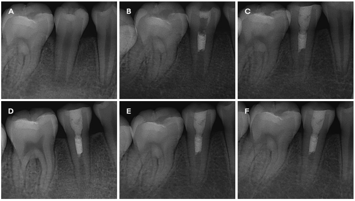
A 10-year-old boy presented to the Department of Pediatric Dentistry, Chonnam National University Dental Hospital with pain in the mandibular right second premolar upon chewing. In clinical examinations, a fractured tubercle, in a dens evaginatus on the occlusal surface, was observed. The tooth was tender to percussion and palpation, with mild mobility. A periapical radiograph revealed incomplete tooth development and periradicular radiolucency (Fig. 1A). The tooth was diagnosed with pulp necrosis and acute apical periodontitis.

Periapical views of case 1. (A) Preoperative radiograph showing an open apex, periradicular radiolucency and dens evaginatus of the mandibular right second premolar. (B) The canal was sealed with MTA. (C) A radiograph at the 1-month follow-up showing root lengthening and narrowing of the canal space. (D, E, F) Follow-up radiographs showing root development at 4, 8 and 11 months, respectively. Periapical radiographs revealed root wall thickening and root lengthening.
Under rubber dam isolation, without local anesthesia, an access cavity was prepared for the mandibular right second premolar. Root canal length was estimated using an endodontic #15 K-file. A 20-gauge needle was situated within 3 mm of the apex, and the canal was copiously and gently irrigated using 10 mL of 2.5% sodium hypochlorite and saline without instrumentation. The canal was then dried with paper points. A triple antibiotic mix, of 250 mg ciprofloxacin, 250 mg metronidazole, and 150 mg clindamycin [12], with sterile saline as a vehicle, was introduced into the root canal below the CEJ as a creamy paste using a Centrix syringe. Hoshino et al. [13] originally recommended the use of minocycline, but, due to its tendency to stain teeth, clindamycin was substituted instead. The access cavity was temporarily sealed using a cotton pellet and Caviton (GC Co, Tokyo, Japan).
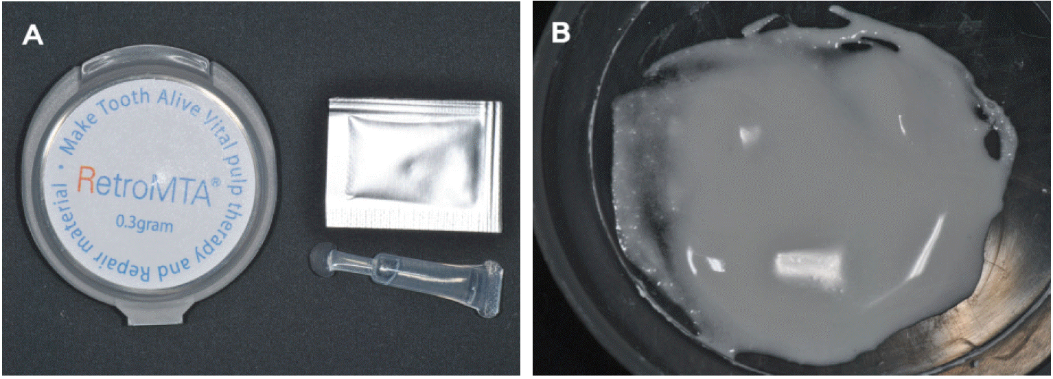
After 3 weeks, the tooth was asymptomatic, with the patient reporting no postoperative pain. Anesthesia, with 3% mepivacaine (Septodont, Cedex, France) without a vasoconstrictor, was given. Following isolation with a rubber dam, the access cavity was reopened, and the canal was irrigated twice. The canal was then dried using paper points. An endodontic #20 K-file was introduced into the canal up to the apex; the apical vital tissue was irritated by gentle scraping, precipitating bleeding into the canal (Fig. 2). A cotton pellet was subsequently placed into the canal, 3 mm below the CEJ, and remained in place for 15 min to ensure clot formation. The presence of the blood clot was confirmed visually. Approximately 3-mm thickness of Retro MTA® (BioMTA, Korea) was then carefully placed over the blood clot, with a moist cotton pellet over the MTA. The tooth was then temporized with Caviton (Fig. 1B, 3A, 3B). The patient was assessed after 1 week; the Caviton was replaced with composite resin restoration and further recall visits were scheduled.
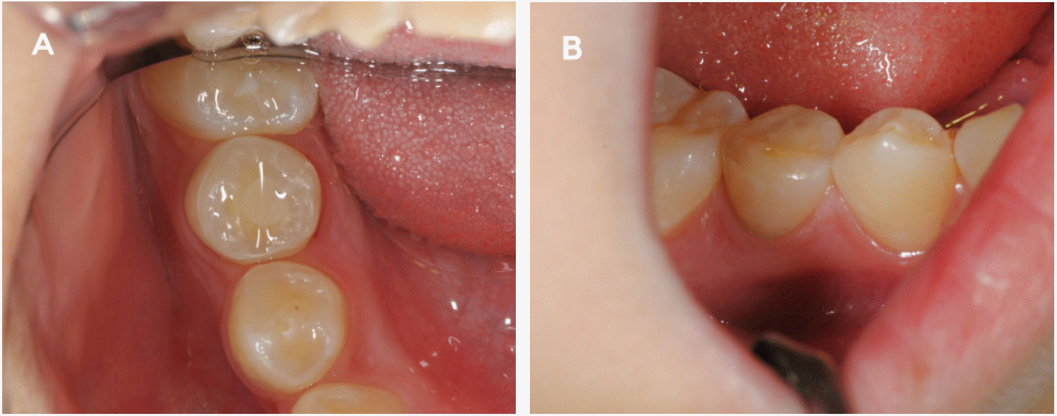
The patient was recalled at 1, 4, 8 and 11 months following treatment. In clinical examinations, the tooth was asymptomatic. In radiographic examinations, the tooth exhibited increased root length and root wall thickness (Fig. 1C-F). Moreover, at 11 months, the tooth displayed no discoloration (Fig. 4).
2. Case 2
A 7.11-year-old boy was referred to the Department of Pediatric Dentistry, Chonnam National University Dental Hospital with an uncomplicated crown fracture in the maxillary right central incisor due to trauma (Fig. 5A, 6A). His maxillary right central incisor was treated by reattaching a fractured tooth fragment using composite resin (Fig 5B). At 2 months, the patient returned due to pain and localized swelling in the anterior region of the maxilla. On clinical examination, swelling and fistula formation in the maxillary right central incisor were observed. A periapical radiograph revealed a radiolucent lesion on the open apex of the maxillary right central incisor (Fig. 6B). The tooth was diagnosed with an infected necrotic pulp and acute apical abscess.

Intraoral views of case 2. (A) Initial examination; maxillary right central incisor with an uncomplicated crown fracture, and (B) view following reattachment of a fractured tooth fragment. The fracture was very close to the pulp, but not involving the pulp.

Periapical views of case 2. (A) Initial visit; maxillary right central incisor with an open apex and uncomplicated crown fracture. (B) At 2 months post-trauma; access cavity opening was prepared with a diagnosis of pulp necrosis. (C) The canal was sealed with MTA over a Teruplug. (D, E, F) Followup radiographs showing root development at 1, 4 and 8 months, respectively. Lengthening of roots, with normal periodontal ligament space, was observed.
Under isolation with a rubber dam, an access cavity was prepared. Root canal length was estimated using an endodontic #15 K-file. The canal was gently irrigated using 10 mL of 2.5% sodium hypochlorite and saline. The canal was then dried with paper points, and a triple antibiotic paste (ciprofloxacin, metronidazole, clindamycin) was introduced into the root canal below the CEJ using a Centrix syringe. The access cavity was sealed using a cotton pellet and Caviton.
After 5 weeks, although the fistula had disappeared, the tooth was tender to percussion and palpation. The access cavity was reopened and the canal was irrigated. The triple antibiotic paste was reapplied to the root canal. The next appointment was scheduled 3 weeks subsequent, at which time the patient reported no pain. Anesthesia, with 3% mepivacaine without epinephrine, was given and the canal was copiously irrigated. An endodontic K-file was introduced into the canal up to the apex to induce bleeding. However, insufficient bleeding into the canal was observed. A collagen sponge (Teruplug™; Terumo Biomaterials Co, Tokyo, Japan), used as a scaffold, was cut into small pieces, soaked with blood, and placed into the canal (Fig. 7A). Approximately 3-mm thickness of Retro MTA was then carefully placed over a collagen plug, and a moist cotton pellet was placed over the MTA (Fig. 6C, 7B). The tooth was temporized with Caviton. The patient visited again after 1 week, to have the Caviton replaced with composite resin restoration.

(A) TeruplugTM (Terumo Biomaterials Co, Tokyo, Japan). (B) MTA filling over a Teruplug of the maxillary right central incisor.
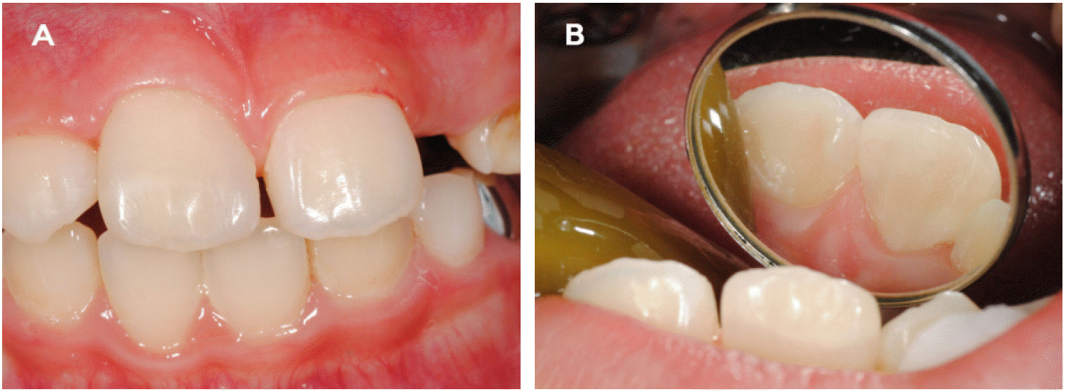
The patient was recalled at 1, 4 and 8 months following treatment. In clinical examinations, the tooth was asymptomatic. In radiographic examinations, the tooth exhibited increased root length and normal periodontal ligament space (Fig. 6D-F). In addition, the tooth exhibited no discoloration (Fig. 8).
Ⅲ. Discussion
Regenerative endodontic treatment enables infected canal spaces to repair or regenerate tissues in the pulp, thereby allowing for resumption of their sensory, immunocompetency, root development, and formation roles [1]. The introduction of stem cells from the apical papilla into the canal by disorganizing the apical papilla tissue with an endodontic file and transferring it into the root canal in accordance with blood clot formation from the periapical tissues has been suggested. When pulp necrosis causes incomplete root development, this endodontic intervention can increase root length and canal wall thickness [2-4]. Significantly higher tooth survival rates were reported when regenerative endodontic treatment (100%) was applied instead of MTA (95%) or CaOH2 apexification (77%). The percentage increase in root length and thickness was also significantly higher using regenerative endodontics instead of either apexification procedure [1]. However, despite these advantages, several drawbacks and unfavorable outcomes are also associated with regenerative endodontic treatment [6,7].
A favorable environment in which pulp and periapical cells can participate in tissue repair and regeneration can be provided by controlling root canal infection following injury [14]. Hoshino et al. [13] demonstrated the effectiveness of a combination of ciprofloxacin, metronidazole and minocycline for eradication of bacteria from the infected root canal. This triple antibiotic paste, when used as an intracanal medication in immature teeth with necrotic pulps, can facilitate further development of the pulp-dentin complex following regenerative endodontic treatment.
Although a triple antibiotic paste is useful for disinfecting the root canal, it can also induce severe discoloration. Kim et al. [8] reported that the major reason for coronal discoloration following treatment was minocycline in the triple antibiotic paste. Sato et al. [14] and Hoshino et al. [13] suggested that minocycline could be replaced by amoxicillin, cefaclor, cefroxadin, fosfomycin or rokitamycin. The combination of metronidazole and ciprofloxacin with any antibiotic has proven equally successful in the sterilization of carious and endodontic lesions [15]. In our protocol, minocycline was replaced with clindamycin [12]: this resolved tooth discoloration, and allowed for simultaneous control of root canal infections.
Several studies reported that following regenerative endodontic treatment, gray MTA can result in discoloration [5,9]. Due to the potential discoloration of teeth treated with GMTA, WMTA has been introduced instead; however, tooth discoloration can still occur using this agent [6,7,10] because even though the concentrations of carborundum (Al2O3), periclase (MgO), and FeO are lower in WMTA compared with GMTA these metal oxides are nonetheless still present [9]. According to Steffen and van Waes [16], bismuth oxide, used as a radiopacifier in MTA, is a possible factor in tooth discoloration. These researchers reported that bismuth oxide is the only difference between Portland cement (PC) and MTA. Further clinical studies of the effects of PC on discoloration are therefore required. Although the material itself may cause discoloration, another possible mechanism has been suggested. Both material and subsequent tooth discoloration might occur with the slow hydrating process of WMTA permitting the absorption and subsequent hemolysis of erythrocytes from the adjacent pulpal tissue [10].
Zirconium oxide represents a possible alternative radiopacifier to bismuth oxide. It can act as an inert filler, and does not participate in the hydration reaction of the PC [17]. However, adding even a minimal amount of radiopacifier to cement can alter its chemistry, biocompatibility and physical properties. Development of cement, using radiopacifier as a component rather than as an adjunct, might be beneficial; in this respect ZrO2-containing calcium aluminate (Ca7ZrAl6O18) cement has excellent potential [18]. Retro MTA® (BioMTA, Korea) is a calcium zirconium aluminate cement containing 60 - 80% calcium carbonate (CaCO3), 5 - 15% silicon dioxide (SiO2), 5 - 10% aluminum oxide and 20 - 30% calcium zirconia complex. According to the manufacturer, Retro MTA has short setting time, contains no heavy metal, possesses no cell toxicity and causes no discoloration, even in the context of blood contamination [11]. Che and Kim [19] reported that Retro MTA has similar properties, in terms of compressive strength and solubility, to ProRoot MTA, and further that the setting time of Retro MTA is only 18 min, shorter than that of ProRoot MTA (at 279 min). In both of our presently reported cases, Retro MTA effected a successful outcome. However, the number of studies pertaining to the various clinical applications of new composition of MTA is very limited. Although the manufacturer claims that a better color stability is achieved with Retro MTA (in comparison to WMTA), no study has investigated color changes using Retro MTA in endodontic procedures. Furthermore, Retro MTA is characterized by certain drawbacks, including difficult in handling, high cost, absence of a known solvent, and difficulty of removal after curing [20].
Other drawbacks associated with regenerative endodontics include failure to produce bleeding and collapse of MTA material into the canal [6,7]. Blood clots allows for the migration of mesenchymal stem cells into the canal, a phenomenon not observed in the absence of blood clots inside a disinfected root canal [3,4]. To induce sufficient bleeding, non-epinephrine local anesthetics could be used [5,6,8], with MTA material placed over the blood clot. MTA has a setting time of between 3 and 4 hours [16]; the blood clot is often insufficiently strong to hold the MTA, resulting in MTA collapse within the root canal [4,6,7]. Placing a collagen matrix above the blood clot can serve as a solid absorbable matrix against which the MTA can be packed [4,6]. However, in one study the amount of bleeding was inadequate, but Teruplug™, an absorbable collagen sponge, allowed for a successful outcome. A recent case report suggested use of platelet-rich plasma instead of a blood clot inside the root canal space to resolve this problem [5].
Ⅳ. Summary
The most significant disadvantage associated with regenerative endodontic treatment is coronal discoloration. The present case reports demonstrate successful regenerative endodontics without coronal discoloration of infected immature permanent teeth using modified triple antibiotics (ciprofloxacin, metronidazole, clindamycin) and Retro MTA. Retro MTA as a aesthetic material can potentially replace WMTA, but further study is required to determine its efficacy.


