소아의 구개부에 발생한 plasmacytoid myoepithelioma의 최소 침습적 제거술
Minimally Invasive Surgery in a Pediatric Palatal Plasmacytoid Myoepithelioma
Article information
Abstract
Myoepithelioma는 타액선에 발생하는 드문 질환이다. Myoepithelioma는 소아 및 청소년보다 성인에서 호발한다. 구개부의 종창을 주소로 8세 여환이 본원으로 내원하였다. 환아의 나이와 상대적으로 큰 종양의 크기를 고려하여, 전신 마취 하에 구개부 점막 조직을 보존하는 보존적인 외과적 절제술이 시행되었다. 수술 부위의 치유가 적절히 일어났으며, 40개월의 관찰기간 동안 재발되지 않았다. 주변의 구개부 조직을 보존하는 최소 침습적 제거술은 소아에서 발생되는 myoepithelioma의 치료시 유용할 것으로 사료되었다.
Trans Abstract
Myoepithelioma is a rare disease in the salivary gland. Myoepithelioma is more common in adults than in children or adolescents. An 8-years-old female patient visited our clinic with a chief complaint of a painless swelling on the palate. Conservative treatment that preserves the overlaying palatal mucosa while surgically excising the tumor was carried out under general anesthesia, because the patient was young and the size of the tumor was relatively large. The surgical wound healed well and there had not been any sign of recurrence during the regular follow-up period of 40 months. Minimally invasive surgical treatment which preserves peripheral palatal tissue can be useful in a pediatric myoepithelioma.
Ⅰ. Introduction
Myoepithelioma is a rare benign neoplasm of the salivary gland composed of myoepithelial cells. It usually represents an asymptomatic and slowly growing submucosal mass [1]. Myoepithelioma may occur frequently in the parotid gland and minor salivary gland of soft palate. It has been reported that there is no gender predilection and it is common in adults between 30 and 50 years old [2]. For the management of myoepithelioma, an excessive surgical excision involving surrounding margin of normal tissue has been recommended [1]. Therefore, it is necessary to consider any possible defects.
To the best of our knowledge, myoepithelioma is extremely rare among children and adolescents, and only few cases with excessive surgical removal of the tumor have been reported [1-4]. Unlike the one in adults, when it is found in young patients, a conservative method with the consideration of the potential future growth and development should be considered.
The aim of this report was to describe a case of myoepithelioma of the palate in an 8-years-old patient, treated with a conservative surgical approach that preserves the overlaying palatal mucosa instead of taking the complete surgical excision involving the surrounding margin of normal tissue. The prognosis has been satisfactory through long-term follow-up.
Ⅱ. Case report
An 8-years-old girl visited our clinic with chief complaint of a painless swelling on the palate. She could not remember the onset of the swelling exactly. She had good oral hygiene and did not have any systemic disease or special familial history. Clinically, well circumscribed firm submucosal nodule was observed on the left side of hard palate (Fig. 1). It was approximately 2.5 cm in diameter. There was not any tenderness to palpation or ulceration on the surface of the lesion. In addition, complications associated with the lesion, such as dysphasia or dysphonia, were not observed. Cone-beam computed tomography revealed well-circumscribed nodule on the hard palate which had no invasion to peripheral tissue (Fig. 2).
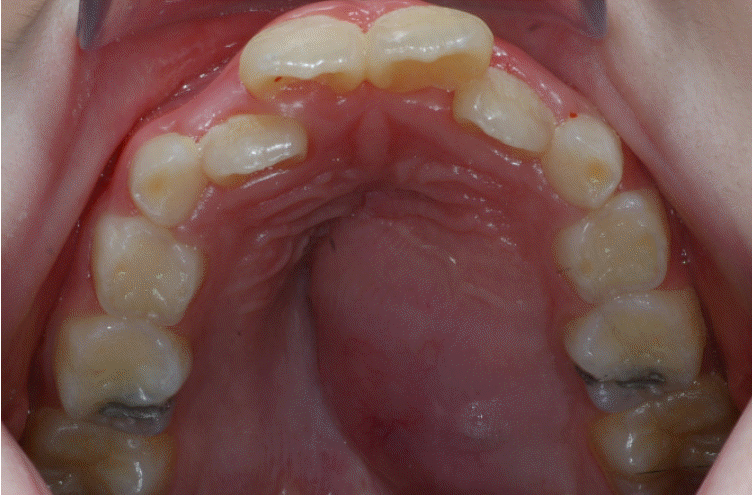
Preoperative clinical photographs. A well circumscribed and firm submucosal nodule (about 2.5 cm in diameter) was observed in the transition between the hard and soft palate on the left.
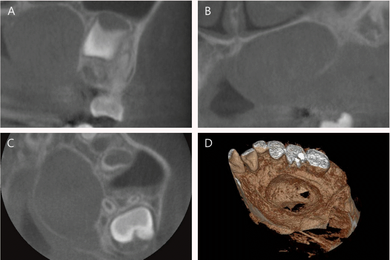
(A), (B) axial CT cuts. (C) coronal CT cut. (D) 3-dimensional CT cut. Computed tomography cuts showed well-circumscribed nodule on the hard palate which had no invasion to peripheral tissue.
After the clinical and radiologic evaluation, it was hypothesized with the tumor of the salivary gland such as pleomorphic adenoma, myoepithelioma, or myoepithelial carcinoma. Thus, surgical excision and excisional biopsy were planned. But it was necessary to modify the traditional surgical method, which removed completely the surrounding margin of normal tissue, because the patient was young and the tumor size was relatively large [1,4]. Preservation the overlaying palatal mucosa was necessary to avoid postoperative pain and scar formation. Tumor was excised and detached from the overlaying palatal mucosa carefully under general anesthesia (Fig. 3). The surgical specimen was submitted to the Department of Pathology in Kyung Hee Medical Center for histopathological assessment.
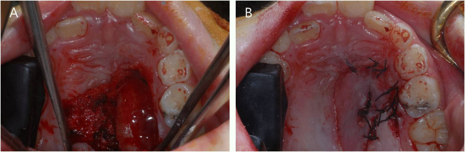
Postoperative clinical photographs. (A) Tumor was surgically excised under general anesthesia, and palatal overlying mucosa above the lesion was preserved. (B) Well limited lesion was separated and removed from the adjacent tissue.
Immunohistochemically, positive reactions on vimentin, S-100 protein, and glial fibrillary acidic protein (GFAP) were observed. Also negative reaction to MSA was found. As a result, the patient was diagnosed with plasmacytoid subtype cells with lack of differentiation as the myoepithelial tumor cell with low-graded differentiation (Fig. 4).
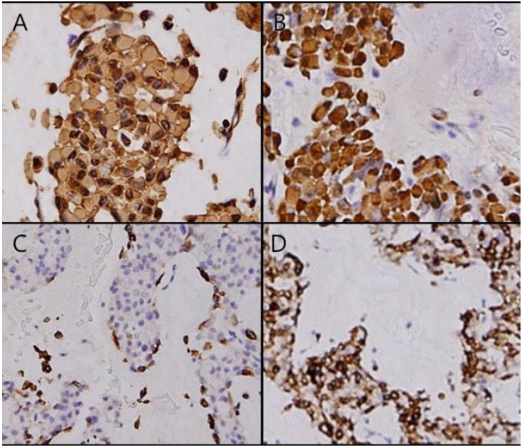
Immunohistochemically, the plasmacytoid cells were diffusely and strongly immunoreactive for cytokeratin, S-100, vimentin and glial fibrillary acidic protein (GFAP). (A) vimentin (×400), (B) S-100 protein (×400), (C) GFAP (×100), (D) Cytokeratin (×100).
The patient became asymptomatic and the surgical wound was healed up postoperatively. And any evidence of tumor recurrence had not been shown during a regular 40-month follow up (Fig. 5).
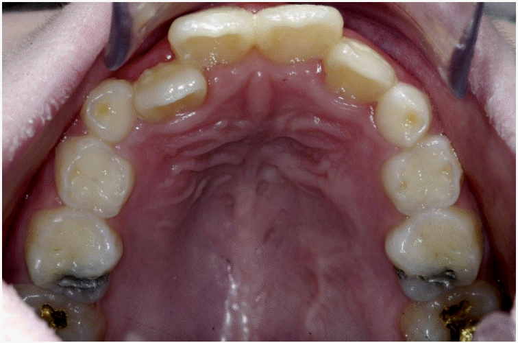
Clinical photograph of 1 year periodic check. The patient has been followed-up 40 months without showing any evidence of tumor recurrence.
Ⅲ. Discussion
Myoepithelioma is a benign tumor rarely occurred in the salivary gland. It had been considered to the subtype of pleomorphic adenoma before WHO classified it as the independent tumor [5]. Diagnosis of myoepithelioma from pleomorphic adenoma can differ by whether a tumor contains less than 5% of ductal and acinar components [6]. Differential diagnosis from other tumors, such as myoepithelial carcinoma, adenoid cystic carcinoma, and benign mesenchymal soft tissue tumor, is necessary because of histological variations of this tumor. It has been reported that more than 10% of Ki-67 label index indicates myoepithelial carcinoma [1]. And other tumors which are not differentiated from myoepitheial cell can be excluded through antibody test to vimentin, S-100 protein, and GFAP [5,6]. There are four types of cells comprising the myoepithelioma by to the shape of the cell of lesion; spindle, plasmacytoid, epitheloid, and clear cell. These cells can be found in a mixed form [4]. Type of myoepithelioma may be classified by reaction to MSA. Plasmacytoid cell type shows negative reaction to MSA [5,6]. Therefore, the patient was diagnosed with plasmacytoid myoepithelioma.
Clinically, myoepithelioma is a slowly enlarging, asymptomatic, solid, and well-circumscribed tumor, usually less than 3 cm in diameter [4,6,7]. The size of tumor varies with an average of 1.9 cm in diameter [1]. Usually the color of this tumor represents white, tan, or gray [7]. Bone destruction or adjacent soft tissue associated with this tumor has rarely been reported [4,7].
Plasmacytoid myoepithelioma is more common in adults than children and adolescents [4]. Thus, few cases of plasmacytoid myoepithelioma have been reported in children and adolescents recently [1-4]. Thus only two cases, including this report, in patient younger than 10 years of age have been reported in the English-literature [1].
The treatment of plasmacytoid myoepithelioma has been proposed to complete surgical excision involving a surrounding margin of normal tissue, because myoepithelioma behaves in a locally aggressive or malignant characteristic [1,8]. Postoperative defects or scar formation is inevitable because of wide dissection. So it may need to reconstruct the defect. There was a report using the buccal fat pad graft for defect closure. Also localized palatal flap, tongue flap or facial artery muscle-myocutaneous flap can be employed for closure of defect [3]. However, soft tissue graft can induce undesirable complications, such as flap failure, infection or additional pain on donor site [9]. Also graft cannot prevent scar formation. There was a report using palatal obturator prosthesis after scar formation [4]. However it has to be removed and inserted whenever feeding and cleaning is needed [9]. Thus, an alternative treatment was necessary because the patient was young and the tumor was relatively large in size. Also, maxillary transverse and dento-alveolar growth may be impeded by scar tissue of the palate [10]. Thus, it was planned to preserve overlaying palatal mucosa while surgically excising the tumor. There was no need of graft or palatal obturator for defect closure through this conservative treatment. The method minimized postoperative pain or scar formation. So we could anticipate that this conservative management would give more favorable effect on future maxilla growth.
Prognosis of myoepithlioma is favorable. And recurrence rate of this tumor is less than pleomorphic adenoma [11]. Although recurrence of this tumor in children and adolescents has rarely been reported, it may be considered that this tumor behaves similar to the tumor seen in adults [4].
Ⅳ. Summary
This clinical report demonstrates a rare case of a plasmacytoid myoepithelioma on the palate in a child. Minimally invasive treatment that preserves peripheral palatal tissue while surgically excising the tumor can be deployed depending on the age of a patient or a tumor size. Postoperative complications were minimized and the prognosis had been satisfactory during the 3-year follow-up.
Acknowledgements
This work was supported by the National Research Foundation of Korea (NRF) grant funded by the Korea government (MEST) (No.2012R1A5A2051384).