Gonial Angle의 단순 회귀 모델: 파노라마 영상과 측모두부 영상간의 비교
The Simple Regression Model of Gonial Angles : Comparison between Panoramic Radiographs and Lateral Cephalograms
Article information
Abstract
본 연구의 목적은 파노라마 영상과 측모두부 영상에서 유의미한 상관관계를 가지는 계측치를 확인하고, 그 관계를 수식화하는 것이다. 99명의 3급 부정교합 환자의 파노라마 영상과 측모두부 영상에서 gonial angle, 하악지 높이, 하악중절치에서 하악하연까지의 길이를 측정하여 평가하였다. 측정된 수치는 각각 대응 표본 T검증, Pearson’s 상관관계 검정, 회귀 분석을 통해 평가하였다.
측모두부 영상에서의 gonial angle 평균값은 127.50°, 파노라마 영상에서의 값은 125.49°, 상관계수는 0.945(p< 0.001)로 나타났으며, 상관관계는 ‘측모두부 영상의 gonial angle = 0.920 × 파노라마 영상의 gonial angle + 12.072’로 나타낼 수 있다. 다른 계측치들 간의 상관관계는 gonial angle에 비해 낮게 나타났다.
Gonial angle은 파노라마 영상과 측모두부 영상에서 모두 재현성 있게 측정 가능하며, 강한 양의 상관관계를 나타낸다. 파노라마 영상 또한 측모두부 영상과 같이 환자의 수직성장패턴을 평가하는데 도움을 주는 방법이다.
Trans Abstract
The aim of this study was to enhancing the panoramic radiograph’s clinical use for assessing mandibular measurements and formulating a function of those measurements from panoramic radiographs and lateral cephalograms in children.
The panoramic radiographs and lateral cephalograms of 99 former orthodontic patients with skeletal class III malocclusion were selected. In each radiograph, gonial angles, ramus heights, and distance between lower incisors and symphysis were measured. The values of the studied parameters were compared by paired t-test, Pearson’s correlation test and regression analysis.
The mean value of the gonial angle in panoramic radiographs was 125.49°, and the value in lateral cephalograms was 127.50°. The Pearson’s correlation coefficient (ρ) between mean values of gonial angle in each radiograph was 0.945 (p< 0.001). The relationship between the gonial angle measurements obtained from each radiographs was represented as ‘Gonial angle (Lateral cephalograms) = 0.920 X Average gonial angle (Panoramic radiographs) + 12.072’ in the linear function. The coefficients of ramus heights, and distance between lower incisors and symphysis portrayed weaker correlations than gonial angles.
A panoramic radiograph could be used to determine the gonial angle as accurately as a lateral cephalogram, and each gonial angle showed a strong positive relation. A panoramic radiograph is a useful tool for examining vertical growth pattern of patients, as well as a lateral cephalogram.
I. Introduction
A panoramic radiograph is a dental X-ray scan that gives a panoramic view of the upper and lower jaws. This type of radiograph is considered as the current standard of care for dental diagnosis and treatment planning. It provides a significant amount of information about teeth and their supporting bone. It is most commonly used to determine congenital missing teeth and the developmental status of permanent teeth; moreover, it is used in orthodontic practices to examine teeth alignments, axial inclinations, developmental stages, and maturation periods [1].
A lateral cephalogram is another radiographic technique used when cephalometric measurements are made. This radiograph provides a lateral view of skull and it is used for orthodontic planning. Although this imaging technique is useful to evaluate patient’s cephalometry, it is not used for evaluating pathology of teeth and surrounding tissues as a panoramic radiograph is used, because in lateral cephalograms, bilateral structures superimpose with each other.
In orthodontics, a gonial angle is used to determine the rotation of the mandible. The gonial angle is a significant indicator to diagnose the vertical growth pattern of patients [2]. The patients with downward and backward rotation of ramus are considered as “high-angle” patients. They display increased gonial angles, whereas “low-angle” patients who show upward and forward direction of mandible display decreased gonial angles [3]. This angle may affect the treatment approach, as it aids in deciding whether class III patients should undergo an orthognathic surgery or not. The patient group that shows unstable orthognathic responses and poor prognoses has a significantly large gonial angle before treatments and the angle is progressively increasing over time [4].
The panoramic radiograph provides a comprehensive overview of the patient’s bony and dental features; however, it has potential shortcomings, especially in the upper anterior maxilla, with its horizontal and vertical distortions. Horizontal distortions are more severe than vertical distortions because of the image making method of panoramic radiographs. On the other hand, the lateral cephalogram does not give a panoramic view. Because of the interference of superimposed images appearing on the lateral cephalogram, reliable measurements of the individual left and right gonial angles are almost impossible.
Even though the panoramic radiograph has image distortions, it is the most useful extraoral radiograph in dental field. Also, the gonial angle can be determined more clearly in a panoramic radiograph than in a lateral cephalogram. The aim of this study was to enhancing the panoramic radiograph’s clinical use for assessing mandibular measurements and formulating a function of those measurements from panoramic radiographs and lateral cephalograms in skeletal class III malocclusion children.
II. Materials and Methods
Panoramic radiographs and lateral cephalograms of 230 patients were obtained from the patient records of the Department of Pediatric Dentistry in Seoul National University Dental Hospital in January 2013 - April 2014. All panoramic radiographs were taken by Orthopantomograph® OP100 (Instrumentarium Imaging Ind. Co. Ltd., Tuusula, Finland), and cephalograms were taken by CX-90SP (Asahi Roentgen Ind. Co. Ltd., Kyoto, Japan). Each radiograph was taken with the same digital machine. Cephalometric data including SNA (Sella–Nasion–A point angle), SNB (Sella–Nasion–B point angle), ANB (A point–Nasion–B point angle), and APDI (Anterior Posterior Dysplasia Indicator) of selected patients, were measured by V-ceph 5.5 (Osstem Implant Co. LTD., Seoul, Korea), to confirm the Angle classification.
For this study, the panoramic radiographs and lateral cephalograms of 99 former orthodontic patients with skeletal class III malocclusion were selected. They were aged between 4.4 and 12.1 years with a mean age of 8.03 years.
Exclusion criteria were as follows: panoramic radiographs and lateral cephalograms not obtained within 6 months; radiographs showing unclear and cropped image; and patients with a systemic disease or a syndrome presenting facial deformity.
In each radiograph, gonial angles, ramus heights, and distance between lower incisors (L1) and symphysis were measured (Table 1, Fig. 1, Fig. 2). All angles and lengths were measured at PiViewStar™ 5.0 (INFINITT Healthcare Co. LTD., Seoul, Korea).
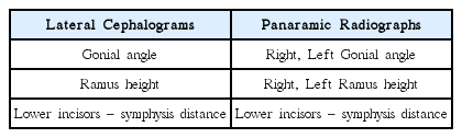
Measured angles and lengths in both radiographs
Measurement of the gonial angle in lateral cephalograms.

Measurement of the gonial angle in panoramic radiographs.
Ethical approval was obtained from the relevant institutions, namely Clinical Dental Research Institute of Seoul National University Dental Hospital. The trial was registered in Institutional Review Board of Seoul National University Dental Hospital (Trail No. : CRI15016).
The normality of the studied parameters was checked using the Kolmogorov-Smirnov test, and because all data displayed a standard distribution, statistical analysis were based on parametric tests. The values of the studied parameters were compared by paired t-test, Pearson’s correlation test and regression analysis. In regards to difference in genders, each value was compared by independent t-test, and the homogeneity of variance was checked with Levene’s test. These analyses were performed using the SPSS 21 (SPSS Inc., Chicago, IL, USA).
III. Results
The study group consisted of 99 subjects (51 males, 48 females, mean age; 8.03 ± 1.69). All studied parameters followed standard distribution and all compared values showed the homogeneity of variance.
Table 2 shows the data of gonial angle values of panoramic radiographs and lateral cephalograms. The mean gonial angle value in females was 127.26° and that in males was 127.73° with no statistically significant difference between two genders (p= 0.611). In the panoramic radiograph, there was no significant difference between right and left gonial angles (p= 0.214). The gonial angle of each gender in panoramic radiographs also showed no statistically significant difference (p= 0.843).
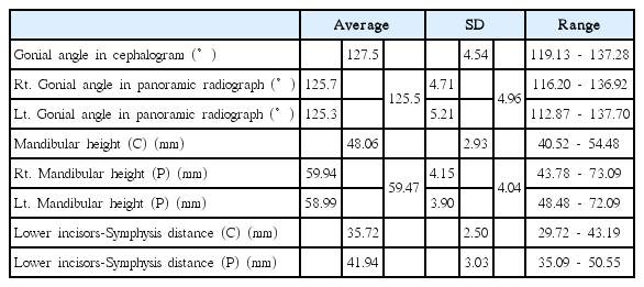
Average, standard deviation and range of panoramic radiographic and cephalometric values
In Table 3, there were significant differences between all values of gonial angles determined by panoramic radiographs and lateral cephalograms. Based on the results of correlation analysis, the Pearson’s correlation (ρ) between average values of gonial angle in panoramic radiographs and lateral cephalograms was 0.945 (p < 0.001), which means that there is a strongly positive relationship between each gonial angle. Also, the coefficient of determination (ρ2) was 0.893. Fig. 3 presents regression analysis between gonial angles. The relationship between the gonial angle measurements obtained from each radiograph was represented in the linear function (GoA (C) : The value of gonial angle (lateral cephalograms), AGoA (P) : The average value of gonial angle (panormic radiographs)).

Differences between the values of gonial angles
The relationship between the gonial angle measurements obtained from each radiograph was represented in a linear function.
GoA (C) = 0.920 × AGoA (P) + 12.072
Range of data : GoA (C) 119.13° - 137.28°, AGoA (P) 114.79° - 136.33°
The data of ramus height and distance between L1 and symphysis in panoramic radiographs and lateral cephalograms in millimeters was listed in Table 2. Data from the lateral cephalogram was presented after corrections of cephalometric magnification. These values also showed no statistically significant difference between two genders. Statistically significant differences were found between the values of ramus height and distance between L1 and symphysis determined by panoramic radiographs and lateral cephalograms (p < 0.001). The Pearson’s correlation coefficients of each value were 0.816 and 0.835, portraying a weaker correlation than gonial angles (Fig. 4, Fig. 5). Relationships between each value from panoramic radiographs and lateral cephalograms could be represented in the following linear equation (MnH (C) = Mandibular height (lateral cephalograms), AMnH (P) = Average mandibular height (panoramic radiographs), IMn (C) = The distance between lower incisors and mandibular symphysis (lateral cephalograms), IMn (P) = The distance between lower incisors and mandibular symphysis (panoaic radiographs)).
The relationship between the ramus height measurements obtained from each radiograph was represented in a linear function.
The relationship between the lower incisor to symphysis length measurements obtained from each radiograph was represented in a linear function.
MnH (C) = 0.619 × AMnH (P) + 11.247
IMn (C) = 0.690 × IMn (P) + 6.798
Range of data : MnH (C) 40.52 mm - 54.48 mm, AMnH (P) 46.13 mm - 72.59 mm
IMn (C) 29.72 mm - 43.19 mm, IMn (P) 35.09 mm - 50.55 mm
IV. Discussion
The panoramic radiograph is usually taken by all dentists because it is considered as an effective tool for screening, diagnosing and treatment-planning. It offers a comprehensive overview of the patient’s bony and dental features, but it also presents a drawback of an image distortion. According to Graber [5], with a panoramic radiograph the distortion varies from 2 to 35%, depending on the area involved. The lateral cephalogram is a valuable method to assess skeletal, dental and soft tissue relationships. It is useful to predict skeletal growth, a response to treatment, and long-term stability after treatment; however, because it does not give a panoramic view, the interference by superimposed images always exists. Both radiographs have such strengths and weaknesses; therefore, dentists have to decide carefully on types of radiographs to be taken in terms of cost and benefit.
Each study about gonial angles in panoramic radiographs and lateral cephalograms has demonstrated controversial results. In the present study, the mean values of external gonial angles were 125.49° and 127.50° respectively in the panoramic radiograph and lateral cephalogram, with a strongly positive correlation between the two radiographs. The gonial angle obtained by panoramic radiograph was generally less than that of lateral cephalogram. The gonial angle in panoramic radiographs showed 2.00° (Range: 1.70-2.31°, 95% confidence interval) less than in lateral cephalograms.
On the other hand, previous studies could not clarify the differences of gonial angles in each radiograph [6-9]. Shahabi [6] concluded that panoramic radiography could be used to determine the gonial angle as accurately as the lateral cephalogam. Based on the obtained results, the mean difference was 0.83°, but there were no statistically significant differences (p= 0.406). When Oksayan [7] also compared the gonial angle, the differences were found to be 0.04° on the right side and 0.02° on the left side with no statistically significant differences. These disparities in results could be results of unspecified samples and study designs. While this study’ s sample age was between 4 and 12 years, Shahabi [6] and Oksayan [7] focused on adult and adolescent (each with age range of 12-29 and 15-30 years). Ghosh [10] concluded that the gonial angle tended to increase with age in the panoramic radiographs. Children’s mandibular body is short, and they have more round mandible than adults, because eruption of the permanent dentition is correlated with an anteroposterior increase in mandibular body length [11]. The corpus divergence of children’s mandible is larger than adults. The direction of an x-ray beam in a panoramic radiograph is perpendicular to a horseshoe shaped focal trough, but the x-ray beam of a lateral cephalogram irradiates perpendicular to the sagittal plane of the head. Therefore, in this study, each gonial angle of children shows statistically significant differences, whereas others did not.
In fact, an image making mechanism of each radiograph is different; and the projection angle of mandibular body in lateral cephalograms is larger than panoramic radiographs. On lateral cephalograms, the mandibular body length is shorter, and the gonial angle is larger than panoramic radiographs. Fisher-Brandies [12] indicated that the value of the gonial angle measured in the panoramic radiograph was 2.2-3.6° smaller than in the lateral cephalograms. Larheim [13] found that the gonial angle from the panoramic radiograph was almost identical to that directly measured on a dried mandible bone. Slagsvold [14] mentioned that lateral cephalograms did not permit reliable registrations of the gonial angle and the distortion was closely associated with the form of the mandible.
Generally, measurements of the gonial angle on panoramic radiographs provide acceptable reliability and reproducibility when the head position is standardized and kept constant. The panoramic image is highly sensitive to the position of the patient in the panoramic machine [15]. Mispositioning of the head could affect horizontal expansions and distortions, and it frequently occurs in practices; therefore, when taking panoramic radiographs, practitioners have to make sure to keep a patient’s head in a standard position, as the mid-facial plane is adjusted vertical to the floor and the Frankfort horizontal plane is kept parallel to the floor.
Based on the correlation analysis, the Pearson’s correlation coefficient (ρ) between the average values of gonial angle in panoramic radiographs and lateral cephalograms was 0.945 (p value < 0.001). It means each value shows a strong positive relationship. As a result, the gonial angle obtained from a panoramic radiograph can help to predict the angle from a lateral cephalogram.
In the panoramic radiograph, the gonial angle of each gender showed no statistically significant difference (Male = 125.59°, Female = 125.40°, p= 0.843), and there was no significant difference between right and left angles (Right = 125.71°, Left = 125.28°, p= 0.214). These findings coincided with the results of previous studies [6,7,13,16]. Akcam [16] stated that there was no statistically significant difference between male and female subjects. Larheim [13] stated that no statistically significant differences were observed between the reproducibility of the right and left gonial angle.
On the other hand, mandibular ramus height and distance between L1 and mandibular symphysis showed a relatively lesser correlation than gonial angles between two radiographs. This result is quite different from previous researches. Ongkosuwito [17] stated a panoramic radiograph had comparable reliability in measuring a mandibular distance condylion to gonion compared with a lateral cephalogram. Oshagh [18] concluded a panoramic radiograph could provide information on vertical and sagittal dimensions. In fact, measuring distances between cephalometric points in panoramic radiographs has not been researched much. A horizontal portion of the distance differs somewhat from the actual distance due to the characteristics of a panoramic image. In panoramic radiographs, image distortion is the smallest at the posterior mandibular region, and the vertical distortion is smaller than horizontal distortion. Therefore, mandibular ramus height was expected to show the smallest difference in distances between two radiographs.
In the present study, however a correlation coefficient of mandibular ramus height was 0.816. This value means a positive relationship. Nevertheless, it is not significant enough when considering the data obtained from same sample. The coefficient of determination was 0.664, and this result came from cumulated weaknesses of both radiographs. Mandible is a three-dimensional structure, and mandibular ramus height consists of sagittal, coronal and axial components. In panoramic radiographs, the horizontal component which consists of sagittal and coronal components makes mandibular ramus height larger than original structure. In lateral cephalograms, the coronal component is not expressed in a radiographic image, so the ramus height in the image is smaller than the original structure. For these reasons, mandibular ramus heights of two radiographs are quite different.
The distance between L1 and mandibular symphysis in panoramic radiographs was also longer than in lateral cephalograms. The value obtained from lateral cephalograms is regarded as the same as real distance if the mandibular asymmetry does not exist. In this study, patients with facial asymmetry were excluded, thus the differences result from not the anatomical structure, but the vertical enlargement of anterior mandibular area in panoramic radiographs. A direction of x-ray beam in panoramic machines is changed. Therefore, there is a difference in the images of each x-ray at the anterior mandibular area. Hayes [19] stated that the panoramic radiograph provided low levels of diagnostic value in the maxillary incisor region.
Some dentists believe that lateral cephalogram is the only useful radiograph to define the skeletal measurements. No two-dimensional radiographs could illustrate a three-dimensional structure as a real size without any distortions [20]. An assessment of three-dimensional structures with twodimensional methodologies has inherent structural limitations. Markic [21] concluded that both panoramic radiographs and lateral cephalograms showed a poor agreement with 3D imaging measurements. Lateral cephalograms are standard radiographic methods for cephalometric analysis, and the finding of this study suggests that a panoramic radiograph is also successful in determining a gonial angle, as well as lateral cephalogram. Skeletal class III malocclusion children with the large gonial angle show unfavorable growth during follow up and unstable results after orthodontic treatment, so they need periodical taking of lateral cephalograms. It is incontrovertible that lateral cephalograms are essential to diagnose the skeletal problem and to study the growth pattern of patients. However, at pediatric dental clinics, panoramic radiographs are taken biennially for many reasons, such as evaluating permanent teeth and trauma to the jaws. The results about relationship between two radiographs could help pediatric dentists to use panoramic radiographs which were already taken. Even though radiation doses during dental examinations are relatively low, patients can be concerned with having a great number of radiographs taken for a dental visit. As radiation has a cumulative effect on the human body, and most orthodontic patients are children in active growth, any reduction in the exposure to radiation is beneficial [22].
A panoramic radiograph could be useful to measure both gonial angles, especially in asymmetry cases. In present study, patients with severe asymmetric jaw were excluded, but nowadays, many patients with facial asymmetry are visiting dental clinics. This radiograph is also useful in cases where the outlines of two sides are not clearly visible, because there are no interferences from superimposed images of anatomical structures as in a lateral cephalogram. Bhullar [23] stated that a panoramic radiograph might be a better choice than a lateral cephalogram for determination of gonial angle without superimpositions.
Many studies suggested that the reproducibility of vertical and angular measurements was acceptable if the patient’s head was positioned properly in the panoramic equipment. In this study, panoramic radiograph was a useful method for the measuring gonial angle, which is an indicator of mandibular steepness and growth direction.
This study focused on the skeletal class III malocclusion children. The shape of the mandible is different from skeletal classification, and it changes as patients grow; therefore, further studies have to classify the sample according to skeletal patterns and specific age ranges. Moreover, this is the first study that established the equation about correlation between panoramic radiographs and lateral cephalograms. To clarify the equation, it is necessary to have an increased the number of samples. In this regard, more studies are still required.
V. Conclusion
Based on the obtained results, this study concluded that a panoramic radiograph could be used to determine the gonial angle as accurately as a lateral cephalogram, because each gonial angle showed a very strong positive relation. The relationship between the gonial angle measurements obtained from each radiograph in skeletal class III malocclusion patients was represented as ‘GoA (C) = 0.920 X AGoA (P) + 12.072’ in the linear function. To clarify the equation, further studies including a great number of and more classified samples are required.