 |
 |
| J Korean Acad Pediatr Dent > Volume 44(2); 2017 > Article |
|
žīąŽ°Ě
žĄ†ž≤úžĻėžôÄ žč†žÉĚžĻėŽäĒ ž∂úžÉĚžčú ŽßĻž∂úŪĖąÍĪįŽāė ž°įÍłįžóź ŽßĻž∂úŪēėŽäĒ žĻėžēĄŽ°ú Žč§žĖĎŪēú Ūē©Ž≥Ďž¶ĚžĚĄ ÍįÄžßÄÍ≥† žěąžúľŽāė, ŽßéžĚÄ Ž∂ÄŽ™®Žď§žĚī žĚīžóź ŽĆÄŪēú ž†ēŽ≥īŽ∂Äž°ĪžúľŽ°ú ž†Āž†ąŪēú žĻėŽ£Ć žóÜžĚī Ž∂ąŪ鳞̥ Í≤™Í≥† žěąŽč§. Ž≥ł žóįÍĶ¨ŽäĒ 2006 - 2015ŽÖĄžóź Í≤ĹŲ̄ŽĆÄŪēôÍĶź žĻėÍ≥ľŽ≥Ďžõź žÜĆžēĄžĻėÍ≥ľŽ•ľ ŽāīžõźŪēú ŪôėžēĄžóźÍ≤ĆžĄú ŽįúÍ≤¨ŽźėŽäĒ žĄ†ž≤úžĻėžôÄ žč†žÉĚžĻėžĚė ŪäĻžĄĪžĚĄ ŪŹČÍįÄŪēėÍ≥†žěź ŪēúŽč§.
ŪôėžēĄŽď§žĚė žĄĪŽ≥Ą, Ž∂ÄŽ™®žĚė ž£ľžÜĆ, žěĄžÉĀž†ĀžĚł ŪäĻžßēÍ≥ľ Ūē©Ž≥Ďž¶Ě ŽįŹ žĻėŽ£ĆŽ≤ēžĚĄ ž°įžā¨Ūēėžó¨ ŪõĄŪĖ•ž†Ā žóįÍĶ¨Ž•ľ žßĄŪĖČŪēėžėÄŽč§. ŪôėžēĄŽď§ ÍįÄžöīŽćį 69%ŽäĒ žĄ†ž≤úžĻėŽ•ľ, 31%ŽäĒ žč†žÉĚžĻėŽ•ľ ÍįÄž°ĆžúľŽ©į Ž™®ŽĎź ŪēėžēÖ ž†ąžĻėžėÄŽč§. žó¨ŪôėžĚī Žā®ŪôėŽ≥īŽč§ ŽßéžēėÍ≥† Žāīžõź ž£ľžÜĆŽ°úŽäĒ Ž∂ÄŽ™®žĚė ÍĪĪž†ēžĚī ÍįÄžě• ŽßéžēėžúľŽ©į Ž≥Ďžõź žĚėŽĘįžôÄ Ž¶¨ÍįÄ ŪéėŽćįŽ≥Ď(Riga-Fede disease) Í∑łŽ¶¨Í≥† žąėžú†žčú ŪÜĶž¶ĚžĚī Í∑ł Ží§Ž•ľ žĚīžóąŽč§. žĻėŽ£ĆŽį©Ž≤ēžúľŽ°úŽäĒ ŽįúžĻėÍįÄ ÍįÄžě• ŽßéžēėžúľŽ©į žĻėžēĄ žú†žßÄžôÄ žĻėžēĄ Žč§Žď¨ÍłįŽŹĄ ŪĖČŪēīž°ĆŽč§.
žĄ†ž≤úžĻėžôÄ žč†žÉĚžĻėŽäĒ Žč§žĖĎŪēú ŪäĻžĄĪÍ≥ľ Ūē©Ž≥Ďž¶ĚžĚĄ ÍįÄžßÄÍ≥† žěąžúľŽĮÄŽ°ú žĻėÍ≥ľžĚėžā¨ŽäĒ žēĄžĚīžĚė ž†ĄŽįėž†ĀžĚł ÍĪīÍįēžĚĄ žúĄŪēī žĚīŽü¨Ūēú žĻėžēĄžĚė Ž≥ĎŽ¶¨žóź ŽĆÄŪēī ž†ēŪôēŪěą žĚīŪēīŪēėŽäĒ Í≤ÉžĚī ž§ĎžöĒŪēėŽč§.
Abstract
Natal teeth that are already present at birth and neonatal teeth that erupt shortly after birth may cause various complications. The purpose of this study is to evaluate the characteristics of natal/neonatal teeth in Korean infants who visited to Kyung Hee University Dental Hospital from 2006 to 2015.
A retrospective review of clinical data, including the sex of the patients, chief complaints of the mothers, clinical appearances and locations of the natal/neonatal teeth, and associated complications and treatments, was collected.
Overall, a total of 75 teeth were found in 48 patients and 69% of the infants had natal teeth, while 31% had neonatal teeth, all of which were mandibular incisors. Females showed more natal/neonatal teeth than males. Major reasons for visiting the dental clinic were parental anxiety, referrals from other clinics, Riga-Fede disease, and pain during breastfeeding. Extraction was the most common treatment choice; observation and grinding were also used.
Eruption of the first tooth is a big event that influences children functionally and psychologically. The first eruption of a tooth usually begins at 6 months of age. However, some teeth show unusual pattern of early eruption. Such teeth are classified as natal teeth, which are present at birth, and neonatal teeth, which erupt within the first 30 days after birth [1].
Natal/neonatal teeth are rare; the reported prevalence is from 1:30,000 to 1:800, and they are more common in females than in males. The occurrence of natal teeth is three times more common than neonatal teeth [1,2]. Because of their rare occurrence, such premature eruptions have become associated with superstition and folklore, believed to be related to good or bad omens [3]. Moreover, early eruption may cause clinical complications in nursing infants and to their mothers, including pain on suckling and refusal to feed; thus, for both the infant and the mother, it requires attention [3].
The purpose of this study was to investigate the clinical characteristics of natal/neonatal teeth in a group of Korean infants.
We analyzed infants younger than 6 months with natal or neonatal teeth retrospectively. All infants in this study visited the Department of Pediatric Dentistry, Kyung Hee University Dental Hospital, between May 2006 and December 2015. The study proposal was reviewed and approved by the Ethics Committee of Kyung Hee Medical Center, Kyung Hee University, Seoul, Republic of Korea (KHD-IRB-1601-4).
After excluding infants with genetic disorders, 48 infants were considered and clinical data and demographic information were collected from medical records. Clinical and radiographic examinations, such as location, clinical features, and complications associated with natal/neonatal teeth were investigated. Periapical radiographs had been taken for all patients.
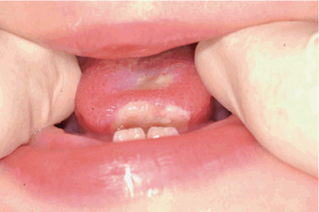
The chief complaints were classified into four categories: parental anxiety, referral from another clinic, pain when breastfeeding, and ulceration of the tongue. The positions of the natal/neonatal teeth were defined by their locations in the jaw: upper and lower incisor, canine, or molar. Clinical features were noted according to the number of affected teeth, presence of increased tooth mobility, enamel hypoplasia, and morphological variations. Teeth were deemed to be ‚Äėmobile‚Äô when the mobility was more than 2 mm in any direction and ‚Äėnormal‚Äô when the mobility was less than or equals to 2 mm. Complications related to natal/neonatal teeth were described using the following categories: none, gingival problems, breastfeeding problems, and ulceration of the tongue, which has been called a Riga-Fede disease (Fig. 1) [4]. The subsequent treatment was categorized as extraction of the affected tooth, observation, and modification of shape.
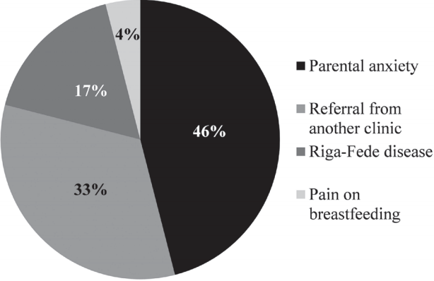
The study population consisted of 21 males and 27 females. 33 (69%) infants had natal teeth and 15 (31%) had neonatal teeth. The most common reason for visiting the dental clinic was parental anxiety, followed by referrals from gynecology clinics, ulceration of the tongue, and pain during breastfeeding (Fig. 2).
The total number of natal/neonatal teeth was 75. More than half of infants (56%) had two natal/neonatal teeth, and the rest (44%) had a single tooth. All of the natal/neonatal teeth were located in the mandibular incisor area. Radiographic examinations of the patients confirmed that all natal/neonatal teeth were deciduous teeth, and none of the affected teeth was a supernumerary tooth.
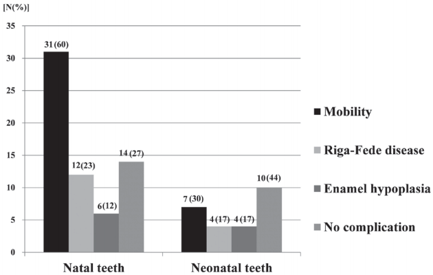
More than half of the teeth showed increased mobility (Fig. 3). Enamel hypoplasia of a natal/neonatal tooth was observed in seven infants, as was morphological variation in the natal/neonatal tooth in one case. Riga-Fede disease was the most common complication (23%), followed by breastfeeding (8%) and gingival problems (4%).
In terms of subsequent treatment, tooth extraction (43%) was the most common treatment of choice, some teeth were maintained and observed (33%), and the rest underwent shape modification (24%; Fig. 4). All modifications involved grinding sharp edges.
After treatment, 13 infants were followed for more than 6 months. Among them, four underwent extractions, and two of them showed space loss. Three patients experienced apical abscesses after grinding.
The occurrence of natal/neonatal teeth has been attributed to several factors (Table 1) [5-11]; however, the etiology of natal/neonatal teeth is not clearly understood. Due to their rarity, most parents have little information on such teeth and this may lead to discomfort for both the mother and infant. A comprehensive understanding the pathology of natal/neonatal teeth is needed for appropriate treatment planning.
In the present study, the prevalence of natal/neonatal teeth was 1:909 and the ratio between natal teeth and neonatal teeth was 2.26:1. The sex ratio between females and males was 1.29:1, and these findings are consistent with previous patterns [1,2].
In previous studies, infants are generally brought to a dental clinic for one of the following reasons: potential risk of aspiration, ulceration of the tongue, difficulty in feeding, interference or discomfort while breastfeeding, and to find out whether a tooth is in pathological or normal status [10,12]. In this study, parental anxiety and referral from a gynecology clinic were the predominant reasons for the dental clinic visit. This might reflect the fact that most Korean women give birth at gynecology clinics, and after delivery, they tend to stay at postnatal care centers associated with gynecology clinics. Given a relatively long stay at a gynecology clinic, physicians may notice the presence of natal/neonatal teeth and recommend a visit to a pediatric dental clinic.
In this study, 44% of the infants had a single natal/neonatal tooth and 56% had two natal or neonatal teeth. Our results support the preference for bilateral occurrence, as in other reports [12-14]. All teeth were found in the anterior portion of the mandible. However, according to King et al. [15], natal/neonatal teeth can be found in various regions of the dentition: most frequently in the anterior mandible, followed by the anterior maxilla, mandibular cuspids or molars, and maxillary cuspids or molars, in descending order. The results and differences with our study are likely due to limited number of samples.
A few infants exhibited sublingual ulcerations, or Riga-Fede disease. This ulceration may be caused by constant traction due to tongue tie, but in our cases Riga-Fede disease was caused by constant trauma from natal/neonatal teeth and may interfere with proper suckling [16]. Insufficient nutrition can prevent infants from gaining weight, putting them at an increased risk of other diseases. In cases of mild tongue irritation, conservative treatments such as smoothing the incisal edge may be the option of choice. However, in cases with a large ulcerated area, even after reducing the incisal edges the teeth can act as irritant to the tongue and interfere with suckling, which can delay healing. In such cases, natal/neonatal tooth extraction is the treatment of choice for rapid resolution of the problem or healing of the lesion [17].
According to Singh et al. [18], if the degree of mobility is more than 2 mm, the natal tooth usually needs to be extracted; we used this as the criterion for mobility. However, unlike previous studies that claimed all natal/neonatal teeth were mobile, about half of the teeth showed increased mobility in our patients [2,10]. This may have resulted from the small sample size and should not be taken as that most Korean infants show no mobility.
In this study, mobility was found more in natal tooth group (Fig. 3). This result can be explained by that natal teeth may have shorter root than neonatal teeth. As described earlier, superficial position of tooth germs may be the reason of natal/neonatal teeth, and natal teeth erupt earlier because they were positioned more superficially at the beginning (Table 1). If natal/neonatal teeth develop same time, and start positions are different, natal teeth will have shorter root when they erupt and eventually show more mobility.
Extraction was the most selected treatment of choice in this study population, due mainly to the increased mobility of such teeth and multiple complicated cases that more than two different complications existed. (Fig. 4). Even though some authors reported natal/neonatal tooth is supernumerary, most other studies have shown that most of such teeth are, in fact, primary teeth [1,13,14,19]. Thus, it would be better to choose maintenance first, rather than extraction.
Apart from extraction, there are other treatments to reduce injury to both the mother and infant. According to Padmanabhan et al. [20], grinding of the incisal edge may be an option to prevent maternal wounding during breastfeeding. Choi et al. [21] described layering the incisal edge with composite resin, which can facilitate rapid healing of an ulcer on the infant’ s tongue. However, unfortunately, most natal/neonatal teeth exhibit immature appearances with enamel hypoplasia, with a limited surface area of enamel available for resin bonding. Also, bonding procedures in these teeth are difficult due to improper moisture control and a lack of cooperation from the infant. Clinicians should also be aware that if the restoration fails, there is a risk that the composite resin could be swallowed [14].
Little information is available about complications after natal/neonatal teeth have been extracted. According to Ooshima et al. [7], after the exfoliation of natal teeth, the retained dental papilla separated from the natal tooth crown can continue its growth and differentiate into another tooth-like structure. Thus, if extraction is used, it is important to also remove the underlying dental papilla and Hertwig’s epithelial root sheath by gentle curettage because root development can continue if these structures are left in situ.
Natal/neonatal teeth should be assessed carefully to determine whether they are supernumerary or normal dentition, so that indiscriminate extractions may be avoided. With the aid of radiographic findings, clinicians should verify the relationship between a natal/ neonatal tooth and adjacent structures including nearby teeth. The presence or absence of a tooth germ in primary teeth will determine whether the tooth belongs to the normal primary dentition or not [22]. Such a comprehensive examination can lead to the maintenance of natal/neonatal teeth of the normal dentition and prevent the premature loss of a primary tooth. Otherwise, a loss of space, collapse of the developing mandibular arch, and subsequent malocclusion in the permanent dentition may occur [23].
After extraction, two of four followed infants experienced space loss. Maintaining primary teeth is important to space maintenance for permanent teeth, and for functional and esthetic reasons. In infants without extractions, the complications included apical abscesses and crown fractures, and two infants’ mothers had problem in breastfeeding. These complications may be related to enamel hypoplasia or morphological variation, which are thought to affect susceptibility to dental caries. Thus, regardless of the treatment, long-term follow-up is essential in the management of natal/neonatal teeth.
The occurrence of a natal/neonatal tooth is a rare phenomenon. When it occurs, the teeth have various clinical characteristics, leading to different complications. Understanding the pathology of natal/neonatal is essential for the overall wellbeing of the child. Careful examination for further treatment planning, and informing parent about all awareness is also important. More efforts should be placed on longitudinal and divergent studies to confirm the etiology and characteristics of natal teeth.
Fig. 1.
Clinical features of Riga-Fede disease. Note the ulceration on the ventral surface of the tongue due to repeated irritation by natal/neonatal teeth.

Fig. 3.
Distribution of natal/neonatal teeth according to clinical features and associated complications. Numbers do not add up to totals because of the presence of multiple complications in several patients.
* Cases in multiple complications : Natal teeth(11), Neonatal teeth(2)

Fig. 4.
Treatment options for natal/neonatal teeth. (A) Distribution of treatments. (B) Relative proportions of treatments according to clinical features and complications.

Table 1.
Hypothetical factors underlying natal/neonatal teeth
References
1. Massler M, Savara BS : Natal and neonatal teeth: A review of twenty-four cases reported in the literature. J Pediatr, 36:349-359, 1950.


2. Kates GA, Needleman HL, Holmes LB : Natal and neonatal teeth: A clinical study. J Am Dent Assoc, 109:441-443, 1984.


3. Magitot E : Anomalies in the erupton of the teeth in man. Br J Dent Sc, 26:640-641, 1883.
4. Slayton RL : Treatment alternatives for sublingual traumatic ulceration (Riga-Fede disease). Pediatr Dent, 22:413-414, 2000.

5. Bjuggren G : Premature eruption in the primary dentition--a clinical and radiological study. Sven Tandlak Tidskr, 66:343-355, 1973.

6. Bigeard L, Hemmerle J, Sommermater J : Clinical and ultrastructural study of the natal tooth: Enamel and dentin assessments. ASDC J Dent Child, 63:23-31, 1995.
7. Ooshima T, Mihara J, Saito T, Sobue S : Eruption of toothlike structure following the exfoliation of natal tooth: Report of case. ASDC J Dent Child, 53:275-278, 1985.
8. ҆tamfelj I, Jan J, Cvetko E, GaŇ°perŇ°ińć D : Size, ultrastructure, and microhardness of natal teeth with agenesis of permanent successors. Ann Anat, 192:220-226, 2010.


9. Jasmin JR, Clergeau-Guerithault S : A scanning electron microscopic study of the enamel of neonatal teeth. J Biol Buccale, 19:309-314, 1991.

11. Alvarez MP, Crespi PV, Shanske AL : Natal molars in Pfeiffer syndrome type 3: a case report. J Clin Pediatr Dent, 18:21-24, 1993.

12. Buchanan S, Jenkins CR : Riga-Fede’s syndrome: Natal or neonatal teeth associated with tongue ulceration. Case report. Aust Dent J, 42:225-227, 1997.


13. Bodenhoff J, Gorlin RJ : Natal and neonatal teeth folklore and fact. Pediatrics, 32:1087-1093, 1963.

14. Brandt SK, Shapiro SD, Kittle PE : Immature primary molar in the newborn. Pediatr Dent, 5:210-213, 1983.

16. Narang T, De D, Kanwar AJ : Riga-Fede disease: trauma due to teeth or tongue tie? J Eur Acad Dermatol Venereol, 22:395-396, 2008.


17. Hedge RJ : Sublingual traumatic ulceration due to neonatal teeth (Riga-fede disease). J Indian Soc Pedod Prev Dent, 23:51-52, 2005.


18. Singh S, Subbba Reddy VV, Dhananjaya G, Patil R : Reactive fibrous hyperplasia associated with a natal tooth: A case report. J Indian Soc Pedo Prev Dent, 22:183-186, 2004.
19. Basavanthappa NN, Kagathur U, Basavanthappa RN, Suryaprakash ST : Natal and neonatal teeth: A retrospective study of 15 cases. Eur J Dent, 5:168-172, 2011.


20. Padmanabhan MY, Pandey RK, Aparna R, Radhakrishnan V : Neonatal sublingual traumatic ulceration - case report & review of the literature. Dent Traumatol, 26:490-495, 2010.


21. Choi SC, Park JH, Choi YC, Kim GT : Sublingual traumatic ulceration (a Riga-Fede disease): report of two cases. Dental Traumatol, 25:48-50, 2009.

- TOOLS
-
METRICS

-
- 0 Crossref
- 0 Scopus
- 4,393 View
- 109 Download
- Related articles
-
A Retrospective Study on the Treatment of Dens Evaginatus for the Last 5 Years2022 May;49(2)


 PDF Links
 PubReader
 ePub Link
 Full text via DOI
 Download Citation
 Print
Print


