맹출 전 치관 내 흡수에 기인한 감염 미성숙 영구치의 치험례
Management of Infected Immature Permanent Tooth with Pre-eruptive Intracoronal Resorption : Two Case Reports
Article information
Abstract
맹출 전 치관 내 흡수(pre-eruptive intracoronal resorption; PEIR)는 미맹출치 치관의 법랑상아경계하방에 위치한 방사선 투과상의 흔치 않은 병소이다. 맹출 전 치아에서의 PEIR은, 흡수 진행속도에 따라 외과적 노출술 후 수복 혹은 지속적 관찰로 치료가능하다. 하지만 맹출 후에는 우식 병소 발달의 위험이 높아 임상가의 대처가 늦을 경우 근관치료 등을 포함하는 심화된 수복치료를 진행하여야 한다. 본 증례는 어린이에서 PEIR로 인해 감염된 미성숙 영구치의 임상적 특징 및 치료에 관하여 보고하고자 한다.
Trans Abstract
Pre-eruptive intracoronal resorption (PEIR) is a rare radiolucent lesion often located within the dentin and adjacent to the dentin-enamel junction, underneath the occlusal aspect of the crowns of unerupted teeth. The treatment approaches for these lesions involved with unerupted teeth have been known as to be relatively simple; depending on the extent of resorption, follow-up or restoration can be performed after surgical exposure. However, once the tooth is exposed to the oral cavity after eruption, it becomes highly vulnerable to the development of carious lesions. Thus, immediate intervention is required in such cases; failure to address it may result in the need for more complex treatments including endodontic therapy. The aim of this case report was to describe the characteristics of PEIR and the clinical management of the impacted immature permanent teeth diagnosed with PEIR.
Ⅰ. Introduction
Pre-eruptive intracoronal resorption (PEIR) appears as a radiolucent lesion, often located within the dentin, adjacent to the dentin-enamel junction and underneath the occlusal aspect of the crowns of unerupted teeth. As the lesion is regarded to be similar to dental caries in appearance, they are referred to as hidden caries, occult caries, developmental caries, or preeruptive caries. In many cases, these lesions are diagnosed incidentally before tooth eruption on panoramic radiographs; the prevalence of the lesions has been reported to be 3 - 6% in children and adolescents and in 0.5 - 2% of teeth [1,2]. Based on previous studies [3-5], PEIR occurs most commonly in first premolars and molars and rarely in mandibular canines.
Detection of the lesion has been known to be difficult at the pre-eruptive stage of PEIR because an unerupted tooth is rarely infected with cariogenic microorganisms and, thus, such lesions are asymptomatic. However, when the affected tooth is exposed to the oral cavity, microorganisms can invade the dentin through fissures, and cause caries in the resorbed area resulting in pulpal inflammation in severe cases [6]. Thus, PEIR is reported to be one of the least common causes of pulpal abscesses, which develop only within a brief window of time during childhood [7].
Previous studies have demonstrated three methods for treating PEIR; the method used depends on several factors, such as the expected size of the lesion at the time of tooth eruption, the rate of progression, the degree of cooperation of the patient, and the risk of caries. The treatment strategies for PEIR include (1) tooth extraction (when restoration is not feasible), (2) restoration after surgical exposure, and (3) restoration (including pulp treatment) after a follow-up examination until tooth eruption [8].
Although PEIR is regarded as a quite rare dental defect, some clinical cases have been described with simple restorations after surgical exposure [6,9-11]. However, cases involving pulp treatment were very rare. The purpose of this report was to describe the clinical symptoms and signs of PEIR and to discuss the management of an infected immature permanent tooth with PEIR.
Ⅱ. Case Reports
Case 1
An 8-year-old girl was referred from a local clinic to Chonnam National University Dental Hospital. The dentist who has referred the patient to the clinic diagnosed PEIR. The patient had visited a local dental clinic 5 months prior to visiting the Department of Pediatric Dentistry and was in pain (Fig. 1). A clinical examination revealed poor oral hygiene, and the left mandibular permanent canine had already erupted through the gingiva, with a notable localized swelling in the buccal mucosa along with sensitivity to percussion and palpation. Although the tooth did not respond to a cold test, pocket depths and mobility were all within the normal range compared with adjacent and contralateral teeth. On radiographic examination, periapical radiolucency and widening of the periodontal ligament (PDL) were evident in relation to the left mandibular canine. An extensive radiolucent area in the crown, suggesting signs of PEIR, was also apparent (Fig. 2A). The lesion seemed to extend from the dentin-enamel junction to the center of the crown, towards the distal surface, and extending to encroach on the pulp. The tooth was diagnosed with pulp necrosis and apical periodontitis. The treatment of choice was regenerative pulp treatment of the immature root. The parent accompanying the patient was provided with detailed explanations of possible treatment options, and consent was obtained to perform revascularization, which was considered to be the most appropriate approach in this case.

Panoramic radiographic image of the patient at the first visit. An incompletely developed apex with periapical radiolucency and widening of the periodontal ligament space of the root of the left mandibular canine are shown.
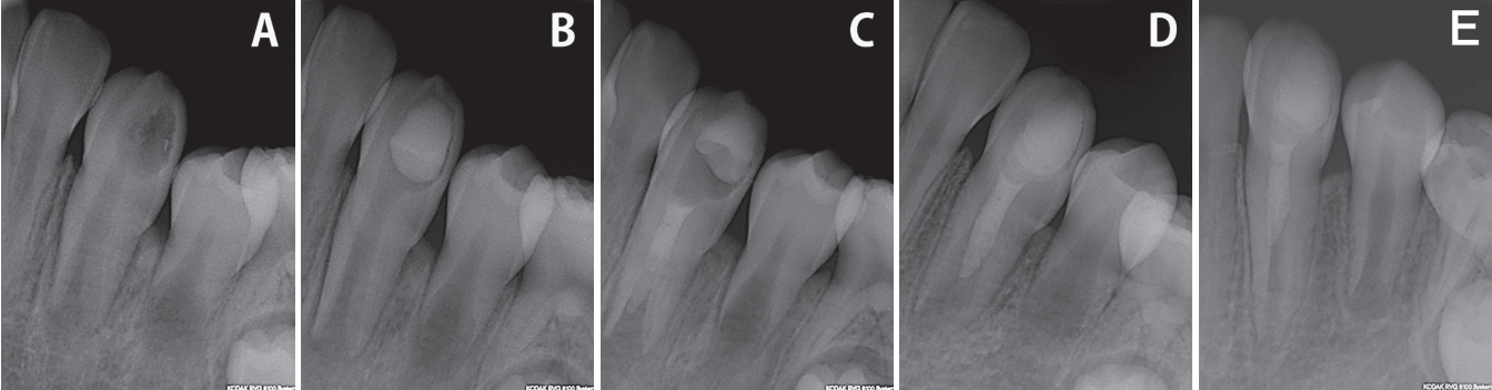
Periapical radiographs of the left mandibular canine. (A) Preoperative state, (B) Bi-antibiotic paste dressing, (C) MTA application, (D) At 6-month follow-up, (E) At 14-month follow-up.
A rubber dam was placed, and both the mantle dentin and the rest of the dentin affected by PEIR caries were removed during the access cavity preparation. Bleeding was not evident in the pulp chamber at the opening of the root canal for endodontic therapy. However, the patient complained of discomfort on insertion of a size #10 K-file.
The root canal was irrigated gently with 10 mL of 2.5% sodium hypochlorite (NaOCl) and dried with paper points. Biantibiotic paste (ciprofloxacin and metronidazole), a material developed after the exclusion of minocycline from the triantibiotic paste because of the discoloring properties of minocycline, was applied as a root canal dressing using a Centrix syringe (Gentrix, USA) to reach the cemento-enamel junction (CEJ). A temporary seal was made using a sterile cotton pellet and Caviton (GC, Japan) (Fig. 2B).
Two and a half weeks later, the gingival swelling had subsided. However, the tooth remained sensitive to percussion, for which root canal irrigation was performed. Bi-antibiotic paste was reapplied.
Three weeks later, the patient reported relief from pain and the tooth was no longer symptomatic or sensitive to percussion. The tooth was anesthetized with 3% mepivacaine (Septodont; Cedex, France) without a vasoconstrictor to facilitate bleeding. After rubber dam isolation and removal of the temporary restoration, the bi-antibiotic paste was removed from the canal with 10 mL of 5.25% NaOCl. The canal was dried with paper points. To minimize discoloration, Clearfil SE bond (Kuraray, Japan) was applied above the level of CEJ at the access opening, and glass-ionomer (GI) cement was used to restore the tooth and reinforce the structure. A #20 K-file was used to induce bleeding by irritating the apical tissue, resulting in over-instrumentation of the root apex. The blood was filled up to 3 mm below the CEJ, and clot formation required approximately 15 minutes. RetroMTA was applied to the clot, in an area with a thickness of 3 - 4 mm, under sterile conditions. Subsequently, a moist cotton pellet was placed, and a temporary seal was created using Caviton (Fig. 2C).
A week later, after a tight coronal seal formed by mineral trioxide aggregate (MTA) was confirmed, a composite resin restoration of the left mandibular canine was performed using resin-modified GI cement (Fuji II LC, Japan) as a base.
At the 6-month follow-up, no particular clinical symptoms were observed, and the previous radiolucent area was no longer present. A normal appearance of the root apex was observed (Fig. 2D). At the 14-month follow-up examination, the patient did not exhibit any particular clinical signs. Furthermore, continued root development, in terms of length and thickness, was evident on the radiograph (Fig. 2E). Additionally, the tooth color remained unchanged (Fig. 3).

Clinical photographs of the patient. No apparent discoloration of the left mandibular canine was observed at the 14-month follow-up. (A) Frontal view, (B) Occlusal view.
Case 2
An 8-year-old male patient visited Chonnam National University Dental Hospital with dental caries on his molar teeth. His medical and dental histories were unremarkable except for mental retardation. A clinical examination revealed deep caries on all permanent first molars and many primary molars. On radiographic examination, there was some distortion on the panoramic radiographic image. However, radiolucency was apparent inside the coronal portion of the left mandibular first premolar, which was not exposed to the oral cavity. An extensive radiolucent area was found in the middle of the crown, suggesting signs of PEIR (Fig. 4A). Because the tooth was impacted below the primary first molar, and due to limitations in compliance, it was challenging to obtain a periapical radiograph for a more accurate diagnosis. The left mandibular first premolar was impacted below the preceding primary molar, making it difficult to intervene. Thus, the plan was made to wait for the tooth to erupt.
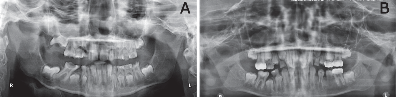
A panoramic radiographic image of the patient. An incompletely developed apex with radiolucency of the crown of the left mandibular first premolar is shown. (A) At first visit, (B) At 1-year follow-up.
A year after the first visit, the left mandibular first premolar had erupted (Fig. 4B). There was no evidence of caries invasion, but a dark area was observed under the enamel layer (Fig. 5). There was no response on percussion, and the tooth did not respond to the cold test or EPT. Panoramic and periapical radiographic images revealed a periapical radiolucency at the apex of the tooth. Pulp necrosis of the immature left mandibular first premolar was diagnosed after clinical and radiographic examinations (Fig. 6A). The parent accompanying the patient was provided with detailed explanations of possible treatment options including apexification and apexogenesis, and consent was obtained to perform revascularization, which was considered to be the most appropriate approach for this case.
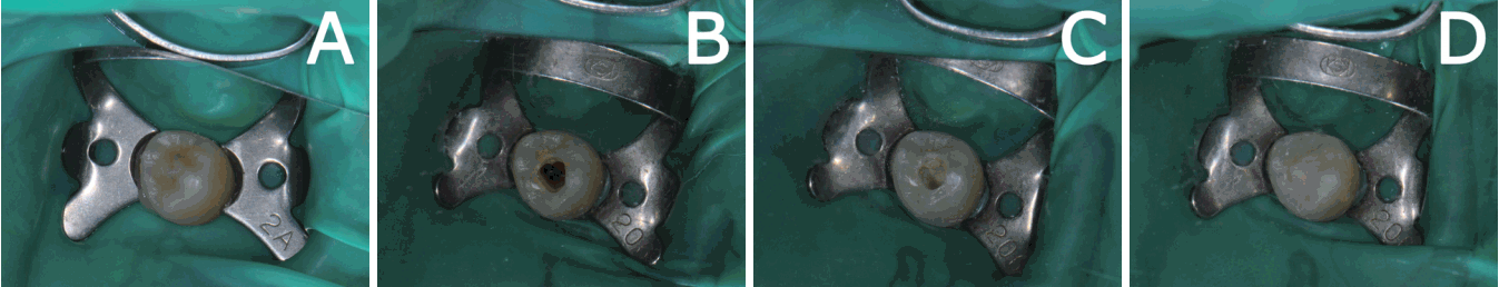
Root canal revascularization procedure. (A) Clinically, the left mandibular first premolar presented with a dark interior, (B) After inducing a blood clot, (C) Placement of MTA in the canal, (D) Final composite resin restoration.

Periapical radiographs of the left mandibular first premolar. (A) Preoperative state, (B) MTA application, (C) At 3-month follow-up.
The patient was treated under midazolam intramuscular sedation with nitrous oxide-oxygen (N2O/O2) inhalation. The gingiva and tooth were anesthetized with 2% lidocaine and 1:100,000 epinephrine. A rubber dam was placed, and using a high-speed bur, sound enamel above the lesion was removed. The mushy dentin was carefully removed with an excavator and a low-speed round bur. There was pulp exposure without evident bleeding in the pulp chamber at the opening of the root canal available for endodontic therapy.
The root canal was irrigated gently with 10 mL of 2.5% NaOCl and dried with paper points. Bi-antibiotic paste was applied as a root canal dressing using a Centrix syringe to reach the CEJ. A temporary seal was made using a sterile cotton pellet and Caviton (Fig. 6B).
Two and a half weeks later, the symptoms and signs had subsided. The antibiotics applied as a dressing in the root canal were removed, and a #20 K-file was used to induce bleeding by irritating the apical tissue, resulting in over-instrumentation of the root apex. The blood was filled up to 3 mm below the CEJ, and approximately 15 minutes were required for clot formation. RetroMTA was applied to the clot, in an area with a thickness of 3 - 4 mm, under sterile conditions. After a tight coronal seal formed by MTA was confirmed, a composite resin restoration of the left mandibular first premolar was performed.
At the 3-month follow-up, no particular clinical symptoms were observed, and the previous radiolucent area was no longer present. A normal appearance of the root apex was observed (Fig. 6C). The patient was scheduled for follow-up at a 3-month intervals.
Ⅲ. Discussion
In the present study, PEIR of immature permanent teeth coincidently diagnosed via radiographic images taken for routine examinations was described. In the first case, the patient was diagnosed with PEIR at a local dental clinic 5 months before visiting the Chonnam National University Dental Hospital. At the time of her visit, the crown had already erupted through the gingiva. However, based on the patient's dental history, in addition to radiographic and clinical examination, consecutive pulp infection and pulp necrosis driven by PEIR were diagnosed. The thin enamel floor was considered to have been caused by the defect in the dentin and a relatively small enamel fracture; appropriate treatment was administered.
In the second case, the patient visited for caries treatment, and the panoramic image unintentionally revealed PEIR of the tooth. At the first visit, the left mandibular first premolar was located below the preceding primary molar, and periodic follow-up was planned. The tooth erupted a year later. Unlike the first case, no enamel fracture or lesion was seen, but intracoronal resorption and radiolucency around the root apex was evident, requiring treatment.
Generally, PEIR of a developing, unerupted tooth have been discovered only through radiographic examination. The pathogenesis of PEIR remains to be fully determined. However, according to histological examination of lesions in previous studies, it might represent a normal resorption process, with accompanying signs such as the presence of osteoclasts at the scalloped dentin-enamel junction. The exact origin of resorptive cells and their progression pathways have not been fully determined [7,8]. Ectopic eruption or prior infection of immature permanent teeth can also be considered as relevant factors [7]. No association has been found between PEIR and race, gender, medical conditions, systemic factors, or fluoride supplementation. Additionally, the medical history of the patient in this case study revealed no history of atypical systemic disease or infection of the deciduous teeth [6].
This type of atypical resorption process is proceeded at a slow pace until the exposure of the crown to the oral environment. However, once the tooth erupts through the gingiva, cariogenic microorganisms invade and progress into the dentin via the resorbed area, and the extensive size of the lesion makes it distinguishable. As the cavity is exposed after full eruption, the lesion is observed to be filled with material that mostly comprises decomposed mantle dentin, or it may become relatively vacant. When the collapse of the cusps of the tooth naturally reveals the cavity, the lesion extends further and is colonized rapidly by microbial flora from the oral cavity [6,10].
Regenerative endodontic therapy enables the infected pulp tissues to regenerate and heal. This includes attaining normal function with regard to sensory innervation, immunocompetency, and root development and formation [12,13]. In the case of pulp necrosis in an incompletely developed root, such treatment can increase both the length and thickness of the root. Regenerative endodontic therapy using calcium hydroxide and MTA has been shown to be more effective than apexification, and it has the same effect regarding the increase in length and thickness of the root [12,13]. In the first case, revascularization was performed to treat a mandibular permanent canine with PEIR, and follow-up at 14 months revealed increased length and thickness of the root compared with the initial radiographic examination. Additionally, no sign or symptom of inflammation was detected. A positive response also was exhibited in electric pulp testing. Unfortunately, a longer follow-up period was required in the second case to adequately evaluate the results, but the signs and symptoms have subsided as did the periapial radiolucency after the treatment.
Because RetroMTA was known not to cause tooth discoloration, it was used to fill the cervical root canal, and minocycline was eliminated from the triple antibiotics used in the canal dressing due to its property of tooth discoloration [14]. Consequently, as in the first case, the color of the treated tooth remained unchanged a year after the treatment.
The study of Hoshino et al. [15] demonstrated that a combination of ciprofloxacin, metronidazole, and minocycline effectively reduced the number of bacteria inside an infected root canal. However, triple antibiotics, including minocyline, were known as the main cause of tooth discoloration after endodontic treatment [14]. To avoid this, amoxicillin, cefaclor, cefroxadin, clindamycin, or bi-antibiotic paste have been used as an alternative [16]. Kim et al. [13] reported a successful regenerative endodontic treatment case without tooth discoloration using RetroMTA and a tri-antibiotic paste that included alternative materials to minocycline. RetroMTA, used instead of ProRootMTA (Dentsply, USA), is a bioceramic material that exhibits similar levels of biocompatibility, as well as angiogenic and odontogenic effects, compared with ProRootMTA. Recently, it has been used in both in vitro and in vivo studies, having been reported as an effective alternative to ProRootMTA. The main advantages of RetroMTA include its reduced setting time and that it contains zirconium oxide as a radiopaque material, resulting in less discoloration than MTAs that contain bismuth oxide (e.g., ProRootMTA and MTA Angelus) [17,18].
Early diagnosis and appropriate management are important in PEIR. The prognosis of a tooth affected by PEIR varies according to several factors, such as the size of the lesion, the degree of pulp involvement, and the developmental level of the root [8]. In particular, regenerative endodontic treatment is needed for a cavity extending to the pulp of an immature tooth with an open apical foramen and as seen in previous studies, the prognosis may not be promising. Thus, treatment was provided after obtaining written informed consent from a parent in both cases.
Despite the early diagnosis of PEIR, endodontic treatment was unavoidable in both cases. In the first case, the patient had a nearly 5-month delay in visiting the hospital, causing progression of pulp inflammation, whereas the patient in the second case showed poor compliance for a surgical approach. Treatment was continued after eruption, but pulpal inflammation and necrosis were observed. PEIR is known to occur when the tooth erupts and becomes susceptible to anachoresis, leading to caries development. It has been reported that application of GI sealant, with early diagnosis, can readily prevent anachoresis [3,6]. Although a longer follow-up period was required for the second case to ensure treatment success, these two cases indicated that regenerative endodontic treatment was an effective method for resolving the lesion around the apex of the root, ensuring continued development of the dental root and avoiding additional clinical symptoms. As a result, the prognosis of a tooth with PEIR is determined by an appropriate treatment plan for the affected tooth that takes into account early diagnosis, lesion progression, caries susceptibility, and the degree of cooperation of the patient regarding oral hygiene measures.
Ⅳ. Summary
PEIR is known to be a rare defect in the dentin of an unerupted tooth, just beneath the dentin-enamel junction, with a prevalence of 0.5 - 2% of teeth. The two cases presented here have shown favorable results of regenerative endodontic therapy for pulpally infected immature permanent teeth with PEIR. Especially, the outcome of the case with the canine was optimistic, showing no coronal discoloration, with normal root development and lack of symptoms within a year.
The findings of our study indicated that prior to performing regenerative endodontic treatment for the immature apex of a root, factors inducing tooth discoloration should be considered. Moreover, an early diagnosis and appropriate treatment strategy for the affected tooth are considered to be crucial to avoid pulp involvement caused by a PEIR defect.