


I. Introduction
Hydrocephalus is a condition in which the volume of inadequately absorbed cerebrospinal fluid (CSF) is abnormally high in the cerebral ventricles and occasionally in the subarachnoid space [1]. This condition results in increased intracranial pressure and often damages the surrounding tissues [2].
Hydrocephalus has multifactorial causes and may present alone or in association with other neurological diseases [2]. The most common causes are intraventricular or subarachnoid hemorrhage, meningitis, tumors, cysts, and inflammation. Congenital hydrocephalus can also be caused by aqueductal stenosis, Dandy-Walker malformation, myelomeningocele, Chiari malformations, and encephalocele [2]. Hydrocephalus is the most common neurosurgical problem in pediatric neurosurgery, with a pediatric incidence rate ranging from 0.4 to 0.8 per 1000 births [3].
The signs and symptoms of pediatric hydrocephalus vary depending on the age, the severity, and the etiology. Infant patients with hydrocephalus exhibit apnea, bradycardia, seizures, vomiting, sunsetting eyes and rapid head growth. The signs and symptoms in children older than 2 or 3 years are headache, nausea, vomiting, irritability, lethargy, delayed development, behavioral disturbance, sunsetting eyes, visual complaints, bradycardia, hypertension and irregular breathing patterns [2]. Because of these signs and symptoms, most hydrocephalus patients require sedation during dental treatment. With growth, shunt-treated patients may exhibit structural differences in the craniofacial region compared with normal people [4,5]. During the pubertal stages, the difference between the level of calcification in the permanent teeth and the chronologic age is higher than for normal healthy children [1].
CSF shunting is the most common treatment for hydrocephalus. Shunting involves transferring the CSF to another body cavity with a ventricular catheter, valve, and distal catheter. There are various shunt treatments in accordance with the drainage location to position the distal catheter and the ventriculoperitoneal shunt (VPS) is the most commonly used and preferred method. If there are any problems in the peritoneal cavity such as an abdominal malformation, postsurgical adhesions, infections, or inadequate reabsorption, ventriculoatrial shunts (VAS) and ventriculopleural shunts are the second and third choices for treatment [6].
The main complications of shunt treatment are infection, overshunting and mechanical problems including migration of the components [2,6]. Vigorous movement by the patients, which may occur during dental treatment, can also displace the distal catheter of the shunt.
Pediatric hydrocephalus is less known and has not been reported in cases about dental management despite the high incidence in pediatrics. Therefore, the aim of this report is to present a case of dental management of pediatric hydrocephalus.
II. Case Report
A 7-year, 6-month old girl presented to the Department of Pediatric Dentistry Gangneung-Wonju National University Dental Hospital for the treatment of facial swelling on her right cheek. She had a history of hydrocephalus and had received VPS surgery 5 years and 3 months earlier. Her mother stated that her daughter felt pain on her lower right posterior teeth about 2 years ago, but had not received any dental treatment because of her strong aversion to a dental visit. During the examination, she exhibited poor cooperation and behavioral disturbance, crying loudly and covering her mouth with her hand. We attempted to obtain a panoramic radiograph, but were unsuccessful because of her constant movements. Therefore, we took only periapical radiographs.
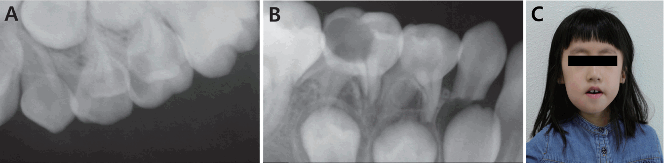
In intraoral and radiographic examination, caries lesions were detected on the left maxillary deciduous second molar and right mandibular deciduous second molar (Fig. 1A, B). Extraoral examination showed swelling on the right mandible, abnormal enlargement of the head, delayed mental and somatic development and blurred vision. (Fig. 1C). The delayed development was recognized due to her limited communication skills and immature adaptive behavior.
The patient was discussed in consultations with the neurosurgery department at the General Hospital to evaluate the medical complications that can occur during invasive dental treatment, sedation, and protective stabilization as well as the need for prophylactic antibiotics. We received a response that the patient could receive the usual dental procedures under sedation with drugs and did not require prophylactic antibiotics.
We chose to perform pulpectomy and restoration with temporary filling material on the right mandibular deciduous second molar under nitrous oxide inhalation sedation as an emergency dental treatment for the following reasons: low correlation between dental infection and shunt infection; difficulty with applying a fixed space maintainer due to partially erupted permanent first molars; prediction of a lack of cooperation for the application of a removable space maintainer; weak effect of oral chloral hydrate used in medical examination; her motherŌĆÖs rejection of intra-muscular injection and general anesthesia; relatively small amount of dental treatment required; limited, but simple communication was possible; minimal movement of the neck past the distal catheter of the shunt.
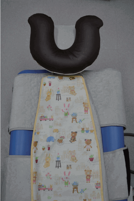
We identified the location of the catheter by palpation of her neck before treatment and a neck pillow was placed under the neck (Fig. 2). Pulpectomy was performed on the right mandibular deciduous second molar under nitrous oxide inhalation sedation preceded by incision and drainage on the right mandibular vestibule. During the treatment, a pedi-wrap was applied loosely and the second assistant gently restricted the head of the patient with both hands to prevent intermittent body movement by the patient and irritability at the sound of the high speed hand piece. Amoxicillin 120mg and acetaminophen 975mg t.i.d. were prescribed for 5 days in addition to 1 bottle of chlorhexidine for the purpose of facial abscess relief. At the 1-week follow-up, the tooth and soft tissues appeared to be in a good healing state and facial swelling was reduced (Fig. 3).
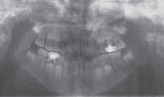
After emergency treatment, dental treatment was performed four times over a period of about a year. Stainless steel crown restoration on the right mandibular deciduous second molar as well as pulpectomy and stainless steel crown restoration on the left maxillary deciduous second molar was performed under local anesthesia and nitrous oxide inhalation sedation for about 20 to 30 minutes. Sealants on the first permanent molars were applied without local anesthesia and nitrous oxide inhalation for about 10 minutes. This patient showed a progressively improved cooperation.
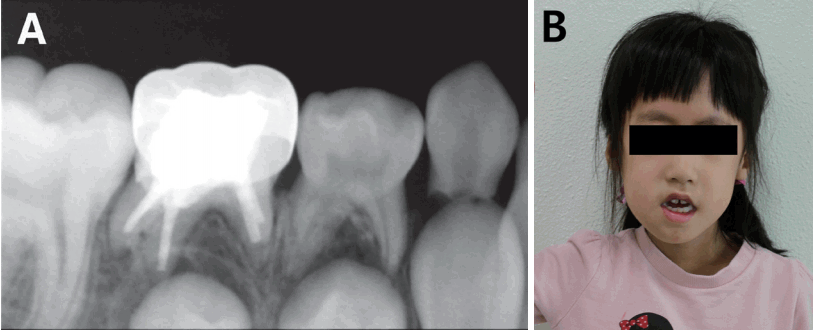
The teeth that had been treated did not appear to have any specific complications until the most recent visit (Fig. 4). This patient was scheduled for the management of overall oral hygiene, development and eruption of permanent teeth, and structural change of the craniofacial region through periodic follow-up.
III. Discussion
Hydrocephalus can occur at any age from infants to adults. However, pediatric hydrocephalus is important in that irreversible changes in the brain occur in the absence of proper treatment and the complications are more fatal than those of adult hydrocephalus (normal pressure hydrocephalus) [2]. Pediatric dentists also require a thorough understanding of hydrocephalus in order to improve the quality of life for patients by providing dental management based on accurate information.
In this case, the patient exhibited macrocephaly, irritability on sound, delayed somatic and metal development, behavioral disturbance and blurred vision as the signs associated with the hydrocephalus (Fig. 1).
A number of patients with intellectual and developmental disabilities experience fear and anxiety during dental care and treatment and most of them require sedation or general anesthesia as behavior management techniques [7]. For hydrocephalus patients, several additional factors should be considered to determine the appropriate method for behavior management.
Shunt-treated patients possess a distal catheter connecting the intraventricular valve and drainage cavity. This catheter is subcutaneously located in the neck of patients and can be easily touched during dental treatment. Pediatric neurosurgeons are advised not to exert excessive force on the catheter as mishandling can lead to physical complications of the shunt system such as improper placement, obstruction, fracture, and migration of the components [2,6]. If dental treatment is planned for shunt-treated patients, it is essential to check the location of the catheter through palpation before treatment. Use of a pillow or cushion such as an anti-bedsore water cushion or a foam pillow may be useful for preventing excessive force and will provide a comfortable position for the patientŌĆÖs head [8].
If a significant amount of dental treatment is required or if the cooperation of the patient is very poor, sedative drugs that can induce deep sedation or general anesthesia may be considered. To date, contraindicated sedative drugs have not been reported in patients with hydrocephalus, with the exception of ketamine, which is not recommended in hydrocephalus patients due to the possibility of intracranial hypertension [9].
Protective stabilization should be carefully applied if required during dental treatment. Carvalho et al. [10] reported that the catheter in a 2-year-old boy migrated to the supraclavicular region from the abdomen after dental treatment, although the specific dental situation was not described. The authors explained that the migration of the catheter was caused by the mechanical forces applied to the shunt and vigorous movements of the patientŌĆÖs head. Patients under mild to moderate sedation may show unexpected reactions during treatment. Therefore, if necessary, proper restriction of the head and shoulders should be employed while a cushion is placed under the neck to prevent excessive neck-movements.
This patient showed improved cooperation regardless of the use of nitrous oxide inhalation sedation after the first visit. This is thought to have been influenced by the adaptation to the dentist, the shorter treatment duration, lack of local anesthesia usage, simplified treatment procedure and reduced use of the high speed hand piece.
The symptoms of hydrocephalus that can interfere with dental treatment include headache, vomiting, seizures, bradycardia, apnea, and irregular breathing patterns, although they were not observed in this patient. Headache, nausea, vomiting, and lethargy are usually present and typically occur in the morning [2]. Thus, early morning appointments for patients with the above symptoms should be avoided. Hydrocephalus patients with bradycardia, systemic hypertension, irregular breathing patterns, and intraventricular hemorrhage require medical care prior to dental treatment [2,11].
Opinions vary about the use of prophylactic antibiotics before dental procedures in shunt-treated patients [2,12-15]. Some studies and the National Health Service in the United Kingdom have reported that there was no relationship between the dental procedure and the infection of shunt systems and that prophylactic antibiotics are not needed [12-14]. However, George et al. [15] and most neurosurgeons recommended using antibiotic prophylaxis in children with VAS and a history of recurring shunt infection although shunt infection is infrequent after dental procedures [2]. There are no reports to date detailing the infection of a shunt with a dental origin. In this case, the medical opinion was that prophylactic antibiotics were not required during dental treatment for the patient, who had a history of VPS surgery. Thus, we recommend a referral to a pediatric neurosurgeon prior to invasive dental treatment for patients with VAS or a history of recurring shunt infection.
The craniofacial structures of shunt-treated patients differ from those of normal people [4,5]. Especially in boys, the angle between the clival and sphenoidal planes may be smaller due to fluctuations in dura stress and the sella may be at a lower position due to mechanical interference from the catheter running along the patientŌĆÖs neck [5]. Unilateral valve-regulated shunt devices may cause craniofacial asymmetry, resulting in the rotation of the maxillary and mandibular horizontal line and the deviation of the maxillary and mandibular interincisal lines on the same side as the inserted device [4]. Possible reasons for this include craniosynostosis, growth restriction caused by scars and fibrosis, or muscular imbalance [4]. Although severe asymmetry requiring additional treatment has not yet been reported, continuous monitoring may be necessary.
Patients with hydrocephalus have also shown a slightly different pattern for the calcification of the permanent teeth. According to Pirttiniemi et al. [1], dental age determined based on the degree of calcification of the permanent teeth was more advanced at the early pubertal stage and delayed at the late stage compared to the chronological age. This phenomenon was observed in both sexes, but was greater in boys and showed a more significant difference at a later state than the early stage [1]. This difference may be explained by the alteration of hormones of the pituitary gland induced by increased pressure and is consistent with the changes of somatic growth patterns in patients with hydrocephalus [1]. The current patient has not exhibited these differences yet, but awareness of this difference will be helpful when dealing with adolescent patients with hydrocephalus.
The patient was admitted to the dental clinic about a year after she first experienced pain due to severe aversion to treatment. If appropriate treatment period is missed, treatment becomes more difficult. Patients with VAS particularly require good oral hygiene to avoid transient bacteremia and shunt infection [2]. Dental clinicians should impress upon the caregiver/parent the significance of a dental home and regular dental checkups.
IV. Summary
Because most hydrocephalus patients show delayed mental development and have shunt devices in their body, they require appropriate behavior management during dental treatment.
In shunt-treated patients, palpation of the catheter running along the patientŌĆÖs neck is necessary before treatment and excessive force to the catheter should be avoided.
For invasive dental treatment of patients with VAS, referral to pediatric neurosurgeons is recommended for prophylactic antibiotics. Hydrocephalus patients with severe symptoms who are medically compromised require medical care prior to dental treatment.
Craniofacial asymmetry or developmental differences of the permanent teeth, which may occur during the growth of hydrocephalus patients, should be evaluated through regular checkups.
 PDF Links
 PubReader
 ePub Link
 Full text via DOI
 Download Citation
 Print
Print


