완전 탈구된 미성숙 영구치의 치수재생치료 증례 보고
Outcome of Regenerative Endodontic Treatment for an Avulsed Immature Permanent Tooth: A Case Report
Article information
Abstract
치아의 완전 탈구는 치조골에서 치아가 완전히 이탈된 것으로 정의되며, 결과적으로 신경혈관 공급의 중단을 야기하는 가장 심각한 치과적 손상 중 하나로 알려져 있다. 완전 탈구는 조직 허혈(tissue ischemia)을 야기하며, 이는 치수 괴사를 초래할 수 있다.
치근단형성술(apexification)은 치수 괴사로 진단된 미성숙 영구치에서 치근단 장벽을 유도하는 전통적인 치료 방법이다. 하지만 치근단형성술로는 치근 길이 및 두께의 증가를 포함하는 치근 발육을 얻을 수 없다.
본 증례는 완전 탈구되어 재식된 이후 치수 괴사로 진단된 치아를 가진 5세 환자에서 ciprofloxacin, metronidazole, cefaclor 및 CollaTape과 Biodentine을 이용하여 시행된 치수재생치료(regenerative endodontic treatment)의 임상적 및 방사선학적 결과를 다루고 있다.
Trans Abstract
Dental avulsion, defined as the complete displacement of a tooth from the alveolar bone with consequent loss of the blood and nerve supply, was reported as one of the most severe dental injuries. Avulsion can cause tissue ischemia, which leads to pulp necrosis.
Apexification is a conventional treatment method that induces an apical calcified barrier in immature roots with pulp necrosis. However, root development characterized by an increase in the root thickness and length cannot be achieved by apexification.
The purpose of this case report was to describe the radiographic and clinical outcomes of regenerative endodontic treatment for the avulsed and necrosed permanent tooth with an immature root after replantation in a 5-year-old girl; the treatment was performed using a mixture of ciprofloxacin, metronidazole and cefaclor, CollaTape and Biodentine.
Ⅰ. Introduction
The treatment of traumatic dental injuries in children is faced with several clinical challenges[1]. Dental avulsion, defined as the complete displacement of a tooth from the alveolar bone with consequent loss of the blood and nerve supply, was reported as one of the most severe dental injuries. The treatment of traumatic dental injuries in children is faced with several clinical challenges[1]. Dental avulsion, defined as the complete displacement of a tooth from the alveolar bone with consequent loss of the blood and nerve supply, was reported as one of the most severe dental injuries. Avulsion can cause tissue ischemia, which leads to pulp necrosis[2]. With regard to avulsed permanent teeth with immature roots, immediate replantation or storage in appropriate media until replantation can aid in achieving pulp revascularization. However, the possibility of pulp necrosis shouldn’t be neglected because it might cause inflammatory root resorption, which is especially rapid in immature permanent teeth[3].
Permanent teeth with immature roots exhibit thin dentin walls, an unfavorable crown-root ratio, and wide apices, which increase the complexity of endodontic and restorative procedures[4]. Apexification or root-end closure is a conventional treatment method including long-term use of calcium hydroxide dressing or placement of a mineral trioxide aggregate (MTA) apical plug. However, root development characterized by an increase in the root thickness and length cannot be achieved by apexification[4,5].
Recently, evidence for pulp regeneration, even in infected immature teeth, was presented[4]. Clinical guidelines for regenerative endodontic treatment (RET) published by the American Association of Endodontics (AAE)[6] and the European Society of Endodontology[7] proposed a two-step protocol as follows. At the first visit, the clinician performs chemical debridement and places intracanal medicaments comprising calcium hydroxide or antibiotic pastes. At the second visit, bleeding is induced, the blood clot is capped, and the access cavity is restored. The goal of two-step RET is the elimination of symptoms, healing of the lesion, promotion of an increase in the root thickness and length, and restoration of pulp vitality indicated by a positive response to vitality testing[6].
The aim of this report was describing the radiographic and clinical outcomes of RET after replantation of an avulsed and necrosed permanent mandibular central incisor with an immature root in a 5-year-old girl.
Ⅱ. Case Report
A healthy 5-year-old girl presented to the emergency room at Wonkwang University Daejeon Dental Hospital with avulsion of the mandibular right central incisor secondary to dental trauma sustained during a fall at home 1 hour back. The tooth had been stored in cold milk until presentation at the emergency department. The need for emergency dental treatment was explained and verbal consent was obtained. The tooth was replanted and splinted using a flexible wire and flowable composite resin (Fig. 2A). Radiographic examination revealed that the tooth root was in the early stage of development (Fig. 1A).
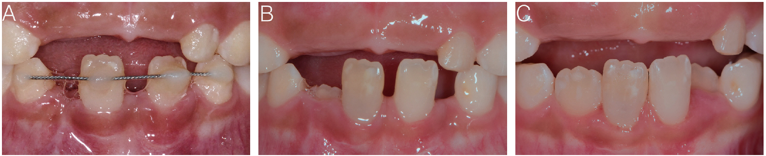
Pre- and postoperative photographs of the patient. (A) Photograph obtained after replantation, (B, C) Photographs obtained at the 7- and 12-month recall visits presented grayish discoloration of the tooth.

Pre- and postoperative radiographs of the patient. (A) Radiograph obtained after replantation presented that the replanted tooth was in the early stages of root development, (B, C) Radiographs obtained at the 7-, and 12-month recall visits presented continued root development and deposition of hard tissue.
During the follow-up period after replantation and splinting, pulp of the tooth became necrosed. Apexification was considered, but the root would not develop. Because the tooth root was extremely immature with pulp necrosis, the long-term prognosis of the tooth with an extremely short root was not favorable, so RET was considered as the treatment of choice. The treatment procedures, risks, and benefits were explained to the patient’s legal guardians and verbal consent for treatment was obtained.
Following the induction of local anesthesia with 2% lidocaine containing 1:100000 epinephrine, the tooth was isolated using a rubber dam and access cavity was prepared. On gaining access, avascular tissue within the canal was observed. The canal was passively irrigated with 1% sodium hypochlorite and sterile normal saline, following which it was dried using sterile paper points. However, these irritated the periapical tissue and induced bleeding. A creamy paste containing equal powdered proportions of ciprofloxacin, metronidazole, and cefaclor mixed with glycerol was applied in the canal using a lentulo spiral. The tooth was temporarily restored using Caviton (GC, Japan).
The patient missed her next appointment and returned after 6 weeks. Abundant bleeding was observed within the canal, so the antibiotic paste was applied again.
After 2 weeks, persistent abundant bleeding within the canal was observed. This was considered as induced bleeding due to overinstrumentation at the first visit. Therefore, Collatape (Zimmer, USA) was adapted on the blood clot above the cementoenamel junction (CEJ). Then, Biodentine (Septodont, USA) was gently adapted on the CollaTape and the cavity was permanently restored with composite resin.
The patient was followed up for 12 months, and remained asymptomatic with no complaint of pain during this period. However, slight grayish discoloration was observed (Fig. 2B, 2C). Periapical radiographs revealed an increase in the root thickness and length and apical narrowing. The root length became similar to that of the contralateral central incisor (Fig. 1B, 1C). At the 12-month recall visit, cone beam computed tomography (CBCT) was taken to evaluate accurate calcification state formed in the root canal space and any reason of discoloration of the crown. An axial view revealed nearly calcified layers between the Biodentine and the preoperative apex and an empty space between the pre- and postoperative apices. The increased root section was completely attached to the original root (Fig. 3).

Axial cone beam computed tomography images obtained at 1-year follow-up. (A) The Biodentine layer, (B) The calcified barrier, (C) The canal space is mostly filled by calcified layers, (D) There is an empty space between the pre- and postoperative root apices. The arrows indicate the affected tooth.
Ⅲ. Discussion
The ideal outcomes of RET include an increase in the root thickness and length and formation of the root apex[8]. However, these outcomes are not always achieved in teeth with immature roots that are subjected to trauma[8-13]. In this report, we described a case involving a 5-year-old girl who successfully underwent RET after replantation of an avulsed mandibular central incisor with an immature root and pulp necrosis.
A wide apical root diameter (≥1 mm) is considered crucial for the successful revascularization of teeth replanted after avulsion[14]. In studies concerning the success of RET, teeth with wide apical diameters (≥1 mm) before treatment tended to exhibit a greater increase in the root thickness and length. The diameter of the apical foramen at initial treatment seems to be a significant predictor of root development after RET[15]. In a previous case report, the outcomes of RET were favorable in an immature permanent tooth in the early stages of root development. Additionally, there was an increase in the root length and thickness observed at the 1-year follow-up visit. The authors reported that hard tissue seemed to deposit more easily in teeth with incomplete root development[16].
Root formation depends on the vitality of Hertwig’s epithelial sheath (HERS)[2]. In animal studies involving monkeys, total or partial removal of HERS before autotransplantation resulted in arrested or little root development as opposed to normal root development in the absence of HERS damage[17]. In recent studies, patients with pulp necrosis for no longer than 6 months showed successful RET outcomes in terms of root development[8]. Continued root development after RET may be associated with the duration of pulp necrosis in cases of apical periodontitis or abscesses, considering that the survival of HERS can be affected by long-lasting pulp necrosis[18].
The consequences of acute trauma, such as severe displacement of immature permanent teeth, can cause direct damage to HERS and lead to partial or total arrest of root development. Furthermore, the root sheath may sustain injury during avulsion and/or extraoral storage and/or the repositioning procedure[2]. In some studies, the outcomes of RET for traumatized immature teeth were not as per expectations, i.e., the root length and thickness did not increase and the apex was not formed[8-12]. Therefore, the vitality of HERS may be a factor affecting the outcomes of RET in patients with dental trauma.
In the present case, the avulsed tooth exhibited a very short root and very thin dentin walls. Therefore, the long-term prognosis was expected to be very poor. The outcomes of RET in replanted teeth are unpredictable and, as mentioned before, depend on the survival of HERS. In the present case, the tooth was stored in cold milk until presentation at the emergency department, and the replantation procedure was properly executed. Therefore, the vitality of HERS was probably maintained. We accordingly decided to attempt RET in anticipation of further root development.
Andreasen et al. [19] reported that the regeneration of tissue in the root canal was categorized into the following four types: type 1, revascularization of pulp with accelerated dentin formation leading to pulp canal obliteration; type 2, ingrowth of cementum and periodontal ligament; type 3, ingrowth of cementum, periodontal ligament, and bone; and type 4, ingrowth of bone and bone marrow. Among these, type 1 was believed to be associated with a good long-term prognosis[19]. In the present case, calcification was accelerated for 12 months after RET. And there were nearly calcified layers between the Biodentine and preoperative apex on an axial CBCT image. Complete pulp canal obliteration was expected.
In this case, there was no mechanical instrumentation of the root canal walls during procedures and antibiotic paste containing equal powdered proportions of ciprofloxacin, metronidazole, and cefaclor was used as protocols by AAE[6] and European Society of Endodontology[7]. But only sodium hypochlorite and sterile normal saline were used as irrigants, which is different from the protocols using EDTA in addition to them.
Tooth discoloration is a common complication of RET that is generally caused by minocycline included in the triple antibiotic paste (TAP)[20]. To prevent discoloration, bonding the coronal dentin before TAP application was suggested[21]. Although this significantly reduced the discoloration, it could not completely prevent it[22]. Therefore, replacement of minocycline with cefaclor was proposed to prevent dentin discoloration[23].
MTA also causes crown discoloration, while Biodentine was reported to exhibit color stability[24-26] or a lower discoloration potential[27,28]. Biodentine is a recently developed calcium silicate-based materials advertised as dentin substitute materials. Because the material was reported to have better physical and biological characteristics, it could be an efficient alternative to MTA[29]. In the AAE guidelines, Biodentine was suggested as an alternative to MTA in teeth with high esthetic requirements[6].
Hemorrhage is a known factor for discoloration. Blood components, particularly red blood cells, can flow into dentinal tubules and discolor the tooth crown[30]. Thus, the presence of blood near the site of MTA application can exacerbate discoloration[31]. Because the setting time for Biodentine is lesser than that for MTA, the former will probably be less affected by blood components compared with the latter[28].
In the present case, although the induced blood clot and Biodentine were placed above CEJ, only mild discoloration occurred; this was not even noticed by the parents and the patient before we pointed it out (Fig. 2B, 2C). Therefore, the use of a ciprofloxacin, metronidazole, and cefaclor mixture instead of minocycline and Biodentine as alternatives to MTA had a considerably positive effect.
Ⅳ. Summary
RET provides more advantages than apexification, particularly in teeth with extremely short roots[15]. However, there is little evidence related to the outcomes of RET for avulsed and replanted teeth with immature roots. Although there is limitation of single case, the findings from this case demonstrate that RET for a replanted tooth with little prior root development can potentially provide excellent treatment outcomes, including an increase in the root thickness and length and apical narrowing. Thus, it can be considered as a first-line treatment for replanted teeth with pulp necrosis, particularly those in the early stages of root development.