불소 민감성 Streptococcus mutans와 불소 저항성 Streptococcus mutans의 우식원성 특성 비교
Comparison of Cariogenic Characteristics between Fluoride-sensitive and Fluoride-resistant Streptococcus mutans
Article information
Abstract
이 연구의 목적은 불소 민감성(fluoride-sensitive) Streptococcus mutans (FS S. mutans) 와 불소 저항성(fluoride-resistant) Streptococcus mutans (FR S. mutans)의 우식원성 특성을 비교하는 것이다. S. mutansATCC 25175 균주를 NaF (70 ppm)를 포함한 trypticase soy broth에서 40일 동안 배양하여 FR S. mutans를 획득하였다. 2% 자당 유무에 따른 FS와 FR S. mutans의 성장과 산 생성 변화를 비교하였고, 타액 세균을 이용하여 FS와 FR S. mutans바이오필름을 형성하여 공초점 레이저 현미경으로 관찰하고 세균 수를 측정하였다. 또한 RT-PCR을 통해 FS와 FR S. mutans의 gtf 유전자 발현 정도를 비교하였다. FR S. mutans는 FS S. mutans보다 자당을 이용한 성장과 산 생성이 모두 낮았다. 타액 세균과 단일 균주 바이오필름의 형성 또한 FR S. mutans가 FS S. mutans보다 적었고, 더 낮은 gtfB, gtfC 및 gtfD 발현을 보였다. 이 연구를 통해 FR S. mutans는 FS S. mutans보다 감소된 우식원성 특성을 가지고 있음을 관찰하였고, 불소의 주기적 사용은 S. mutans세균의 활성을 낮추어 우식 발생을 줄이는데 도움이 될 수 있을 것이다.
Trans Abstract
The aim of this study is to compare cariogenic characteristics of fluoride-sensitive Streptococcus mutans [fluoride-sensitive (FS) S. mutans ] and fluoride-resistant Streptococcus mutans [fluoride-resistant (FR) S. mutans] in the presence of sucrose, and to evaluate its effect on cariogenic biofilm formation. S. mutansATCC 25175 was continuously cultured in trypticase soy broth (TSB) containing NaF (70 ppm) for 40 days to generate FR S. mutans. FS and FR S. mutanswere inoculated in TSB with or without 2% sucrose, and optical density and pH were measured every hour. An oral biofilm was formed using saliva bacteria and analyzed through confocal laser scanning microscopy and CFU count. Finally, the expression of glucosyltransferases genes of both S. mutanswas investigated through RT-PCR. FR S. mutansexhibited slower growth and lower acidogenicity in the presence of sucrose compared to FS S. mutans. Both cariogenic and single species biofilm formation was lower in the presence of FR S. mutans, along with reduced number of bacteria. FR S. mutansshowed significantly low levels of gtfB, gtfC, and gtfD expression compared to FS S. mutans. On the basis of results, FR S. mutansmay be less virulent in the induction of dental caries.
Ⅰ. Introduction
Dental caries is caused by the breakdown of the demineralization/remineralization balance as a consequence of frequent sugar intake and changes in the oral environment due to low pH conditions[1]. When sugar is supplied frequently or salivary secretion is too scarce to neutralize the acid produced, pH decrease in the biofilm may enhance the acidogenicity and acidurance of the non-mutans bacteria, resulting in establishment of a more acidic environment. As these changes disrupt microbial homeostasis, an ecological shift occurs in the oral biofilm toward more proportion of cariogenic bacteria such as mutans streptococci and lactobacilli, along with gradual decrease of non-mutans and further accelerating caries lesion progress[1]. This caries etiology is known as ‘ecological plaque hypothesis’, emphasizing the dynamic stability of the biofilms[2]. Among cariogenic bacteria, Streptococcus mutans is considered to be closely related with the formation of cariogenic biofilms, as it possesses caries-related virulence factors, such as acidogenicity, aciduricity, and adhesin[3].
S. mutans produces abundant lactic acid by metabolizing carbohydrates using lactate dehydrogenase (LDH) and, thereby, decreasing the environmental pH value in biofilms[4]. Further, as S. mutans presents aciduricity (acid tolerance) mediated by a membrane bound F-ATPase (F-type adenosine triphosphatase) proton pump[5], it is competent at metabolizing carbohydrates into lactic acid even though the external pH of its environment is decreased[5,6]. Furthermore, this bacterium contributes to bacterial adhesion and biofilm formation by synthesizing glucan from sucrose using its glucosyltransferases (Gtfs)[3].
The sucrose-dependent mechanism of S. mutans to biofilm formation is by producing extracellular polysaccharide (EPS) with three types of glucosyltransferases (GtfB, -C, and -D) encoded by gtf genes[3,7]. GtfB and GtfC mainly synthesize water-insoluble glucan, also called mutan, comprising α(1-3) glucosidic bonds, whereas GtfD synthesizes water-soluble glucan, also called dextran, comprising α(1-6) glucosidic bonds[8]. GtfB is absorbed to enamel and binds other oral microorganisms, GtfC is absorbed to pellicle, and GtfD forms readily metabolizable polysaccharides while also acting as a primer for GtfB. Overall, the Gtfs contribute to cariogenic biofilm formation and accumulation, resulting in dental caries[9].
Fluoride is commonly used to prevent dental caries due to its remineralization effects and antimicrobial activity against bacteria and biofilms[10]. Fluoride has been shown to inhibit the growth and metabolism of cariogenic bacteria even at sub-minimum inhibitory concentrations (1 mmol/L)[11,12]. At sub-millimolar levels, fluoride can directly inhibit bacterial metabolism by binding to a variety of enzymes in the form of F-, Hydrogen fluoride(HF), or metal-F complexes. In addition, at micromolar levels, HF acts as a transmembrane proton carrier to enhance the proton permeability of the cell membrane, resulting in cytoplasm acidification, reduction of acid tolerance, and inhibition of glycolysis[13,14].
However, prolonged use of fluoride prophylaxis has resulted in the emergence of fluoride resistance in bacteria[15]. Accordingly, studies on fluoride-resistant S. mutans strains have been conducted to identify its cariogenic characteristics, but the results have been inconsistent[16], requiring more studies on characteristics of fluoride-sensitive (FS) and -resistant (FR) S. mutans regarding the effects of changes in gtf expression and formation of cariogenic biofilms.
This study aimed to compare the cariogenic characteristic of FR S. mutans with that of FS S. mutans in the presence of sucrose and to determine its effect on cariogenic biofilm formation using salivary bacteria.
Ⅱ. Materials and Methods
1. Bacterial strain and culture conditions
S. mutans ATCC 25175 was used in the following experiments. The bacteria were cultured in trypticase soy broth (TSB; BD Biosciences, Franklin Lakes, NJ, USA) in the presence and absence of fluoride at 37℃ in anaerobic conditions. For cariogenic biofilm formation, salivary bacteria were cultured in the presence of FS and FR S. mutans in brain-heart infusion (BHI; BD Biosciences, Franklin Lakes, NJ, USA) medium containing 2% sucrose.
2. Preparation of fluoride resistant S. mutans
To decide fluoride concentration of the culture media to make fluoride resistance, the susceptibility of S. mutans for fluoride had been investigated by serial dilution method from 1000 ppm of fluoride. The 50% inhibitory concentration of fluoride against S. mutans growth was found to be between 62.5 and 125 ppm, and 70 ppm of fluoride concentration was decided for the study. 10,000 ppm of NaF solution was filtered with 0.22 μm polyvinylidene fluoride (PVDF) membrane and added to TSB media while adjusting the concentration to 70 ppm. Isogenic fluoride-resistant S. mutans from parental S. mutans strains was generated by sequential culture in TSB containing 70 ppm of NaF for 40 days. In order to isolate FR S. mutans , sequentially cultured-S. mutans was streaked on trypticase soy agar (BD Biosciences) containing 200 ppm of NaF, and a single colony was picked and inoculated into TSB containing 70 ppm of NaF. The identity of S. mutans was determined by gram staining and polymerase chain reaction (PCR) using specific primers follows as 5′-CTC AAC CAA CCG CCA CTG TT-3′ and 5′-GTT TAA CGT CAA AAT TAG CTG TAT TAG C-3′. All isogenic strains were kept frozen at -85℃.
3. Comparison of cariogenic related factors
In order to investigate growth and acidogenicity, the concentration of FS and FR S. mutans was adjusted to 0.4 optical density at 660 nm wavelength using fresh medium, and 5 mL of FS and FR S. mutans media were each inoculated into 200 mL of BHI broth with or without 2% sucrose. The bacterial growth and pH were measured every hour. The growth of S. mutans was evaluated by optical density (OD) measurement at 660 nm using a spectrophotometer (Biotek instrument, Winooski, VT, USA) after transferring 200 µL of bacterial suspension into a 96-well plate (SPL bioscience, Gyeonggi, Korea). The pH of the culture media was measured using a pH meter (Orion A211; Thermo Fisher Scientific, Waltham, MA USA) after transferring 7 mL of culture media into a fresh 50 mL conical tube (SPL bioscience).
4. Preparation of conditioning plate for biofilm formation
Pooled saliva of 10 healthy donors were centrifuged at 8,000 × g for 10 min at 4℃. The supernatant was filtered with a 0.22 μm polyvinylidene fluoride (PVDF) membrane and diluted to twofold with phosphate-buffered saline (PBS, pH 7.2). The prepared saliva was added to 8-well chamber glass slip and polystyrene 24-well plate. The chamber and the plate were dried at 40℃ in a drying oven and sterilized in an UV sterilizer, and these procedures were repeated five times.
5. Biofilm formation and observation
Salivary biofilm was formed according to the method described by Lee[17]. Unstimulated saliva was collected from 10 healthy donors and pooled. The pooled saliva was mixed with BHI broth containing 2% sucrose and centrifuged at 1,200 × g for 10 min at 4℃ to remove debris. The supernatant was transferred into two fresh tubes, and then FS or FR S. mutans (1 × 10⁶ cells) was added respectively.To analyze biofilm formation, the mixture was vortexed for 15 s, and 1 mL and 400 μ L of the mixtures were inoculated into a conditioned polystyrene 24-well plate and a 8-well glass chamber. The plates were incubated in an anaerobic chamber at 37℃ for 36 hours to form biofilm. The biofilm formed polystyrene 24-well plate was washed three times with PBS to remove planktonic bacteria, and the biofilm was physically detached with a scraper (Corning Co., Corning, NY, USA). The bacterial suspensions were serially diluted from 10 to 107 with BHI and inoculated on BHI agar plates to count whole bacteria or mitis-salivarius bacitracin (MSB) agar plates to count S. mutans , respectively. For the observation of the biofilm formation, 8-well glass chamber was washed three times with PBS and stained with SYTO 9 (Invitrogen, Eugene, OR, USA) according to the manufacturer’s instructions. The cariogenic biofilm including FS and FR S. mutans was observed with a confocal laser scanning microscope LSM 700 (CLSM; Zeiss, Carl-Zeiss, Oberkochen, Germany). A zstack of three randomly chosen locations from 0 to 30 µm was taken per sample and the 3D biofilm image was obtained using ZEN software (Carl-Zeiss).
6. Investigation of glucosyltransferase expression
RT-PCR was performed to investigate the expression of gtfB, gtfC, and gtfD genes from FS and FR S. mutans cultured in BHI including 1% sucrose. Total RNA from sequentially culturedFS and FR S. mutans was isolated with a TRIzol® Max bacterial RNA isolation kit (Invitrogen Life Tech, Carlsbad, CA) according to the manufacturer’s instructions. cDNA was synthesized by mixing total RNA (1 µg) and MaximeTM RT premix with random primers (iNtRON, Gyeonggi, Korea) in a 20 µL reaction volume and incubating the mixture at 45 ℃ for 1 h. cDNA were mixed with 25 µL of SYBR Premix Ex Taq and 0.4 mM of each primer pair in a 50 µL final volume and subjected to 40 PCR cycles (95℃ for 15 s, 60℃ for 15 s, and 72℃ for 33 s) on an ABI PRISM 7500 Fast RT-PCR system (Applied Biosystems, Foster City, CA, USA). The PCR products were investigated for nonspecific amplification using a dissociation curve of amplification.
The primer sequences used for RT-PCR were as follows: 5′ -AGC AAT GCA GCC AAT CTA CAA AT-3′ and 5′-ACG AAC TTT GCC GTT ATT GTC A-3′ for gtfB gene; 5′-CTC AAC CAA CCG CCA CTG TT-3′ and 5′-GTT TAA CGT CAA AAT TAG CTG TAT TAG C-3′ for gtfC gene; 5′-CAC AGG CAA AAG CTG AAT TAC A-3′ and 5′-AAT GGC CGC TAA GTC AAC AG-3′ for gtfD gene; and 5′-CTA ATT CAC CTG TAC GAG-3′ and 5′-CCG AAT CTT CTG TAA G-3′ for the 16s rRNA gene. 16s rRNA of S. mutans , which is a house keeping gene, was used as a reference to normalize the expression levels of gtf genes. The process was performed four times.
7. Statistical analysis
All statistical analyses were performed by Mann-Whitney test using SPSS 21 (SPSS Inc., Chicago, IL, USA). The data are presented as mean ± standard deviation or as median and interquartile range. p values less than 0.05 were considered statistically significant.
Ⅲ. Results
1. Growth of FS and FR S. mutans
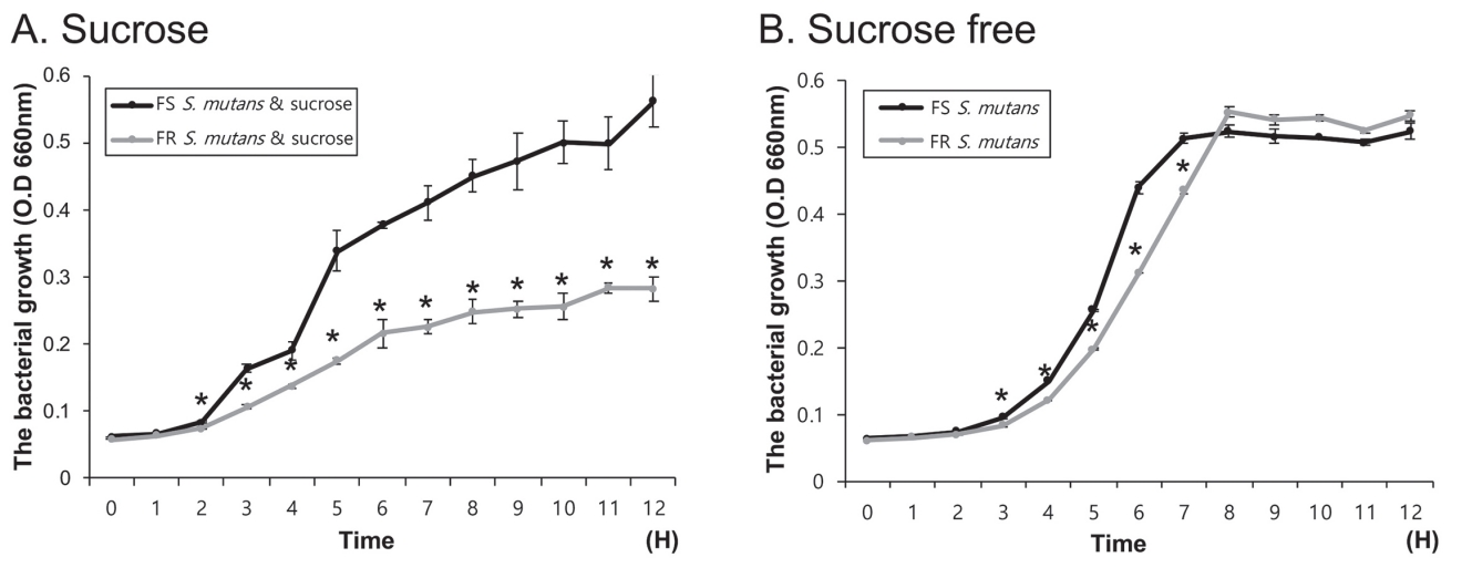
Growth of FR S. mutans was found to be significantly lower than that of FS S. mutans after 3 hours to the end point in the presence of sucrose (p < 0.05). At the end point, the growth of FR S. mutans was about the half of FS S. mutans (Fig. 1A). In sucrose-free condition, no significant difference was found between FS and FR S. mutans at the end point of growth (Fig. 1B). However, there was a significant difference from 3 to 7 hours after inoculation. The growth of FR S. mutans from 3 to 7 hours was lower than that of FS S. mutans , indicating that FR S. mutans required longer time to reach the stationary phase.
2. Acidogenicity of FS and FR S. mutans
Acid production by S. mutans was evaluated through pH changes in spent culture media in the presence or absence of 2% sucrose (Fig. 2). As shown in Fig. 2A, the pH value of spent media of FS S. mutans was significantly lower than that of FR S. mutans from 5 hours in the presence of sucrose (p < 0.05). No significant difference was found in the end pH value between FS and FR S. mutans in the sucrose-free condition (Fig. 2B). However, a significant difference in pH value after 6 and 7 hours showed longer time for FR S. mutans to reach a pH below 5.5.
3. Comparison of salivary and single species biofilm formed with FS or FR S. mutans
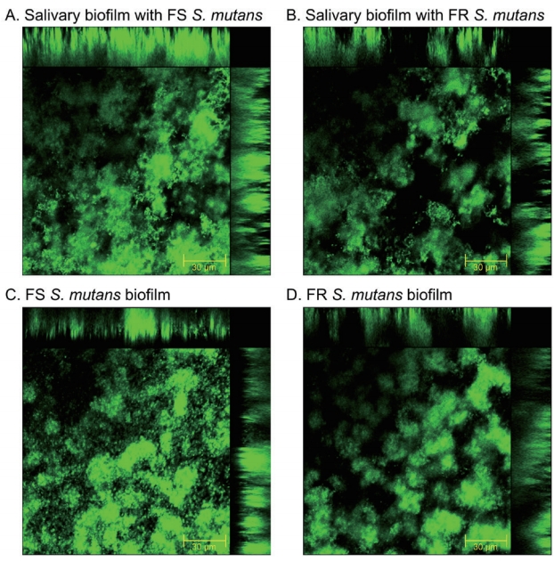
CLSM results showed that FR S. mutans formed less biofilm in both salivary biofilm, and single species biofilm compared to FS S. mutans (Fig. 3). Bacterial levels in salivary biofilm and single S. mutans biofilm were determined and the results are shown in Fig. 4. The number of total bacteria and S. mutans was significantly lower in the salivary biofilm with FR S. mutans (p < 0.05). BHI and MSB agar were used to determine the total bacteria and S. mutans counts in the salivary biofilm, respectively.

Confocal laser scanning microscopic images biofilm formed with FS and FR S. mutans. (A) Salivary biofilm formed with FS S. mutans, (B) salivary biofilm formed with FR S. mutans, (C) FS S. mutans biofilm, (D) FR S. mutans biofilm. CLSM was performed with a Zeiss LSM 700 microscope using a 40x/1.3 oil immersion lens. Bar 30 µm.
4. Comparison of gtf expression between FS and FR S. mutans
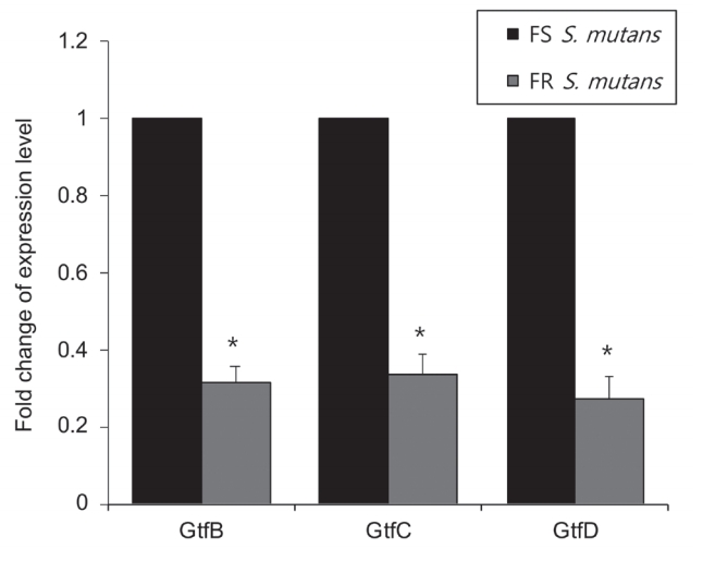
As biofilm formation exhibited a difference between FS and FR S. mutans, Gtfs production as a biofilm formation related factor in FS and FR S. mutans was compared (Fig. 5). The expression of gtfB, gtfC, and gtfD was significantly reduced to about 30%, in FR S. mutans compared to FS S. mutans (p < 0.05).

Comparison of expression of gtf genes in FS and FR S. mutans. After isolation of total RNA, gtfB, gtfC, and gtfD expression in FS and FR S. mutans was analyzed by RT-PCR. The experiment was performed four times, and the gene expression data were shown as the average fold change with means and their standard deviations.
p value from Mann-Whitney U test.
Ⅳ. Discussion
Fluoride has been recognized as a major factor for decreasing caries due to its antimicrobial and remineralization effects[18]. However, prolonged use of fluoride has been reported to cause mutation of S. mutans and adaptation to fluoride[19]. Studies on FR S. mutans have reported different characteristics of FR S. mutans compared to the FS parental strain[16], and phenotypic and genotypic changes in FR S. mutans have been evaluated[20-22]. In this study, the cariogenic properties of FR S. mutans were compared with those of FS S. mutans in the presence of sucrose, and the effect of FR S. mutans on salivary biofilm formation was assessed.
In previous study, FR S. mutans was mostly generated through a stepwise procedure to select colonies that grew in the presence of 400 - 600 ppm of fluoride under laboratory conditions, and its characteristics were compared with those of FS S. mutans [22]. However, due to different experimental designs and different strains used, no conclusive result was obtained[16]. Our study generated FR S. mutans by long term exposure of FS S. mutans to a 50% inhibitory concentration of fluoride, and fluoride resistance was verified by streaking on 200 ppm trypticase soy agar.
Sucrose is considered the most cariogenic dietary carbohydrate as its fermentation induces low pH in biofilms and it serves as a substrate for the formation of extracellular and intracellular polysaccharides[23]. Acidogenicity and growth of FS and FR S. mutans was compared with and without 2% sucrose in planktonic conditions. The acidogenicity of S. mutans was compared by evaluating pH changes. When FR S. mutans was cultured without sucrose, the end pH and growth value of spent media was not significantly different from that of FS S. mutans . However, media containing FR S. mutans took longer time to reach a pH below 5.5, which is the critical pH for the demineralization to occur[24]. A significant difference between FS and FR S. mutans was found in the sucrose added condition. In FR S. mutans , end growth was decreased to half, and the end pH value was high compared to that of FS S. mutans . From the results, it seems that sucrose acts as an inhibitor for growth and acid production in FR S. mutans . Under stress conditions, S. mutans show stress mechanisms, such as alteration in F-ATPase activity, catabolic pathways, and gene expressions[25]. The results of the present study suggest the modification of sucrose metabolic activity in FR S. mutans , resulting in a shift to decreased cariogenicity. This is in line with a previous study that lower cariogenicity of FR S. mutans was found in rats[26].
As prevention and treatment of dental caries focus on the microbial environment as well as on the cariogenic bacteria[1], cariogenic and single species biofilm formation with FS or FR S. mutans was observed. FR S. mutans formed reduced mass of salivary biofilm and single species biofilm compared to FS S. mutans , and the number of total bacteria and S. mutans was also significantly reduced. Oral biofilms are complex polymicrobial communities embedded within an extracellular matrix, present as a highly organized 3D structure adhered to the tooth[27,28]. It is consists of various microbial constituents including bacterial strains and EPS, which is one of the major components of biofilms and virulent factor in cariogenic biofilm formation[23]. Water soluble and water insoluble glucans are synthesized from sucrose by Gtfs in S. mutans and contribute to sucrose-dependent adhesion and biofilm formation[9].
When gtf expression of FS and FR S. mutans was compared through RT-PCR, the expression of gtfB, gtfC, and gtfD of FR S. mutans was significantly reduced compared to that of FS S. mutans. Reduction of gtfB, gtfC, and gtfD expression in FR S. mutans indicates that both water insoluble and soluble glucan were produced at lower levels by FR S. mutans than FS S. mutans, resulting in reduced adhesion to the surface, cell adhesion, and biofilm formation[9]. Expression of gtf genes in S. mutans can be changed in the presence of other oral bacteria[27], and fluoride did not seem to have an inhibitory effect on the Gtf activity of Streptococcus species in previous studies[29,30]. However, our results showed that FR S. mutans expressed significantly lower levels of gtf gene expression and formed a reduced mass of cariogenic biofilm compared to FS S. mutans with the salivary bacteria.
The concentration of fluoride in plaques can be elevated by the use of fluoride-enriched dentifrice[13,31] and topical NaF[32]. Although fluoride seems to decrease after initial exposure[33], it persists for a long time and even sub-millimolar levels of fluoride can affect the physiology of microbial cells[34]. Generally, daily mouthwash of 0.05% NaF (approximately 230 ppm F) for 1 min is recommended to prevent dental caries[35]. However, lower concentration of NaF (10, 50, and 125 ppm F) has also been suggested to show inhibitory effects on virulence factors and composition of S. mutans biofilm[29], and fluoride concentration in biofilm is known to increase along with duration of fluoride exposure[32]. This suggests that frequent use of low concentrations of fluoride would be effective in preventing dental caries by direct antimicrobial effects of fluoride and generation of FR S. mutans .
However, the limitation of this study is that as stresses from the oral environment are periodic rather than continuous and natural variations occur in the pH of plaques, generation of FR S. mutans in the oral environment is not easy[10]. Also, investigation of the stability of the resistance acquired form long term exposure of fluoride, and the affects of FR S. mutans to other microbial species should be considered in the future studies.
Ⅴ. Conclusion
In this study, long term exposure of S. mutans to 50% inhibitory level of fluoride generated FR S. mutans . Sucrose metabolic activity and acid production were reduced in FR S. mutans. Moreover, decreased salivary biofilm formation was observed along with decreased gtf gene expression. These findings suggest that FR S. mutans may be less virulent than FS S. mutans in inducing dental caries and may be effective in maintaining oral microbial homeostasis and reducing dental caries.


