


ŌģĀ. Introduction
Standard care of dental caries involves removing the demineralized tissues and replacing them with a filling material[1]. Once restorations are placed, their lifetimes are influenced by an array of factors and vary enormously[2]. Over time, the lack of marginal sealing increases the risk of secondary caries[3]. Secondary caries occurs along the margins of the enamel or dentin and is one of the most common reasons for replacing composite resin restorations[4]. Attempts have been carried out to utilize antibacterial agents such as fluoride in the dental restorative materials[5].
Fluoride interferes with the caries process by reducing demineralization and enhancing remineralization of the enamel and dentin[6]. The fluoride content in restorative materials should be as high as possible without deteriorating the physical properties and the fluoride release should be as large as possible without excessive degradation of the materials[7]. Glass ionomer (GI), the first-developed fluoride-releasing restorative material, has excellent potential to release fluoride, but has poor mechanical properties[8]. To overcome these limitations, materials combining the properties of composite resin have been developed[9], and giomer and alkasite restorative material (ARM) were recently developed.
Giomer contains a pre-reacted glass ionomer filler to release fluoride ions[10]. Studies on its properties have reported a smooth surface finish, excellent esthetics, and clinical stability, but low rate of ion release[10,11].
ARM is a tooth-colored, basic filling material for direct restorations, which is considered to release substantial levels of fluoride ions owing to its patented alkaline filler[12]. A study on the mechanical properties of the restoration showed that the strength of ARM was similar to that of a composite resin[13].
Since giomer requires an adhesive system for adhesion to the tooth structure, an adhesive layer is required between the tooth and the restorative material. Although the ARM can be used without an adhesive system, applying an adhesive system has the advantages of enabling minimally invasive treatment and minimizing microleakage[14].
Fluoride should be released through the dentin adhesive layer to exert an anticariogenic effect on the interface between the dentin and restorative materials. It is known that the surface coating of restorations acts as a physical barrier, significantly reducing the amount of fluoride release[15]. Wiegand et al.[9] mentioned that an intermediary material layer, such as an adhesive hybrid layer, might hamper fluoride uptake. However, there are few studies directly evaluating the amount of fluoride release through the adhesive layer, and there has been no research on ARM yet.
The aim of this study was to compare the fluoride release of ARM and giomer and to evaluate the effect of an adhesive on the fluoride release by measuring the amount of fluoride released through the adhesive layer.
ŌģĪ. Materials and Methods
1. Materials
In this study, Cention┬« N (Ivoclar Vivadent, Schaan, Liechtenstein) and Beautifil Injectable (Shofu Inc., Kyoto, Japan) were used as ARM and giomer, respectively. A composite resin without fluoride, FiltekŌäó Z350XT flowable (3M ESPE, St. Paul, MN, USA), was employed as the control. A 4th-generation dentin adhesive containing no fluoride, ScotchbondŌäó Multi-Purpose adhesive (3M ESPE, St. Paul, MN, USA) was used to form the adhesive layer. All materials, as well as their compositions and manufacturers, are summarized in Table 1.
ARM, giomer, and composite resin groups were termed the ARM, GM, and CR groups, respectively. Groups in which an adhesive was applied to each specimen were termed the ARM-AD, GM-AD, and CR-AD groups. A total of 60 specimens, 10 for each group, were prepared.
2. Specimen preparation
Every specimen was prepared in a metal mold with a diameter of 7.0 mm and a height of 2.0 mm. After interposing a celluloid strip between the bottom of the metal mold and the glass slab, the material was filled inside the mold. A celluloid strip was placed on the filled material, and the top of the mold was covered by a glass slab and pressed by hand to form a flat surface. The materials were light-cured for 18 sec each on the upper and lower surfaces.
The light source was a blue light-emitting diode (B&LiteS, B&L Biotech, Ansan, Korea) with a round tip of 10.0 mm diameter. In the light-curing procedure, the round tip was in contact with the glass slab and the output power was 1200 mW/cm2. After separating the specimens from the mold, the adhesive was applied according to the group.
3. Application of the dentin adhesive
For the ARM-AD, GM-AD, and CR-AD groups, the dentin adhesive was applied immediately after preparing the specimens.
A 15.0 ╬╝L drop of the adhesive was released on the upper surface of the specimen using a micropipette. A celluloid strip was positioned to ensure an even spread of the adhesive, and a slide glass was placed to form a flat adhesive layer, which was light-cured for 9 sec. The same procedure was performed on the lower surface of the specimen.
Next, 50.0 ╬╝L of the adhesive was evenly applied to the sides of the specimen using a 1.5 mm diameter microbrush. The sides of the specimen were divided into 4 parts and lightcured for 9 sec each.
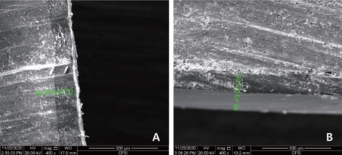
4. Evaluation of uniformity of the adhesive layer
An additional 18 specimens, 6 specimens for each restorative material, were prepared for the evaluation of the adhesive layer. The dentin adhesive was applied only to the upper and lower surfaces of the 9 specimens. The remaining specimens were only treated with the adhesive on the sides. All specimens were coated with platinum particles, and the adhesive layer thickness was measured by observing the specimen with a field emission scanning electron microscope (SEM, Inspect F, FEI, USA).
5. Measurement of fluoride release
The prepared specimens were placed in polyethylene tubes containing 2.0 mL deionized water and stored in a 37.0Ōäā water bath for the duration of the study. The amount of released fluoride was measured on the 1st, 3rd, 7th, 14th, 21st, and 28th day after storage. The deionized water was replaced after each measurement. To stabilize the ion strength, the solution to be measured was mixed with the same amount of TISAB ŌģĪ (Total Ionic Strength Adjuster Buffer ŌģĪ, Thermo ScientificŌäó OrionŌäó, Beverly, MA, USA), and the amount of fluoride release was measured using a pH/ISE meter (920A+, Thermo ScientificŌäó OrionŌäó, Beverly, MA, USA) and a fluoride ion selective electrode (9609BNWP, Thermo ScientificŌäó OrionŌäó, Beverly, MA, USA). The combined electrode was calibrated using 0.1 ppm, 1.0 ppm, and 10.0 ppm fluoride standard solutions (Thermo ScientificŌäó OrionŌäó, Beverly, MA, USA) on every measurement day.
6. Statistical analysis
The average and standard deviation of the amount of released fluoride and the total cumulative fluoride release were calculated. The total cumulative fluoride release in the study groups, except the control groups, was tested using the Mann-Whitney test. For statistical analysis, the SPSS software version 25 (SPSS Inc., Chicago, IL, USA) was used.
Ōģó. Results
1. Evaluation of the adhesive layer
Evaluation of the adhesive layer of the specimen using scanning electron microscopy revealed that the thickness of the upper and lower adhesive layers was between 80 - 100 ╬╝m, with a thickness of 80 - 110 ╬╝m on the sides regardless of the restorative material (Fig. 1).
2. Measurement of fluoride release
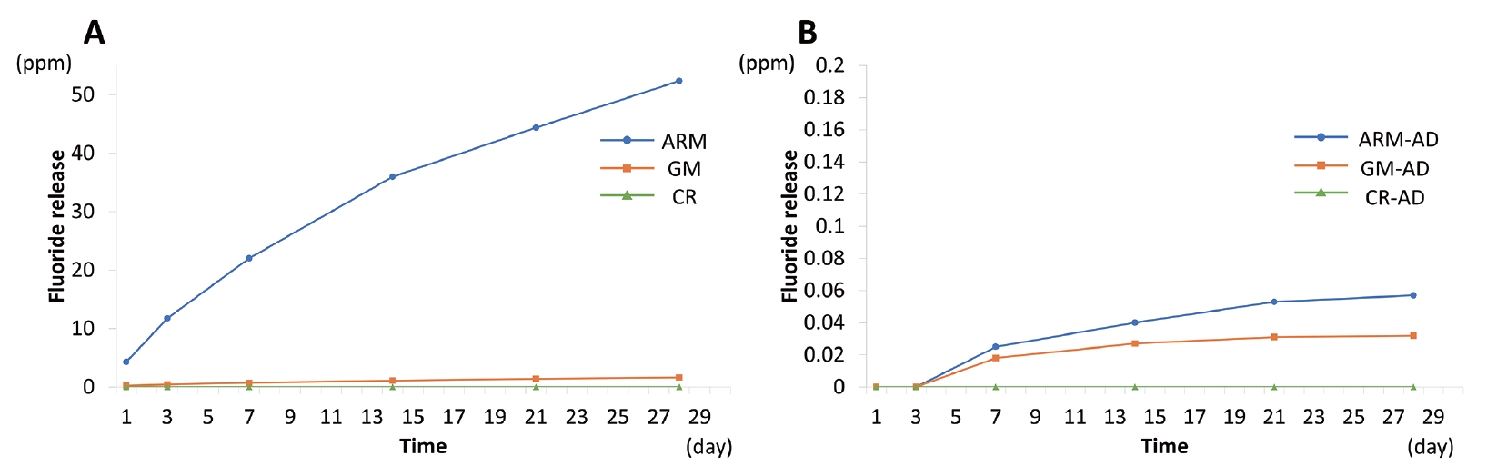
The amount of released fluoride per group is shown in Table 2. Fluoride release was identified in all study groups, except in the control. The daily fluoride release was obtained by dividing the amount of released fluoride by the measurement period (Fig. 2). In both the ARM-AD and GM-AD groups, fluoride release was detected from the 7th day onward.
In comparison with the ARM group, the daily fluoride release in the GM group was significantly lower. The fluoride release in the ARM group was the highest on the 1st day and then sharply decreased and remained roughly constant from the 21st day. The daily fluoride release of the GM group was also the highest on the 1st day, decreased until the 3rd day, and remained without significant changes from the 7th day.
The cumulative fluoride release in the ARM group for 28 days was 52.37 ppm and that in the GM group after 28 days was 1.629 ppm (Table 3). The ARM-AD group showed a 99.9% decrease equal to 0.059 ppm, and the GM-AD group showed a 97.9% decrease, equal to 0.034 ppm. On the Mann-Whitney test, the ARM group showed a significantly higher amount of cumulative fluoride release amount when compared with the GM group (p=0.000). The ARM-AD and GM-AD groups showed decreased fluoride release when compared with the ARM and GM groups, and it was confirmed that fluoride release was significantly lower according to the adhesive application (p=0.000). The amount of fluoride release was higher in the ARM-AD group than in the GM-AD group (p=0.011). The cumulative fluoride release over 28 days is shown as a graph (Fig. 3).
ŌģŻ. Discussion
The restorative materials used in this study is a fluoride-releasing composite resin. Beautifil Injectable, the giomer used in this study, contains a surface pre-reacted glass ionomer (S-PRG) as a fluoride component[10], while Cention┬« N, an ARM, releases fluoride due to the presence of three inorganic glasses known as ŌĆ£alkasite fillersŌĆØ. When both giomer and ARM are placed in a moist environment, they lead to water absorption in the fillers, which can then release calcium, aluminum, and fluoride ions[16].
Various factors such as the fluoride amount present in the cement, powder-liquid ratio, and the capacity for water diffusion in the materials affect fluoride release[17,18]. In this study, the amount of fluoride released from ARM was significantly higher than that from giomer. ARM and giomer differ in the filler content. The filler content of Cention® N was 78.4% and whereas that of Beautifil Injectable was 50 - 60% according to the manufacturer. The hydrophilicity of monomers can affect the ion release owing to water diffusion in the material. Cention® N contains PEG-400DMA, and Beautifil Injectable contains TEGDMA as the hydrophilic monomer. The differences in water resorption of the materials could be explained by the differences in matrix composition[19]. In addition, ARM may contain more air-filled voids within the matrix which may cause more moisture diffusion because ARM is a hand-mixing type restorative material whereas giomer is provided with a pre-mixed syringe.
Fluoride was released the most on the 1st day and then gradually decreased in both materials in this study. The pattern of fluoride release was similar to GI. An initial high fluoride release from GI over the 1st day is due to the burst of fluoride released from the setting reaction of the glass particles and the polyalkenoic acid[9]. Although there is no acid-base reaction in the fluoride-releasing composites, short-term high elution of fluoride release is possible because of the surface wash-off process[20]. An initial fluoride burst effect is advantageous, because it reduces the viability of bacteria in the inner carious dentin and induce remineralization of enamel and dentin[7]. After the initial high release, constant fluoride release occurs in the subsequent days because of the capability of fluoride to diffuse through cement pores[10]. The steady release of fluoride from ARM and giomer reduces microbial attachment, neutralizes the acidic environment, and prevents caries in adjacent teeth, thereby reducing the occurrence of secondary caries.
The amount of adhesive was determined through a pilot study so that it can be uniformly applied while having an adequate thickness similar to the clinical application. It was mentioned that an ideal adhesive layer thickness should be between 50 and 150 ╬╝m to provide adequate stress relief[21]. The thickness of the adhesive layer in this study observed by SEM was 80 to 110 ╬╝m, which was similar to the thickness actually used in the clinic.
This study confirmed the fluoride release through the adhesive layer. Previous studies also reported that when adhesives were applied to various restorative materials, fluoride was released through the adhesive by permeation[22-24]. Tay et al.[25] suggested that the adhesive coating acts as a semipermeable membrane, which allows water transport from the outside into the interface. The osmotically-induced permeability of the adhesive may transport not only water molecules but also small solutes[26]. It was suggested that fluoride is released because moisture that penetrates through the adhesive layer reacts with the alkaline and S-PRG fillers.
The amount of fluoride released through the adhesive layer was significantly lower than in the group without adhesive. On applying adhesive under the same conditions, the cumulative fluoride release amount decreased by 99.9% and 97.9% in the ARM and giomer groups, respectively. Mazzaoui et al.[22] reported that the reduction rate of the fluoride released through the adhesive layer is 43 - 74% for the GI and 91 - 96% for the fluoride-releasing composite resin. This difference is considered to be due to the different release mechanisms of the materials. The major mechanism of fluoride ion release by GI is the acidbase reaction in the setting reaction. Giomer and ARM release fluoride ions by an ion exchange process when the materials are exposed to moisture[16,27]. Since the release of fluoride from ARM and giomer is more water-exposure dependent, the adhesive layer is expected to exhibit a greater barrier effect. Although the barrier effect was slightly higher in the ARM-AD group than the GM-AD group, the cumulative amount of fluoride release for 28 days was higher in the ARM-AD group. In clinical situations where fluoride release is desired, the use of ARM may be more advantageous.
A trace amount of fluoride < 0.012 ppm promotes enamel remineralization[28], but a greater amount of fluoride is needed to enhance the remineralization of dentin when compared with that of the enamel[29]. Cate et al.[30] deduced that dentin demineralization was inhibited to a clinically relevant percentage only at fluoride > 1 ppm. This study confirmed that a sufficient amount of fluoride required for dentin remineralization was released in ARM. Francois et al.[16] classified ARM as a bioactive composite because this material is capable of inducing remineralization of the underlying hard tissue with which it is in contact. However, when ARM was used with a dentin adhesive, the amount of fluoride released over one month in this study was insufficient for promoting dentin remineralization. Although the amount of released fluoride is much smaller, both materials are expected to exhibit a considerable effect by employing fluoride-recharging properties[11,31], and further research into this aspect is needed.
The main limitation of this study is the use of a single type of adhesive. Water permeability varies depending on the monomer composition, residual solvent, and degree of polymerization[32,33]. It is necessary to study the amount of fluoride released through various types of adhesives. In addition, this study was conducted for only 28 days, and a long-term study is needed given the steady release of fluoride from ARM and giomer.
Ōģż. Conclusion
Fluoride release through the adhesive layers of ARM and giomer was evaluated and it was confirmed that in both the ARM and the giomer, fluoride was released through the adhesive layer. The amount of fluoride release was significantly lower when the dentin adhesive was applied. The cumulative amount of fluoride release was higher in ARM than in giomer.

 PDF Links
 PubReader
 Full text via DOI
 Download Citation
 Print
Print


