


Ⅰ. 서 론
유치는 어린이의 성장과 발달에 있어 중요한 기능을 담당한다[1]. 치아 우식은 유치에서 치수병변을 일으키는 주요 원인으로, 유치 치수 치료를 올바르게 시행하여 유치를 건전하게 유지하기 위해서는 유치의 근관에 대한 이해가 필수적이다. 그러나 일반적으로 영구치 근관계에 대한 연구는 비교적 많이 이루어지고 있는 반면, 유치 근관계에 대한 연구는 많지 않다[2-10]. 유치의 근관계는 영구치에 비해 비교적 덜 복잡하며, 유구치의 경우 하방의 계승 영구치가 존재하여 분리된 치근을 가지면서 이개도가 크다는 점에서 계승 영구치와 차이를 보인다. 따라서 영구치 근관계에 대한 이해를 유치에서 온전히 적용하는 것은 한계가 있다[1,11-13].
유치 근관계에 대한 연구는 다양한 방법으로 시도되어 왔다. 이 중 치근단 방사선 사진[14-17]은 3차원적인 치아 구조를 2차원적으로만 보여주며, 3차원적인 근관계 이해를 위해 발치된 치아를 이용한 연구 방법들은 다소 침습적이라는 한계가 있다. 최근 Cone beam computed tomography (CBCT) 및 3D Computerized tomography (CT)를 이용하여 유치의 치근과 근관 형태를 3차원적으로 이해하려는 시도가 이루어지고 있으나, 유치는 영구치와 달리 생리적 치근 흡수가 특징적이기 때문에 온전한 치근 상태의 표본 수를 갖는 연구가 많지 않고[1,18-20], CBCT 및 3D CT를 활용한 연구로는 치근 내부 즉, 근관 형태를 3차원적으로 이해하기에는 다소 제한적이다.
이 연구에서는 서울대학교 치과병원 소아치과에서 CBCT와 3D CT를 촬영한 268명의 환아를 대상으로 상악 유구치의 치근과 근관 형태를 분석하고, 3D CT로 촬영된 DICOM (Digital imaging and communications in medicine) format file을 활용, Mimics 및 3-Matics software를 사용하여, 치근부 치수의 3차원적인 모습을 비침습적인 방법으로 재현하고자 하였다.
Ⅱ. 연구 대상 및 방법
1. 연구 대상
이 연구는 서울대학교 치의학대학원 생명윤리심의위원회 승인 하에 이루어졌다(IRB File No. : S-D20200030).
2006년 1월부터 2020년 4월까지 과잉치 혹은 매복치 진단을 위해 서울대학교 치과병원에서 CBCT 또는 3D CT를 촬영한 3 - 7세의 건강한 환자 4,216명 중, 영상의 질이 우수하고 치아 우식은 법랑질 부위에 제한적이고, 치근 흡수와 치수 병변이 없으며 치수치료 병력이 없는 상악 제1, 2유구치를 갖는 268명(CBCT 152명, 3D CT 116명)을 대상으로 하였다. CBCT 영상은 CS9300 (Carestream Dental, USA), 3D CT 영상은 SOMATOM (Siemens Healthineers, Germany)으로부터 얻어졌다.
2. 연구 방법
1) 치근의 수, 치근당 근관의 수
268명의 상악 제1, 2유구치 중 치아 우식은 법랑질 부위에 제한적이고, 치근 흡수와 치수 병변이 없고 치수치료 병력이 없는 1,002개(상악 제1유구치 490개, 제2유구치 512개)의 치아를 대상으로 축면(Axial) 영상을 이용하여 치아당 치근의 수와 치근당 근관의 수를 조사하였다(Fig. 1).
2) 치근과 근관의 형태, 치근의 이개각도, 치근과 근관의 길이 및 근관의 단면 형태
서울대학교 치과병원에서는 268명의 환자 중 CBCT 촬영이 이루어진 152명의 경우 DICOM format file을 보관하지 않아 OnDemand3D software (Cyvermed, Korea)를 활용한 분석이 이루어질 수 없었다. OnDemand3D software에서 재현이 가능한 DICOM format file이 보존되어 있는 환자는 3D CT로 촬영한 116명 중 60명이었다. 이 60명의 상악 제1, 2유구치 중 치아 우식은 법랑질 부위에 제한적이고, 치근 흡수와 치수 병변이 없고 치수치료 병력이 없는 218개의 치아(상악 제1유구치 106개, 제2 유구치 112개)를 OnDemand3D software를 사용하여 치아의 해부학적 형태를 2D와 3D 이미지로 구축한 후, 치근과 근관의 형태, 치근의 이개 각도, 치근과 근관의 길이 및 근관의 단면 형태를 분석하였다.
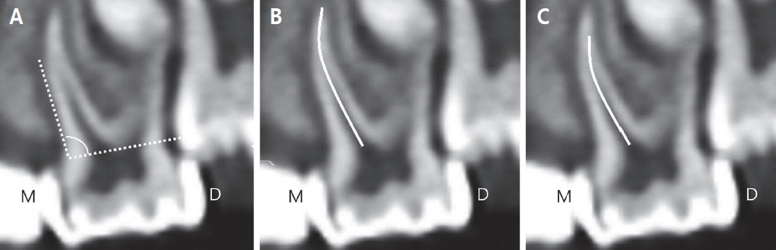
(1) 치근과 근관의 형태는 직선형, 곡선형, S자형 3가지로 분류하였다(Fig. 2).
(2) 치근의 이개각도는 백악법랑 경계를 협설로 연결한 선에서 가장 돌출된 치근 부위까지의 각도를 측정하였다(Fig. 3A).
(3) 치근의 길이는 백악법랑 경계에서 근첨부까지의 길이 중 가장 긴 부위를 측정하였다(Fig. 3B).
(4) 근관의 길이는 백악법랑 경계에서 근단부 근관까지의 길이 중 가장 긴 부위를 측정하였다(Fig. 3C).
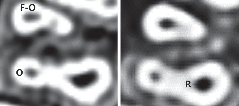
(5) 근관의 단면 형태는 치근의 치관(cervical), 중간(middle), 치근단(apex)에서 단면의 모습을 round, ovoid, flat-oval 3가지로 분류하였다(Fig. 4).
3) 치근 치수 부피, 치근 치수 표면적, 치근-치수 비
(1) Mimics software에서 이미지 분석
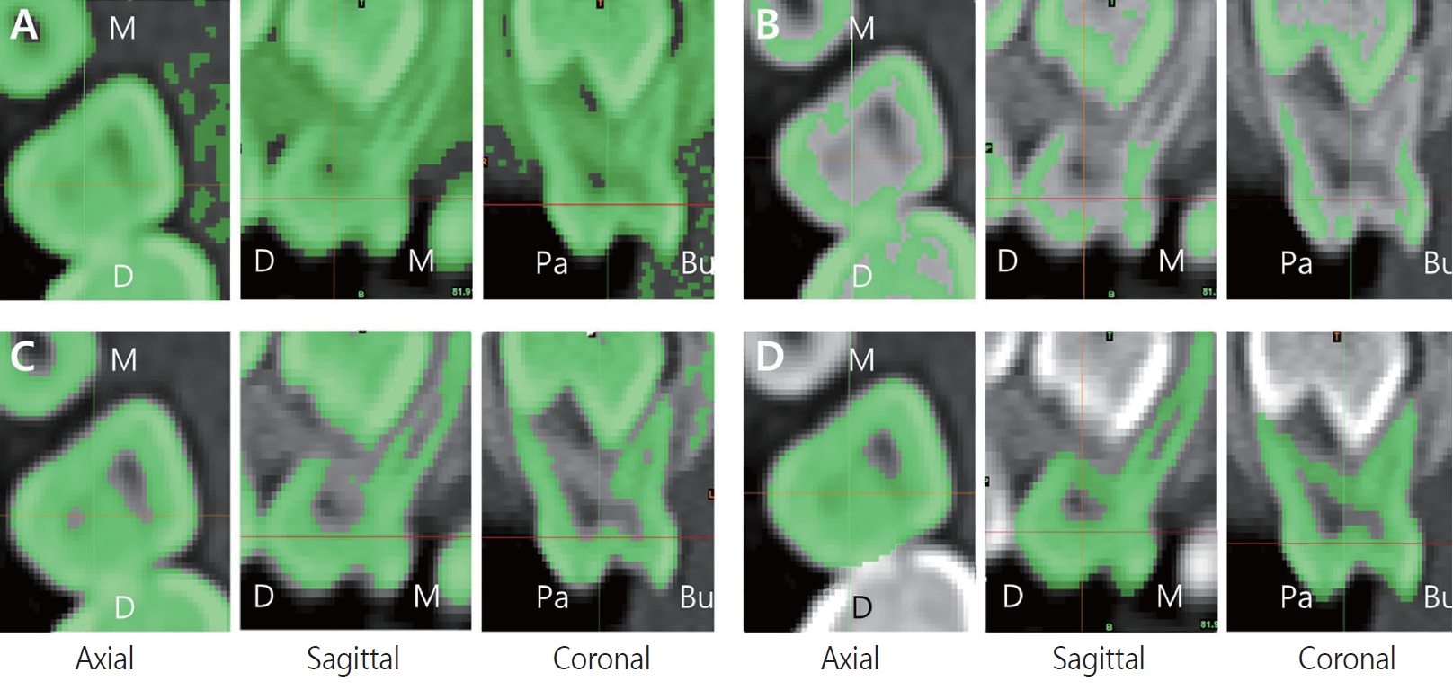
3D CT DICOM format file이 제공된 60명의 환자 중 MATERIALISE MIMICS software (Materialise, Belgium, version 21.0)를 사용해 3D로 재현하였을 때 총 12명(4 - 7세)의 48개 치아(상악 제1유구치 24개, 상악 제2유구치 24개)를 대상으로 하였다(Fig. 5). 연령 분포는 4 - 5세 5명, 6 - 7세 7명으로 이루어졌다. 노출 정도(치근 HU: 905-1196, 치수 HU: 205-495)는 환자와 치근 그리고 치수에 따라 다르게 적용하였다.
Threshold 값이 너무 낮게(86 Hu) 설정되어 치아와 주변 구조물 간의 분리가 어려운 경우(Fig. 5A), Threshold 값이 너무 높게(1792 HU) 설정되어 치아 외형을 적절히 포함하지 못한 경우(Fig. 5B), Threshold 값이 비교적 적절하여(1182 HU) 치아와 주변 구조물 간의 분리가 용이하고, 치아 외형을 적절히 포함시킨 경우(Fig. 5C), Software상에서 치아와 주위 구조물을 보다 명확히 구분하기 위해서 개별적으로 수정(Fig. 5D)한 경우를 나타내었다.
(2) 3-Matics software에서 이미지 분석
Mimics software를 이용하여 3D 모델로 재현된 상악 유구치 치수와 치아 외형을 3-Matics software (Materialise, Belgium, version 13.0)로 분석하여, 치근 치수의 부피와 표면적 그리고 치근 전체 부피 중 치근부 치수 부피가 차지하는 비율인 치근-치수 비를 측정하였다(Fig. 6).
3. 통계 분석
치근의 수, 치근당 근관의 수 항목에 대해 좌우 대칭성 여부를 측정하기 위해 Cohen’s kappa coefficients가 계산되었고, 대칭성 검사에서 좌우측 중 결손이 있는 경우(상악 제1유구치: 34개, 상악 제2유구치: 20개) 대칭성 검사에서 제외되었다(Table 2). 치근의 이개각도, 치근의 길이, 근관의 길이 항목에서 한 치아 내 치근 사이에 차이가 있는지 확인하기 위하여 일원 변량 분산분석(Oneway repeated ANOVA)을 사용하였다(Table 3). 치근 치수의 부피와 표면적, 치근-치수 비 항목에서는 한 치아내 치근 사이에 차이가 있는지 확인하기 위해 일원 변량 분산분석을 사용하였다. 치근에 2개 이상의 근관이 있는 경우 가장 긴 치근과 근관의 길이 및 각도를 통계에 사용하였다. 통계 분석은 SPSS 23.0 (Statistical Package for Social Sciences, IBM Corp., USA)을 이용하여 분석하였다.
Ⅲ. 연구 성적
1. 조사자 내 일치도
치근의 수, 치근당 근관의 수 그리고 치근과 근관의 형태와 근관의 단면 형태 항목에 대한 ICC 값은 모든 항목에서 0.98 이상으로 나타났고, 치근의 이개각도, 치근의 길이, 근관의 길이, 치근 치수의 부피와 표면적 그리고 치근에서 치근부 치수 부피가 차지하는 비율인 치근-치수 비 항목에 대한 ICC 값은 모든 항목에서 0.84 이상으로 나타났다.
2. 치근의 수, 치근당 근관의 수
상악 제1유구치에서는 3개의 치근(57.9%)과 2개의 치근(41.9%)을 가지는 비율이 높았으며, 상악 제2유구치에서는 3개의 치근(79.2%)을 가지는 비율이 그 외 유형들 보다 높았다. 상악 제1유구치에서는 1개의 치아만이 3개의 협측 치근과 1개의 구개측 치근을 가져 4개의 치근을 보였으며, 상악 제2유구치에서는 2.7%(14개)의 치아에서 4개의 치근을 보였고, 협측과 구개측에 각각 2개의 치근을 가지는 경우로 나타났다. 치근당 근관 수에 있어, 상악 제1유구치의 경우 3개의 치근에 3개의 근관을 가지는 경우(56.3%)가 가장 많았고, 2개의 치근에 3개의 근관을 가지는 경우(41.1%)가 그 뒤를 이었다. 상악 제2유구치의 경우 3개의 치근에 3개의 근관을 가지는 경우(63.5%)가 가장 많았고, 3개의 치근에 4개의 근관을 가지는 경우(15.7%)가 그 뒤를 이었다. 3개의 치근에 4개의 근관을 가지는 경우 근심 협측 치근에 2개의 근관을 가지는 비율(15.3%)이 원심 협측 치근에 2개의 근관을 가지는 비율(0.4%) 보다 높았다(Table 1).
Table 2에서는 같은 환자의 좌우측 상악 유구치 치근당 근관 유형의 대칭성 정도를 보여주고 있다. 상악 제1유구치와 상악 제2유구치 치근당 근관 유형의 Cohen’s kappa coefficients는 각각 0.632, 0.639로 나타났다.
3. 치근과 근관의 형태, 치근의 이개각도, 치근과 근관의 길이 및 근관의 단면 형태
치근과 근관 형태의 경우 상악 제1유구치에서는 직선형이 많이 관찰되었고, 상악 제2유구치는 곡선형이 더 많이 관찰되었다. S자형은 상악 제2유구치의 근심 협측 치근과 구개측 치근에서 관찰되었다(Table 3).
치근의 이개 각도는 상악 제2유구치의 치근 이개각도가 대응되는 상악 제1유구치에 비해 큰 것으로 나타났고(근심 협측: 2.5o, 원심 협측: 2.2o, 구개측: 3.0o), 같은 치아 내에서는 구개측 치근의 이개 각도가 가장 크고, 원심 협측, 근심 협측 치근 순서로 나타났다(Table 3).
길이는 상악 제2유구치의 치근 길이가 상악 제1유구치에 비해 길었으며(근심 협측: 1.0 mm, 원심 협측: 1.0 mm, 구개측: 1.1 mm), 근관의 길이 또한 상악 제2유구치에서 대응되는 제1유구치의 근관 길이에 비해 길었다(근심 협측: 1.2 mm, 원심 협측: 0.9 mm, 구개측: 1.2 mm). 같은 치아 내에서는 구개측 치근과 근관의 길이가 다른 치근들의 치근과 근관 길이에 비해 긴 것으로 나타났다(Table 3).
4. 치근 치수 부피, 치근 치수 표면적, 치근-치수 비
치근 치수의 부피와 표면적 그리고 전체 치근에서 치근부 치수가 차지하는 비율인 치근-치수 비는 상악 제1, 2유구치 모두 구개측 치근에서 가장 큰 값을 보였다(Table 6).
Ⅳ. 총괄 및 고찰
유치의 치근과 근관의 형태를 이해하는 것은 성공적인 치수 치료를 시행하는데 중요한 요소이다. 유치 치근과 근관 구조를 이해하기 위해 치근단 방사선 사진을 연구하였으나, 이러한 방법은 3차원적인 근관의 구조를 2차원적으로 재현하여 실제 근관의 모습을 정확하게 표현해 줄 수 없었으며, 염료 주입, micro CT와 같은 방법들은 다소 침습적인 방법으로 연구되어야 한다는 한계점을 보여주었다. 이와 달리 CT 촬영을 이용한 방법은 3차원적인 근관의 모습을 다소 비 침습적이면서도 비용과 소요되는 시간면에서 보다 효과적이라는 점에서 최근 널리 사용되고 있다.
상악 제1, 2유구치의 경우 대부분 이개된 3개의 치근을 가지는 것으로 보고되었다[1,12,13,20-22]. 이 연구에서도 상악 제1유구치는 분리된 3개의 치근(57.9%) 혹은 융합된 2개의 치근(41.9%)의 비율이 높았고, 상악 제2유구치는 분리된 3개의 치근(79.2%)의 비율이 높아, 기존 연구와 일치하는 결과를 보였다. 상악 제1, 2유구치에서 4개의 치근을 가지는 경우는 각각 1개(0.2%), 14개(2.7%)가 관찰되었다. 한국 소아 환자를 대상으로 보고된 Sim과 Mah[18]의 연구에 따르면 상악 제1유구치에서는 분리된 3개의 치근(58.7%)과 융합된 2개의 치근(41.3%)의 비율이 이번 연구와 비슷한 결과를 나타냈으나 상악 제2유구치에서는 융합된 2개의 치근(42.0%)과 분리된 3개의 치근(54.9%) 비율로 나타나 이번 연구와 상악 제2유구치에서 차이를 보였다. Choi 등[19]의 연구에서는 상악 제1, 2유구치 모두 분리된 3개의 치근만 관찰되었다. 이번 연구에서는 상악 제1, 2유구치에서 분리된 4개의 치근이 관찰되었으나, Sim과 Mah[18]의 연구에서는 상악 제2유구치에서만 분리된 4개의 치근(3.1%)이 관찰되는 것으로 보고되었다. 이는 기존 연구와 달리 연구의 표본수가 많아 보다 다양한 형태가 관찰된 것으로 생각된다.
상악 유구치에서는 대부분 치근당 하나의 근관을 가지는 것으로 나타났고, 근심 협측 치근에서 2개의 근관을 가지는 경우는 상악 제2유구치(21.2%)에서 상악 제1유구치(2.2%)에 비해 높은 비율로 나타났다. 상악 유구치 치근의 융합은 원심 협측 치근과 구개측 치근에서 나타났고, 이 경우 개별 근관을 가지는 것으로 나타났다. 마찬가지로 분리된 구개측 치근을 갖는 경우에도 개별 근관을 보였는데, 이는 Sim과 Mah[18]의 연구와 일치하는 결과이다.
치근과 근관의 형태는 주로 직선형 혹은 곡선형이었고, S자형은 상악 제2유구치의 근심 협측과 구개측 치근에서만 관찰되었다. Ozcan 등[1]의 연구에서는 상악 유구치 치근과 근관의 형태는 주로 곡선형이었고, S자형은 상악 유구치 모든 치근(근심 협측, 원심 협측, 구개측)에서 관찰되었다. Reddy 등[20]의 연구와 한국 소아 환자들의 유구치 치근을 대상으로 분석한 Choi 등[19]의 연구에서는 원심 협측 치근에서 S자형은 관찰되지 않았다. 직선형 치근에 비해 만곡된 치근에서의 근관 치료시 치근 천공과 같은 부정적인 예후를 초래할 수 있으므로, 치근과 근관의 형태에 대한 이해는 이러한 합병증을 줄이는데 도움이 될 수 있다[7,8,20].
이 연구에서 상악 제1, 2유구치 치근의 이개 각도는 각각 105.7°, 109.8°, 115.9° 와 108.2°, 112.0°, 118.9°로 나타났으며, 한 치아내 각 치근은 유의미한 이개각도 차이를 보였다. 그러나 Reddy 등[20]의 연구에서는 상악 제1유구치에서는 구개측 치근(84.17°)이, 상악 제2유구치에서는 원심 협측 치근(84.24°)의 이개 각도가 가장 큰 것으로 나타났다. 한국 소아 환자들의 유구치 치근을 대상으로 분석한 Choi 등[19]의 연구에서는 상악 제1, 2유구치 구개측 치근에서 각각 114.2°, 118.4°로 가장 큰 이개 각도를 보였으나 상악 제1유구치에서 근심 협측 치근과 원심 협측 치근의 이개 각도에서는 통계적으로 유의미한 차이를 보이지 않았다. 이는 인종과 연령, 표본수의 차이들과 관련되어 있을 것으로 보인다.
상악 제1유구치의 평균 치근 길이는 근심 협측, 원심 협측, 구개측에서 각각 7.6 mm, 6.5 mm, 8.0 mm로 구개측 치근의 길이가 가장 길었다. Fumes 등[13]의 연구에서는 7.9 mm, 6.7 mm, 5.9 mm로 구개측 치근의 길이가 가장 짧은 것으로 나타났고, 한국 소아 환자들의 유구치 치근을 대상으로 분석한 Choi 등[19]의 연구에서는 각각 7.7 mm, 6.7 mm, 7.9 mm로 이번 연구에서와 비슷한 경향성을 보였다. 이번 연구에서 상악 제2유구치의 평균 치근 길이는 근심 협측, 원심 협측, 구개측에서 각각 8.6 mm, 7.5 mm, 9.1 mm로 상악 제1유구치에서와 마찬가지로 구개측 치근의 길이가 가장 길었다. Fumes 등[13]의 연구에서는 8.5 mm, 6.5 mm, 7.4 mm로 근심 협측 치근의 길이가 가장 긴 것으로 나타났고, 한국 소아 환자들의 유구치 치근을 대상으로 분석한 Choi 등[19]의 연구에서는 각각 8.5 mm, 7.6 mm, 8.8 mm로 이번 연구와 비슷한 경향성을 보였 보였는데, 이는 Choi 등[19]의 연구와 달리 Fumes 등[13]의 연구에서는 인종에 따른 차이가 관련되어 있을 것으로 보인다.
연구에서 측정된 상악 제1유구치의 평균 근관 길이는 근심 협측, 원심 협측, 구개측에서 각각 6.4 mm, 5.4 mm, 6.8 mm로 관찰되었다. Ozcan 등[1]의 연구에서는 5.4 mm, 4.8 mm, 6.3 mm로 구개측 근관의 길이가 가장 긴 것으로 나타났고 이는 이번 연구에서와 비슷한 경향성을 보였다. 한국 소아 환자들의 유구치 치근을 대상으로 분석한 Choi 등[19]의 연구에서는 각각 6.6 mm, 5.7 mm, 6.9 mm로 마찬가지로 이번 연구에서와 비슷한 경향성을 보였다. 이번 연구에서 상악 제2유구치의 평균 근관 길이는 근심 협측, 원심 협측, 구개측에서 각각 7.6 mm, 6.3 mm, 8.0 mm로 상악 제1유구치에서와 마찬가지로 구개측 근관의 길이가 가장 길었다. Ozcan 등[1]의 연구와 Choi 등[19]의 연구에서도 각각 6.1 mm, 5.9 mm, 6.5 mm 와 7.3 mm, 6.5 mm, 7.6 mm로 이번 연구에서와 비슷한 경향성을 보였다. Ozcan 등[1]의 연구가 Choi 등[19]의 연구 및 이번 연구에 비해 상악 제1, 2유구치 평균 근관 길이에 있어 다소 짧은 것으로 관찰되었는데, 이는 인종과 연령에 따른 차이로 보이며, 경향성에 있어서는 큰 차이가 없는 것으로 나타났다.
근단부에서 근관의 단면 형태에 있어 ovoid형은 49.8% (274/550), Round형은 42.0% (231/550) 관찰되어, Ozcan 등[1]의 연구에서 상악 유구치 근단부에서 ovoid형은 41.9% (221/527), round형은 41.2% (217/527) 관찰된 것과 유사한 결과를 보였다. 그러나 Fumes 등[13]의 연구에서는 원심 협측 근관에서 round형은 관찰되지 않았다. Wang 등[21]의 연구에서는 상악 유구치 근단부에서 79.7% (47/59)정도는 ovoid형이 관찰되었다. 이번 연구에서 cervical에서 apex에 이르기까지 근관 단면 형태의 일관성에 있어서는 상악 제1유구치 59.5% (150/252), 제2유구치 45.6% (136/298)로 나타났는데 이는 상악 제1유구치 76.0% (19/25), 제2유구치 76.5% (26/34)로 관찰된 Wang 등[21]의 연구와 차이를 보였다. 이 연구들[1,13,21]의 차이는 인종과 연령, 표본수의 차이들과 관련되어 있을 것으로 보인다. 근관 치료에 회전식 니켈-티타늄(Nickel-titanium) 파일 시스템이 도입되면서 근관 치료에 있어 성형 및 세정 능력에 발전이 이루어졌으나 flat oval 그리고 oval 형태의 근관 외형은 여전히 근관내 기구가 닿지 않는 부분이 남아 있을 수 있으며[23-25], 이번 연구에서도 근관 단면 형태의 일관성이 높지 않아 상악 유구치 근관 치료시 근관의 성형과 세정에 주의를 기울여야 할 것으로 보인다.
이번 연구에서는 최근 치의학 분야에서 치아를 이용한 연령 추정, 하악골 성장 양상, 교정용 미니스크류, 악안면외과 수술 등[26-34]에서 적용되고 있는 Mimics 및 3-Matics software를 사용하여 상악 유구치의 치근과 근관 형태를 재현하고 분석해 보았다. 치근 치수 부피는 한 치아내에서 구개측 치근의 치수에서 상악 제1유구치 4.2 mm3, 상악 제2유구치 8.5 mm3로 가장 큰 부피를 보였고, 치근 치수의 표면적 또한 구개측 치근 치수에서 상악 제1유구치 21.2 mm2, 상악 제2유구치 36.7 mm2로 가장 큰 것으로 나타났다. 또한 치근에서 치수가 차지하는 비율은 구개측 치근에서 상악 제1유구치 9.8%, 제2유구치 10.5%로 다른 치근에 비해 높은 것으로 나타났으며, 통계적으로도 유의미한 차이를 보였다. 상악 유구치 근관 치료시 구개측 치근 치수에서 보다 많은 치수 잔사 제거가 필요할 수 있음을 염두해 두어야 할 것으로 보인다. Fumes 등[13]의 연구에서는 상악 유구치의 구개측 치근 치수의 부피가 상악 제1유구치 2.9 mm3, 상악 제2유구치 5.4 mm3로 다른 전체 치근 치수에 비해 큰 값을 보였으나, 치근 치수의 표면적에 있어서는 상악 제1유구치의 경우 근심 협측 치근 치수에서 24.5 mm2, 상악 제2유구치의 경우 구개측 치근 치수에서 31.8 mm2으로 가장 큰 값을 보였다. Peters 등[32]은 발거된 영구치 상악 대구치를 대상으로 치근 치수의 부피와 표면적을 측정하였는데, 치근 치수의 부피는 근심 협측에서 2.8 mm3, 원심 협측에서 1.5 mm3, 구개측에서 4.3 mm3로 이번 연구에서 측정된 상악 제1유구치에 비해서는 크지만, 상악 제2유구치의 치근 치수들에 비해서는 다소 작은 것으로 관찰되었고, 치근 치수의 표면적은 근심 협측에서 25.5 mm2, 원심 협측에서 13.2 mm2, 구개측에서 23.3 mm2으로 이번 연구에서 측정된 상악 제2유구치 원심 협측 15.3 mm2, 구개측 36.7 mm2 치근 치수의 표면적을 제외하고는 나머지 치근 치수 표면적 보다 큰 것으로 관찰되었다. 일반적으로 상악 대구치의 평균 치근 길이가 상악 제2유구치 치근에 비해서 긴 것으로 알려져 있으므로[2], 상악 제2유구치 근관 치료시 보다 많은 근관 치수 잔사 제거가 고려되어야 할 것으로 보인다.
치수 절제술의 주요 목적중 하나는 근관내 감염된 유기물을 제거하는 것을 의미한다[35]. 유치는 내부 및 외부 형태에 있어 영구치와 차이를 보이기에 유치에서 효과적인 근관 치료를 위해서는 무엇보다 유치 근관에 대한 이해가 뒷받침되어야 한다[11,12,36,37]. 많은 연구자들이 근관계에 대한 이해를 도모하고자 치아 구조를 파괴하거나 다소 제한적인 방법으로 연구해왔다. CBCT와 3D CT는 이러한 연구들에 비해 다소 비 침습적이면서도 3차원적 정보를 제공해 주고 있다. 이번 연구에서 사용된 Ondemand software는 3D CT 영상 정보로 치아의 외형을 3차원적으로 재현 가능했다. 하지만, 이 프로그램만으로는 근관계 모습을 온전히 3차원적으로 보여주기에 한계가 있다. 따라서 이번 연구에서 Mimics 및 3-Matics software를 사용하여 근관계에 대한 3차원적인 재현을 통해 상악 유구치 치근과 치수에 대한 이해를 도모하고자 하였다. 단, 3D로 재현 가능한 고화질 DICOM file 환자 수가 적었다는 한계가 있어, 추후 더 많은 치아를 대상으로 한 추가적인 연구가 필요할 것으로 보인다.
Ⅴ. 결 론
이 연구는 상악 유구치 치근과 근관의 형태학적 특징을 분석하기 위해 268명의 환아 CBCT 및 3D CT 영상을 평가하여 다음과 같은 결과를 얻었다.
상악 유구치에서는 주로 단일 치근에 단일 근관 형태로 관찰되었고, 1개의 치근에 2개의 근관은 근심 협측과 원심 협측 치근에서 발견되었다. 치근과 근관 형태의 경우 상악 제1유구치에서는 직선형이, 상악 제2유구치에서는 곡선형이 가장 많이 관찰되었고, S자형은 상악 제2유구치에서만 관찰되었다. 이개 각도는 구개측 치근에서 가장 큰 각도를 보였고, 치근의 길이와 근관의 길이 또한 구개측 치근에서 가장 길었다. 상악 유구치 치근단 부위에서 근관의 단면 형태는 근심 협측 치근에서는 주로 ovoid형이, 원심 협측 치근과 구개측 치근에서는 주로 round형이 관찰되었다. 치근 치수의 부피와 표면적, 치근-치수 비는 상악 제1, 2유구치 모두 구개측 치근 치수에서 가장 큰 것으로 관찰되었다.


 PDF Links
 PubReader
 Full text via DOI
 Download Citation
 Print
Print


