Smart Chromatic Technology 기반 복합 레진의 폴리싱 이후 표면 거칠기 및 우식원성 미생물 부착
Surface Roughness and Cariogenic Microbial Adhesion after Polishing of Smart Chromatic Technology-based Composite Resin
Article information

Abstract
본 연구는 “Smart chromatic technology”를 기반으로 새롭게 개발된 복합 레진인 Omnichroma의 폴리싱 이후 표면 거칠기와 우식원성 미생물 부착을 서로 다른 필러 조성을 가진 기존의 2가지 복합 레진과 비교 평가하였다. 세 종류의 복합 레진: Omnichroma (nano-spherical), Filtek Z350XT (nano-fill), Tetric N-Ceram (nanohybrid)을 사용하여 한 군 당 48개, 총 144개의 시편을 디스크 형태로 제작한 후, 텅스텐 카바이드 버를 이용하여 피니싱 처리하였다. 이후 대조군과 SofLex 디스크, PoGo 버에 따른 3가지 하위군으로 분류하였다. 표면 관찰은 원자 힘 현미경과 주사 전자 현미경을 이용하여 정량적, 정성적으로 분석하였다. 우식원성 미생물 부착은 Streptococcus mutans를 시편에 24시간 배양 후 표면에 부착한 집락 형성 단위를 측정하여 평가하였다. Omnichroma는 피니싱 처리만으로도 매끄러운 표면을 얻을 수 있었으며, 다른 두 가지 복합 레진보다 유의하게 낮은 표면 거칠기를 보였다. 그러나, 폴리싱 후에는 연마 방법과 상관없이 세 종류의 복합 레진의 표면 거칠기 사이에 유의한 차이가 없었다. 세 종류의 복합 레진 모두에서 SofLex 하위군은 대조 하위군에 비해 훨씬 매끄러운 표면을 나타냈다. 그러나, Tetric N-Ceram을 제외한 다른 두 복합 레진에서 대조 하위군과 PoGo 하위군의 표면 거칠기는 통계적으로 유의한 차이가 없었다. 미생물 부착은 세 종류의 복합 레진 각각의 하위군 사이에서 유의한 차이가 없었다. 그러나, Omnichroma는 다른 두 종류의 복합 레진에 비해 전반적으로 더 높은 미생물 부착을 보였다.
Trans Abstract
This study compared the surface roughness and microbial adhesion characteristics of Omnichroma, a novel composite resin developed using “smart chromatic technology”, with those of two other conventional composite resins with different filler compositions. A total of 144 specimens were fabricated using 3 types of composite resins: Omnichroma (nano-spherical), Filtek Z350XT (nanofill), and Tetric N-Ceram (nanohybrid) and, divided into 3 groups of 48. Finishing was performed using tungsten carbide burs. Specimens were then divided into 3 subgroups using different polishing methods: Control, SofLex, and PoGo. Surface roughness was analyzed quantitatively and qualitatively using an atomic force microscope and a scanning electron microscope. Microbial adhesion was assessed by culturing Streptococcus mutans on the specimens for 24 hours and then measuring colony-forming units attached to the upper surface. The surface roughness (Ra) of Omnichroma was 0.123 μm after finishing, and it exhibited a smooth surface compared to the other resins. However, after polishing, there were no significant differences in the surface roughness between the three composite groups, regardless of the polishing methods. The surfaces of the Control subgroups were significantly rougher than those of the SofLex subgroups in all 3 composite groups. However, except for Tetric N-Ceram, there were no significant differences between the Control and PoGo subgroups in the other composite groups. Microbial adhesion assessment showed no significant differences between any of the 3 composite resin subgroups; however, Omnichroma exhibited higher microbial adhesion than the other two composites. No significant correlation was observed between surface roughness and microbial adhesion.
Introduction
Recently, the esthetics of dental restorations has become a crucial concern as patients’ demands for esthetics have greatly increased. It has been challenging to find restorative dental materials that perfectly suit each patient, and many dentists have made significant efforts to match the shade of the restoration to the color of the tooth. In response to these needs, there have been significant advancements in esthetic restorative materials [1].
Composite resins are prevalently employed in restorative dentistry owing to their excellent bonding ability and mechanical properties, as well as their good esthetics. However, there are still limitations resulting from their non-crystalline forms, presence or lack of fluorescence, and low translucency [2], which hinder the perfect matching of the colors of the tooth and the restoration.
A novel resin composite called Omnichroma (OM, Tokuyama Dental, Tokyo, Japan) was recently introduced. This single-shade resin was developed based on “smart chromatic technology”, which uses the principle of structural color, and can be applied to all teeth with shades ranging from A1 to D4, creating harmonious colors.
Structural color is the color produced by the reflection, scattering, and diffraction of light caused by the physical structure of a material, such as the shape or arrangement of the particles. In other words, it is not a color produced by pigments or dyes but a color generated by the ultrastructure of the object itself [3]. Structural color, therefore, is not an inherent color but the visible color resulting from the light interference caused by the microstructure [4]. Examples include the colors of soap bubbles and compact discs.
OM is the first composite resin to use structural color as the main color mechanism in composite dentistry. Unlike other conventional composites, OM generates structural color via 260 nm uniform spherical fillers and does not require any additional dyes or pigments. According to the manufacturer, the red-to-yellow structural color produced by the filler itself blends with the shade of the surrounding tooth, creating an outstanding color-matching effect covering the A1 to D4 shades. By using such structurally colored, single-shade composite resins, excellent esthetics can be achieved without excessive effort from the dentists. Chair time can be greatly reduced, and it can also be cost-effective, as there is no need to stock up on composite resins of multiple colors [1].
For the clinical success of dental restorations, it is important to understand not only the esthetics or mechanical properties but also the surface properties [5,6]. The rough surfaces in composite restorations can increase plaque deposition, which can cause the inflammation of gingival tissues, discoloration of the restoration, and secondary caries [7-9]. The surface quality of the composite resin can be affected by its physical characteristics, such as volume, quantity, hardness, surface treatment of filler particles, and resin matrix structure [10].
In contrast to traditional composite resins, OM has a unique filler shape and size, and as per the manufacturer, these features contribute to its excellent polishability. Therefore, this study compared the surface roughness (Ra) of OM with those of 2 other conventional composite resins, and its correlation with microbial adhesion was analyzed.
Materials and Methods
1. Study design
A total of 144 specimens were fabricated with 3 different composite resins and divided into 3 groups: OM, a novel resin composite with nano-spherical filler; Filtek Z350XT (FT, 3M ESPE, St. Paul, MN, USA), a conventional nanofill composite resin; and Tetric N-Ceram (TC, Ivoclar Vivadent, Schaan, Liechtenstein), a conventional nanohybrid composite resin (n = 48 for each group). Table 1 lists the characteristics of the 3 composites tested. The 3 groups were further divided into 3 subgroups (n = 16 for each subgroup): Control (unpolished), SofLex, and PoGo. Microbial analysis was performed on 10 specimens from each subgroup, and surface analysis was performed on the remaining specimens.
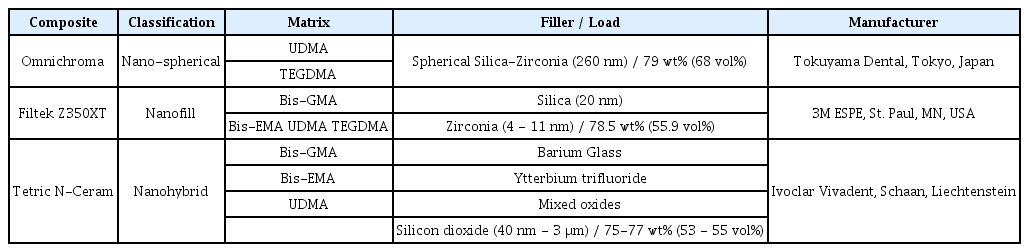
Characteristics of the composite resins used in this study
2. Specimen preparation
Each specimen was fabricated using a metal mold that was 7.0 mm and 2.0 mm in diameter and height, respectively. A mylar strip was placed over the glass slab, and the upper surface was flattened with a resin applicator. Each specimen was polymerized using an LED curing light (VALO™, Ultradent, South Jourdan, UT, USA), with 1400 mW output power and a 10.0 mm-diameter round tip, following the manufacturer’s instructions. The upper surface of the specimen was designated as the surface on which the mylar strip was located.
All specimens underwent the finishing step after light curing. The finishing was performed using a tungsten carbide bur (Komet, Stuttgart, Germany) with a high-speed handpiece at 200,000 rpm for 30 seconds. After the finishing, polishing was performed. The Control subgroup was kept unpolished. The SofLex subgroup was polished using SofLex™ discs (3M ESPE) for 20 seconds in each step corresponding to 3 different abrasive grades: medium, fine, and superfine. The PoGo subgroup was polished using a Pogo™ bur (Dentsply Caulk, Milford, DE, USA) for 60 seconds. The polishing was done at 25,000 rpm using a low-speed handpiece. The characteristics of the instruments used for finishing and polishing are summarized in Table 2. To minimize variability, the same operator performed all procedures.
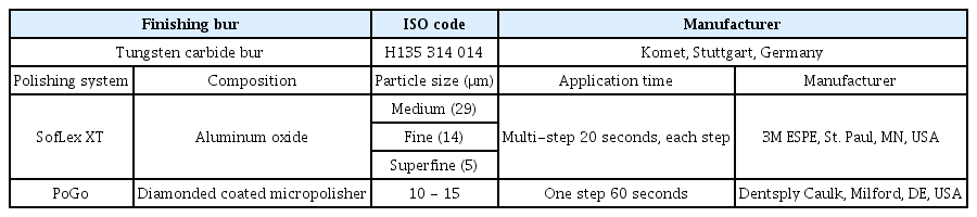
Finishing and polishing materials used in this study
Streptococcus mutans ATCC 25175 was cultivated in brain heart infusion broth (BHI broth; Becton, Dickinson and Company, Sparks, MD, USA) for 18 hours at 37°C under aerobic conditions supplemented with 5% CO2. After measuring turbidity using a spectrophotometer, S. mutans was diluted to 1 × 10⁹ colony-forming units (CFU)/mL. A total of 30 specimens from each group (10 for each subgroup) were inoculated with a bacterial solution containing 1980 μL of BHI broth + 1% sucrose, and 20 μL of S. mutans . The final concentration of S. mutans was set at 1 × 10⁷ CFU/mL. The specimens were then stored at 37°C in a 5% CO2 incubator for 24 hours.
3. Surface analysis
The surface roughness (Ra) of 15 specimens from each group (5 for each subgroup) was assessed using an atomic force microscope (PSIA XE-100, Park System, Suwon, Korea). The roughness of 3 randomly selected spots in the middle of each specimen was measured, and the Ra was calculated by averaging these values. For the remaining specimens, a scanning electron microscope (SEM, JSM-IT500, JEOL, Tokyo, Japan) was used to evaluate the qualitative surface roughness.
4. CFU count
Specimens were washed with phosphate-buffered saline (PBS), and a bacterial suspension was obtained after 20 seconds of sonication (VC 100, Sonics & Materials Inc., Danbury, CT, USA). After sonication, 50.0 μL of the suspension diluted with PBS was spread on a blood agar plate and cultivated at 37°C under aerobic conditions supplemented with 5% CO2 for 72 hours. Bacterial colonies were counted with the naked eye.
5. Statistical analysis
IBM SPSS version 25.0 (SPSS Inc., Chicago, IL, USA) was used for the statistical analysis. Analysis was performed using the Kruskal-Wallis test, and Bonferroni’ s post-hoc test was used if there was a significant difference between the groups.
Results
1. Surface roughness
Table 3 and Fig. 1 show the means and standard deviations of Ra. The Control subgroup of OM showed a significantly lower Ra than the other 2 composite groups. The Ra of the SofLex subgroups of all composite resins was significantly lower than that of the Control subgroups. However, no significant differences were observed between the SofLex subgroups of the 3 composite resins. Similarly, no statistically significant differences were found between the PoGo subgroups of the 3 composites. In the TC group, the Ra of the PoGo subgroup was lower than that of the Control subgroup, but there was no significant difference between them in OM and FT groups. Fig. 2 shows the SEM images of the surfaces of the specimens from each group.
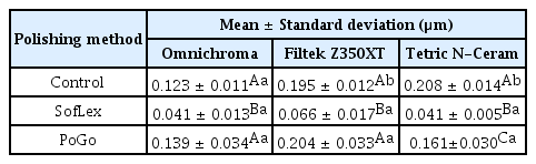
Surface roughness (Ra) values of specimens
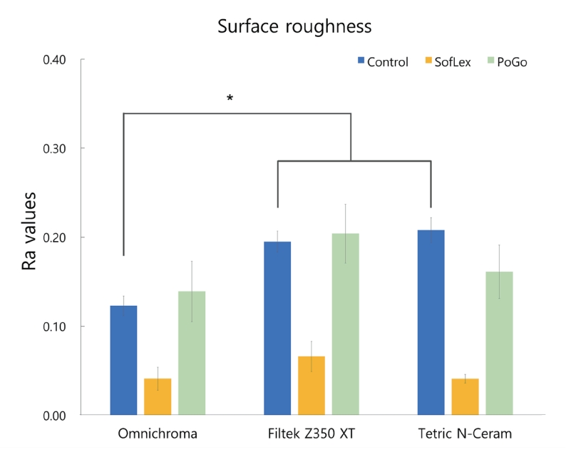
Surface roughness values of the groups.
*: Means significant differences between the Control subgroups by Bonferroni’s post-hoc test.
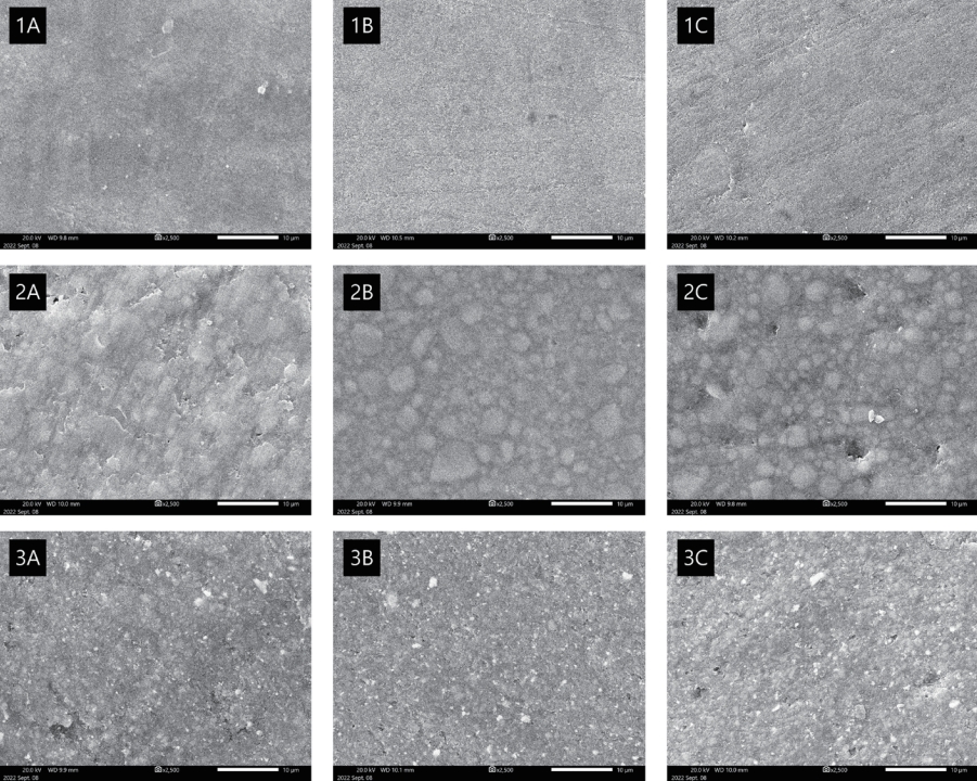
Scanning electron microscope images of specimens in each group.
(1) Omnichroma (2) Filtek Z350 XT (3) Tetric N-Ceram
(A) Control (B) SofLex (C) PoGo
2. CFU count
No significant differences were found between any of the three subgroups of the composites (Fig. 3, Table 4). However, OM generally showed higher microbial adhesion than those of the other 2 composites. No association was found between surface roughness and microbial adhesion.

Bacterial adhesion of the groups.
*: Means significant differences between the Control subgroups by Bonferroni’s post-hoc test.

Colony-forming unit counts of Streptococcus mutans
Discussion
The surface quality of the composite resin is an important characteristic directly related to clinical success. The smooth, polished surface of the composite resin increases esthetics, facilitates hygiene, and makes patients comfortable, all of which are associated with a high clinical success rate [11-14]. In contrast, rough surfaces cause discoloration, irritate gingival tissues, increase plaque accumulation and secondary caries, and are associated with a high risk of fracture [5,15,16].
The surface roughness of the composite resin influences the initial adhesion of bacteria, and once attachment occurs, the bacteria accumulate independently on the surface [17]. The threshold of surface roughness (Ra) for clinical success is known to be 0.2 μm [18]. Ra values of 0.2 μm or above have been reported to provide a retention area to which bacteria can attach [8], whereas the attachment of S. mutans has been reported to decrease at values below 0.15 μm [19]. Another study found that patients could notice Ra changes of 0.3 μm with the tip of their tongue [20].
Recognizing the color of a tooth is a complex process that is influenced by multiple variables, including lighting conditions, light scattering, transparency, and the visual pathway from the eye to the brain[21]. Instead of utilizing pigments or dyes to represent colors, the newly developed single-shade OM controls optical properties using structural colors. This approach, also called “smart chromatic technology”, reflects specific wavelengths inside the tooth color space, producing an excellent color-matching effect [1,22]. OM uses spherical fillers with a uniform size of 260 nm to achieve this structural color. The surface quality of the composite resin is determined by many factors, such as the structure of the resin matrix, filler size, shape, or loading, and finishing/polishing procedures [23]. Owing to the unique filler shape, size, and arrangement of OM, the surface properties may be different from those of conventional resin composites. Therefore, this study evaluated the surface roughness of OM compared with that of conventional nanofill and nanohybrid composite resins.
The smoothest surface of the composite resin can be obtained using a mylar strip [17,18]. However, finishing and polishing procedures are necessary for practically all clinical restorations; thus, it is reasonable to evaluate surface roughness after the finishing and polishing steps. A tungsten carbide bur was used for the finish in this study because the previous study[4] found that it was the most effective in producing the smoothest surfaces of composite materials.
In this study, OM showed a significantly smoother surface than the other 2 types of composites after finishing. According to the manufacturer, uniform nano-sized spherical fillers and consistent spacing between filler particles contributed to the superior polishability of OM. This smooth surface can also be observed in the SEM image in Fig. 2. The SofLex™ system was found to be effective in polishing all tested composites in this study. The Ra of the SofLex subgroups was significantly lower than those of the Control subgroups in all composite groups; however, there were no statistically significant differences between the SofLex subgroups of the 3 composites. This result agrees with the previous study showing that there was no significant difference in the surface roughness after polishing OM and other nanohybrid resins [4]. Kaizer et al. [24] also reported no significant differences in the surface roughness between nanofill, nanohybrid, and micro-hybrid composites.
In this study, SofLex™ discs provided a much smoother surface than the PoGo™ bur, which is in line with the previous systemic review [11] demonstrating that multistep polishing systems are more effective than one-step systems, and aluminum oxide particles provide the smoothest composite surfaces. Fruits et al. [25] stated that the motions applied during the polishing procedure also affect the surface quality of the restoration and found that among the 3 motions of rotary, planar, and reciprocal, the planar motion produced the smoothest surface. SofLex™ is a representative disc-type instrument using this planar motion.
Unlike previous studies that found PoGo™ to be an efficient polishing method [26-28], this study found no significant differences between the Control and PoGo subgroups of OM and FT. This is assumed to be related to the difference in the applied revolutions per minute (rpm) during polishing. In this study, both SofLex™ discs and PoGo™ burs were used at the same speed of 25,000 rpm. However, based on other studies [29,30], PoGo™ appears to have an efficient polishing effect at lower rpm.
It is generally known that the rough surface of a composite resin is associated with increased bacterial adhesion. However, there were no statistically significant differences in CFUs of the attached S. mutans in the Control, SofLex, and PoGo subgroups of each of the tested composites. This result is consistent with the previous finding [8] that bacterial accumulation no longer decreases at a Ra below 0.2 μm. Bacterial adhesion in all OM subgroups was generally higher than that in the other composite resins, and this difference was statistically significant. This high bacterial adhesion may be attributed to the resin matrix composition that constitutes OM.
FT and TC are Bis-GMA-based composite resins, whereas OM is composed of UDMA and TEGDMA. Kim et al. [31] found that UDMA has no significant effect on either the planktonic growth or cellular viability of S. mutans, whereas Bis-GMA has been demonstrated to considerably retard its planktonic growth and decrease its viability [32]. They also reported that UDMA promotes biofilm formation by increasing the surface adhesion of S. mutans, which is in line with the results of this study. Another study found that a composite resin that did not contain Bis-GMA showed significantly higher adhesion to S. mutans than other Bis-GMA-based resins [33]. Nevertheless, Bis-GMA should not be regarded as the only factor affecting bacterial adhesion, as all components of the composite resin can interact with each other, and the combinations can also be one of the influencing factors.
Conclusion
OM showed clinically acceptable smooth surfaces after finishing and polishing procedures and demonstrated smooth surfaces compared to the other 2 types of composites after finishing. However, the adhesion of S. mutans to OM was slightly higher than that of the other tested composite resins. No significant correlation was found between surface roughness and microbial adhesion. Further studies on the other mechanical properties of OM are required for its effective clinical application.
Notes
Conflict of Interest
The authors have no potential conflicts of interest to disclose.