어린이에서 저속 상악 확장에 따른 골격성, 치아치조성, 기도 변화에 대한 3차원적 평가

Three Dimensional Skeletal, Dentoalveolar and Airway Space Changes after Slow Maxillary Expansion in Children

Article information

J Korean Acad Pediatr Dent. 2023;50(2):155-167
Publication date (electronic) : 2023 May 31
doi : https://doi.org/10.5933/JKAPD.2023.50.2.155
Department of Pediatric Dentistry and Institute of Oral Bioscience, School of Dentistry, Jeonbuk National University, Jeonju, Republic of Korea
김나운orcid_icon, 이대우orcid_icon, 김재곤orcid_icon, 양연미orcid_icon
전북대학교 치과대학 소아치과학교실 및 구강생체과학연구소
Corresponding author: Yeonmi Yang Department of Pediatric Dentistry, School of Dentistry, Jeonbuk National University, 20, Geonji-ro, Deokjin-gu, Jeonju, 54907, Republic of Korea Tel: +82-63-250-2212 / Fax: +82-63-250-2131 / E-mail: pedo1997@jbnu.ac.kr
Received 2023 February 8; Revised 2024 March 7; Accepted 2023 March 13.

Abstract

이 연구의 목적은 상악 확장 속도에 따른 치아치조성, 골격성 효과 및 상기도에 대한 효과를 CBCT를 통해 3차원적으로 분석하여 저속 상악 확장의 효과를 확인하는 것이다. 전북대학교 소아치과에서 Banded hyrax를 이용하여 상악 확장을 시행한 23명(평균 8.93 ± 1.61세)의 환자가 포함되었다. 확장 속도에 따라 저속 상악 확장군과 급속 상악 확장군으로 분류되었다. 치료 전후의 치아치조성, 골격성, 기도 부피 변화를 평가하기 위해 치료 전(T0)과 치료 종료 및 유지 후(T1)에 촬영한 CBCT를 사용하였다. 상악 확장 결과 두 군 모두에서 치아치조성, 골격성 측정 값 및 상기도 부피의 유의한 증가가 관찰되었다. 또한 모든 측정 값에서 저속 상악 확장과 급속 상악 확장 간의 유의한 차이를 보이지 않았다. 이 연구는 혼합치열기 어린이에서 저속 상악 확장의 효과에 대해 확인하였다. 저속 상악 확장은 치아치조성, 골격성 측정 값 뿐만 아니라 기도 부피, 상악동 기체 부피에서도 유의한 효과를 보였다. 또한, 급속 상악 확장의 효과와 비교하였을 때 유의한 차이를 보이지 않았다. 따라서 소아치과의사는 성장기 어린이의 치주 조직의 생리적 측면, 불편감에 따른 협조도 등을 고려하여 급속 상악 확장과 저속 상악 확장 중 적절한 방법을 선택할 수 있을 것이다.

Trans Abstract

The aim of this study was to investigate the effects of slow maxillary expansion (SME) on the dentoalveolar, skeletal, upper airway, and maxillary sinus using cone-beam computed tomography (CBCT). Twenty-three orthodontic patients (mean age 8.93 ± 1.61 years) who were treated with maxillary expansion using banded hyrax in the Department of Pediatric Dentistry at Jeonbuk National University Dental Hospital were included. According to the expansion speed applied, they were divided into two groups: SME (12 subjects, mean age 8.92 ± 1.45 years) and rapid maxillary expansion (RME, 11 subjects, mean age 8.94 ± 1.84 years). CBCT were obtained before (T0) and after (T1) the treatment and were analyzed with InVivo5 software (Anatomage, San Jose, CA, USA). Descriptive statistics showed no significant differences between the two groups in age, sex, or skeletal maturity. There were significant increases in maxillary width at the dentoalveolar and skeletal levels for both groups. Upper airway volume revealed a significant increase of 38.59% in the SME group and 28.72% in the RME group. However, there was no significant difference between SME group and RME group in all measurements. This study suggested the efficacy of SME in growing patients. SME was effective in increasing not only dentoalveolar and skeletal measurements but also airway volume. Therefore, pediatric dentists should select an appropriate expansion method considering the physiological aspects of periodontal tissues and discomfort in growing children.

서론

두개안면은 경조직과 연조직의 긴밀한 상호관계로 이루어져 있으며, 성장에 따라 지속적인 변화를 일으킨다[1]. 상악 협착은 두개안면 부위의 가장 흔한 골격성 기형 중 하나이며, 관련된 병인으로 선천적 원인 외에도 빨기 습관, 비강 호흡 장애 등이 알려져 있다[2,3]. 상악 협착은 상하악의 횡적 부조화를 발생시키며, 이는 치아와 악골의 성장과 발달에 장기적인 영향을 미칠 수 있다[4,5]. 또한 비정상적인 하악 움직임(mandibular shift)을 유발하여 턱관절과 저작계에 영향을 미칠 수 있으며, 경추와 머리 자세에도 영향을 미친다[6,7]. 따라서, 성장기 어린이의 정상적인 두개안면 발달을 위해 상악 협착에 대해 상악 확장과 같은 조기 개입이 필요하다[8,9].

상악 확장 치료의 효과는 주로 급속 상악 확장(Rapid maxillary expansion, RME)을 사용하여 연구되었다. 급속 상악 확장시 확장량은 상방에서 하방으로 갈수록, 후방부에서 전방부로 갈수록 크며, 치조골의 휘어짐과 치아의 협측 경사 이동량은 전방부에서 후방부로 갈수록 더 크다[10-12]. 그러나 최근 CBCT를 이용한 연구에서는 상악 확장이 장치 및 개인에 따라 확장 양상이 다소 다를 수 있으며, 그 원인으로는 연구마다 다른 장치 디자인, 환자마다 다른 골 저항력 등이 제시되었다[12-14].

상악 확장은 구개뿐만 아니라 비강의 폭을 유의하게 증가시킬 수 있으며 이는 비강의 부피 증가를 일으킨다[10,15]. 이러한 비강에 대한 영향은 비강의 기도 저항을 감소시키고, 비인두의 부피를 증가시켜 환자의 구호흡, 코골이, 수면무호흡증 등의 개선을 일으킬 수 있다[16-18]. McNamara 등[19]은 급속 상악 확장이 안면부의 기능적, 구조적 불균형을 초래할 수 있는 원인들을 차단하여 성장기 환자에서 긍정적인 효과를 보인다고 하였다.

상악 확장은 고정원에 따라 골 지지성과 치아 지지성으로 분류될 수 있으며, 골 지지성 상악 확장이 치아 지지성 상악 확장에 비해 치아치조성 부작용이 적고, 비강 및 상악의 골격성 확장과 비강 기도 저항 감소에 더 효과적이라고 보고되었다[20,21]. 하지만, 혼합치열기 어린이에서 골 고정원은 미맹출 치아 및 미성숙골 때문에 사용이 용이하지 않다. 골 고정원 사용시 나이는 중요한 고려 요소이며 교정 치료에 적절한 골밀도는 초기 청소년기까지 얻어지지 않으므로, 골 고정원은 대부분의 혼합치열기 치료에 사용될 수 없다. 또한 혼합치열기 어린이에서는 작은 힘으로도 골격성 확장이 비교적 쉽게 발생하므로, 주로 치아 지지성 상악 확장을 통해 상악 협착을 개선한다[4].

급속 상악 확장은 빠른 속도로 확장력이 봉합부에 전달되기 때문에 골격성 이동으로 봉합부가 개방되며 확장이 일어난다. 따라서 확장 초기에는 골격성 확대가 주로 일어나지만, 유지 기간 동안 구개 하방부에서 골격성 재발에 의한 내측 이동이 일어나 치아치조성 확장이 꾸준히 일어나게 된다. 반면 저속 상악 확장(Slow maxillary expansion, SME)은 생리적인 속도로 천천히 확장하기 때문에 확장 초기부터 골격성 확장과 치아 치조성 확장이 비슷한 비율로 일어나며, 확장이 완료될 때까지 같은 비율이 유지된다[4,22,23]. 상악 확장에 대해 보고된 이전의 많은 연구들은 급속 상악 확장이 치아치조성 및 골격성 횡적 확장 효과 뿐 아니라, 비기도의 저항을 감소시켜 구호흡 및 수면호흡장애의 개선 등에 대해 보고한 반면, 저속 상악 확장에 대한 연구는 적다. 특히 저속 상악 확장의 비강 및 기도와의 관련성에 대한 연구는 드물며 국내에서 초기 혼합치열기 어린이를 대상으로는 거의 연구되지 않았다. 따라서 이 연구의 목적은 CBCT를 이용하여 상악 확장 속도에 따른 치아치조성, 골격성 효과 및 상기도에 대한 효과를 분석하여 저속 상악 확장의 효과를 확인하는 것이다.

연구 재료 및 방법

이 연구는 전북대학교병원 생명의학연구윤리심의위원회(Institutional Review Board, IRB)의 승인을 받아 시행되었다(IRB No: CUH 2020-04-058-004).

1. 연구 대상

2011년 1월부터 2020년 4월까지 전북대학교 치과병원 소아치과에서 상악 확장을 포함한 교정 치료를 진행한 환자를 대상으로 하였다. 전자의무기록 분석을 시행하여 선별 및 제외 기준에 따라 최종적으로 23명의 환자가 연구에 포함되었다.

1) 선별기준

1) 상하악 제1대구치가 맹출 완료된 초기 혼합치열기

(2) 상악 확장 시 Banded hyrax를 이용한 경우

(3) 상악 확장 후 최소 3개월 이상의 유지를 한 경우

(4) 교정 치료 전후 CBCT 촬영을 시행한 경우

2) 제외 기준

(1) 구강악안면에 영향을 미치는 전신 질환을 가진 경우

(2) 이전 교정 치료 경험이 있는 경우

2. 연구 방법

1) 상악 확장 속도에 따른 분류

23명의 어린이는 전자의무기록에 기록되어 있는 상악 확장의 속도에 따라 저속 상악 확장군(SME group, 2 turns/week, 0.5 mm/week)과 급속 상악 확장군(RME group, 2 turns/day, 3.5 mm/week)으로 분류하였으며, 각각의 확장 기간 및 유지 기간을 분석하였다.

2) Cone-beam computed tomography (CBCT) 영상의 분석

CBCT는 Alphald VEGA (Asihi Roentgen Ind. Co., Kyoto, Japan)로 촬영되었고, 관전압 80 kV, 관전류 8 mA, 촬영 시간 17초, 154 mm × 154 mm의 field of view, 300 μm의 voxel size로 설정되었다.

CBCT 영상을 분석하기 위해 InVivo5 software (Anatomage, San Jose, CA, USA)를 이용하였다. 상악 확장 전(T0)과 확장 및 유지 후(T1) CBCT에서 비교 가능한 영상을 얻기 위해 Frankfort horizontal plane (FH plane)이 지표면과 평행하도록 설정하고, 재현 가능한 기준점을 이용하여 CBCT 영상의 방향을 변경하였다(Fig. 1). CBCT 영상의 계측시 1명의 연구자가 동일한 날 총 2회 측정하여 평균치를 기록하였다.

Fig 1.

Reorientation of CBCT image using InVivo5 software. (A) Sagittal section, (B) Axial section, (C) Coronal section. Reoriented as parallel to the FH plane (Sagittal section), passing through the center of furcation area of both maxillary first molars (Axial section), and parallel to the hard plate (Coronal section).

(1) 횡적 변화

치료 전후 상악의 치아치조성, 골격성 횡적 변화를 분석하였다. 치아의 측방 확장량을 분석하기 위해 양측 상악 견치 사이의 가장 짧은 거리인 견치간 폭경(intercanine width)과 양측 상악 제1대구치 사이의 가장 짧은 거리인 구치간 폭경(intermolar width)을 계측하였으며, 치축 변화를 확인하기 위해 상악 제1대구치의 협측 교두와 구개측 치근을 이은 선을 기준으로 각도(intermolar angulation)를 계측하였다. 치조골 및 상악골 확장량을 분석하기 위해 상악 기저부(maxillary base), 구개 폭경(palatal width), 비강 폭경(nasal width), 치조골 폭경(alveolar width)를 측정하였다(Fig. 2).
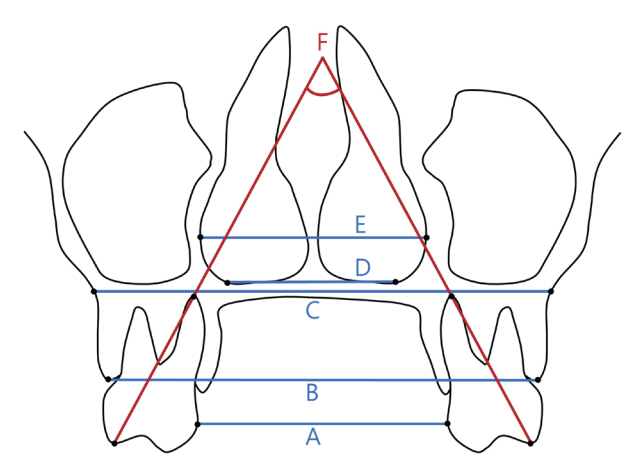
Fig 2.

The transverse measurement in the coronal section. (A) Intermolar width (mm): The shortest width between the crowns of maxillary first molars on both sides, (B) Alveolar width (mm): Line between the lowest point of the alveolar process on both sides, (C) Maxillary base (mm): Line between the jugular process for maxillary width on both sides, (D) Palatal width (mm): Line between the junction of the palatal cortical alveolar bone and the lateral cortical nasal bone on both sides, (E) Nasal width (mm): Width at the widest portion of the nasal aperture, (F) Intermolar angulation (°): Angle between the line joining the tip of the buccal cusp to the apex of the palatal root of the maxillary first molars on both sides.

(2) 상기도 부피의 변화

치료 전후의 상기도 부피 평가를 위해 총 4개의 기준선을 설정하였다(Fig. 3). Posterior nasal spine (PNS)에서 FH plane에 내린 수선, FH plane과 PNS 사이를 이등분하는 점에서 FH plane과 평행한 선, 제3경추의 최전하방점에서 FH plane과 평행한 선은 각각 상기도의 전방 경계, 상방 경계, 하방 경계로 설정되었다. InVivo5 소프트웨어에서 상기도를 해당 기준선을 이용하여 구획하고 3차원적인 상기도 이미지를 추출하였다. 이후 PNS와 제1경추의 최전하방점을 이은 선을 기준으로 상기도를 retropalatal airway와 retroglossal airway로 구분하여, total upper airway volume, retropalatal airway volume, retroglossal airway volume을 측정하였다.
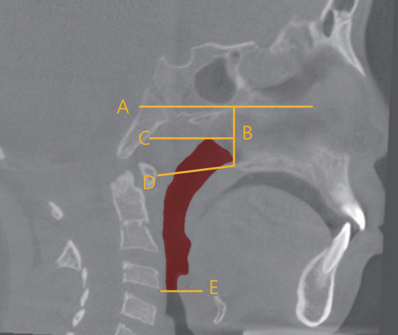
Fig 3.

The planes for delimiting the upper airway. (A) FH plane, (B) Anterior border. Plane perpendicular to the FH plane on PNS, (C) Superior plane. Plane parallel to the FH plane on the point of the (B) line, (D) Border of the retropalatal and retroglossal airway sections. Anterior lowest point of C1-PNS, (E) Inferior plane. Plane parallel to the FH plane on the anterior lowest point of C3.

(3) 상악동 내 기체 부피의 변화

치료 전후의 상악동 기체 부피를 측정하기 위해 CBCT 영상을 inverse image로 변환하여, 부비동 및 상기도의 3차원적 이미지를 재구성하였다. 상악동을 상악동 구(ostium)를 경계로 분리하고, 3차원적 이미지의 선명도를 높여 경계를 명확하게 한 후 각각의 부피를 측정하였다(Fig. 4).

Fig 4.

Airway space of the maxillary sinus in a 3D image. CBCT images were converted into inverse images to reconstruct 3-dimensional images of the paranasal sinuses and upper airway. The maxillary sinus was separated with the ostium as the boundary, and each volume was measured.

3. 통계 분석

상악 확장 방법과 치료 전후에 따른 치아치조성, 골격성, 기도부피의 변화의 통계적 유의성을 검증하기 위해 Wilcoxon signed rank test를 시행하였다. 통계 분석은 SPSS (version 23.0, IBM, Chicago, IL, USA)를 이용하였다.

연구 성적

1. 환자의 특성과 확장 기간

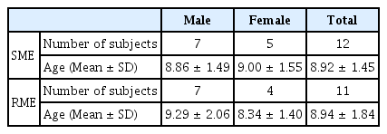
연구에 포함된 총 23명의 환자의 나이는 8.93 ± 1.61세였으며 이중 남자는 14명(9.07 ± 1.74세), 여자는 9명(8.70 ± 1.44 세)이었다. 상악 확장의 속도에 따라 저속 상악 확장군 12명, 급속 상악 확장군 11명으로 분류되었다(Table 1). 확장 기간은 저속 상악 확장군은 평균 14.27주, 급속 상악 확장군은 평균 2.07 주였으며, 유지 기간은 저속 상악 확장군은 평균 3.47개월, 급속 상악 확장군은 평균 3.62개월이었다.

Classification of subjects according to maxillary expansion rate

2. 상악 확장 치료 전후 횡적 변화

1) 치아치조성 변화

상악 확장 치료 전후 상악 제1대구치의 치축 변화를 비교했을 때, 저속 상악 확장군은 3.88 ± 6.35°, 급속 상악 확장군은 8.01 ± 4.39° 증가하였으며, 급속 상악 확장군에서만 통계적으로 유의한 차이를 보였다(p < 0.01). 상악 제1대구치간 폭경은 저속 상악 확장군은 5.96 ± 1.45 mm, 급속 상악 확장군은 6.41 ± 1.49 mm 증가하였다(p < 0.01). 치조골 수준에서의 횡적 변화는 저속 상악 확장군은 견치 부위에서 5.45 ± 1.20 mm, 대구치 부위에서 4.25 ± 1.51 mm 증가하였으며, 급속 상악 확장군은 견치 부위에서 5.65 ± 1.36 mm, 대구치 부위에서 4.74 ± 1.26 mm 증가하였다(p < 0.01, Table 2).

Transverse changes in dentoalveolar measurement before and after maxillary expansion

2) 골격성 변화

상악 확장 치료 전후 nasal width는 저속 상악 확장군은 견치, 대구치 부위에서 각각 1.91 ± 0.79 mm, 1.69 ± 1.09 mm, 급속 상악 확장군은 견치, 대구치 부위에서 각각 2.51 ± 0.82 mm, 1.98 ± 1.08 mm 유의하게 증가하였다. Palatal width는 저속 상악 확장군은 견치, 대구치 부위에서 각각 1.86 ± 1.78 mm, 1.45 ± 1.24 mm 증가하였으며, 급속 상악 확장군은 견치, 대구치 부위에서 각각 2.61 ± 0.65 mm, 2.34 ± 1.71 mm 증가하였으나 견치 부위에서만 통계적으로 유의한 차이를 보였다(p < 0.01). Maxillary base는 저속 상악 확장군에서 2.06 ± 1.59 mm 증가하였고, 급속 상악 확장군에서 2.53 ± 0.77 mm 증가하였다(p < 0.01, Table 3).

Transverse changes in skeletal measurement before and after maxillary expansion

3. 상악 확장 치료 전후 상기도 부피의 변화

저속 상악 확장군의 total upper airway volume은 상악 확장 치료 전 9.20 ± 2.04 cc, 상악 확장 치료 후 12.75 ± 2.55 cc 였으며 치료 전후 38.59%의 유의한 증가를 보였다(p < 0.01). Retropalatal airway와 retroglossal airway는 각각 53.85%, 25.63% 증가하였고, retropalatal airway만이 통계적으로 유의한 차이를 보였다(p < 0.01).

급속 상악 확장군의 total upper airway volume은 상악 확장 치료 전 12.60 ± 3.20 cc, 상악 확장 치료 후 16.22 ± 3.13 cc 였으며 치료 전후 28.72%의 유의한 증가를 보였다. Retropalatal airway와 retroglossal airway는 각각 28.44%, 28.97% 증가하였으나, 모두 통계적으로 유의하지는 않았다(Table 4).

Volumetric changes in upper airway space before and after maxillary expansion

4. 상악 확장 치료 전후 상악동 기체 부피의 변화

상악 확장 치료 전후 상악동 내 기체 부피는 저속 상악 확장군에서 좌우측 각각 3.32 ± 4.00 cc, 2.60 ± 3.93 cc 유의하게 증가하였다. 급속 상악 확장군에서는 좌우측 각각 1.82 ± 2.22 cc, 1.26 ± 1.50 cc 증가하였으나, 통계적으로 유의한 차이는 없었다(Table 5).

Volumetric changes in airway space of maxillary sinus before and after maxillary expansion

5. 상악 확장 속도에 따른 확장 효과의 비교

상악 확장 치료 전후 저속 상악 확장군과 급속 상악 확장군 간의 확장 효과 차이를 분석하였을 때, 횡적 변화(치아치조성, 골격성), 상기도 부피, 상악동 기체 부피의 모든 측정값에서 저속 상악 확장군과 급속 상악 확장군 간의 통계적으로 유의한 차이를 보이지 않았다(Table 6).

Comparison of expansion effects between SME and RME

총괄 및 고찰

상악 확장 치료는 1860년대에 상악골의 횡적 부조화를 개선하기 위해 최초로 도입되었으며, 빠른 속도로 악궁을 확장시키는 급속 상악 확장 방법으로 연구가 이루어졌다[24]. 급속 상악 확장은 악궁 확장, 크라우딩 해소 등의 치아치조성, 골격성 효과 외에도 비인두, 비인강, 부비동의 기체 부피를 증가시키고 호흡 양상을 개선시키는 효과를 보인다[19,25-27]. 급속 상악 확장과 저속 상악 확장의 효과를 비교한 연구에서, Lo Giudice 등[28]은 급속 상악 확장과 저속 상악 확장 모두에서 비강 부피가 증가하였으나, 실험군의 수가 적어 두 군 간의 통계분석은 어려웠다고 하였다. 또한 Lanteri 등[29]은 혼합치열기 환자에서 저속 상악 확장이 인두 기도와 상악동의 부피를 증가시키는데 효과적이며, 급속 상악 확장과 유의한 차이가 없다고 보고했지만, 해당 연구는 치료에 사용된 구내 장치가 다르다는 한계가 있었다. 따라서 이 연구에서는 급속 상악 확장과 저속 상악 확장 두 군 간의 장치를 banded hyrax appliance만으로 제한하고, 확장 속도를 단일 변수로 설정하여 확장 속도에 따른 상악 확장 효과를 비교하였다.

이 연구의 상악 확장 치료 전후 저속 상악 확장군과 급속 상악 확장군의 횡적 변화에 대한 분석에서 치아치조성, 골격성 계측치는 모두 양의 값을 보이며 증가하였다. 저속 상악 확장군과 급속 상악 확장군의 확장량을 비교하였을 때 유의한 차이를 보이지 않으며 유사한 치아치조성, 골격성 확장 효과를 보였다. 그러나 치아치조성, 골격성 확장 효과에 대해서 상반된 결과를 보이는 연구들이 있다. Proffit 등[4]은 저속 상악 확장과 급속 상악 확장이 확장의 속도가 다를 뿐이며 확장량은 유사하다고 하였으며, 다만 저속 상악 확장이 보다 생리적인 확장 방법이라고 하였다. Luiz Ulema Ribeiro 등[23]은 평균 8.18세 환자에서 급속 상악 확장이 저속 상악 확장보다 치아치조성, 골격성 확장 효과가 뛰어나다고 보고하였으며, Zhou 등[30]은 메타 분석을 통해 저속 상악 확장이 대구치부 확장의 경우 급속 상악 확장보다 더 뛰어나며, 이외의 계측치에서는 저속 상악 확장과 급속 상악 확장 간의 유의한 차이가 없다고 보고하였다. 또한 메타분석에 포함된 여러 연구간 결과의 불일치는 적은 수의 고품질 연구, 연구간의 다양한 치료 전략, 분석시 측정 계측점의 차이 등에서 기인한다고 하였다. 따라서 표준 측정 방법과 통합된 치료 전략을 통해 고품질의 근거를 나타내는 연구가 시행되어야 함을 제시하였다.

여러 연구에서 급속 상악 확장이 상기도 폭과 용적을 증가시키는 간접적인 효과가 있으며, 이는 흡기시 비강 및 기도의 압력을 감소시킴으로써 호흡 기능의 개선을 일으킨다는 보고가 있다[31,32]. 이 연구에서는 상기도의 총 부피가 저속 상악 확장시 38.59%, 급속 상악 확장시 28.72%씩 유의하게 증가하였다. 상기도를 retropalatal airway와 retroglossal airway로 구획하여 분석하였을 때는 저속 상악 확장시 retropalatal airway 만이 통계적으로 유의한 증가를 보였다. 또한 상악동 저류 기체는 저속 및 급속 상악 확장 모두에서 증가 양상을 보였으나 저속 상악 확장만이 통계적으로 유의한 증가를 보였다. 하지만 두 군을 비교하였을 때 상기도 및 상악동에 대한 모든 값에서 유의한 차이를 보이지 않아 저속 상악 확장과 급속 상악 확장이 유사한 효과를 나타냄을 확인하였다. 기존 연구에서 밝혀진 바와 같이 상기도 및 부비동의 부피 증가는 호흡 기능의 개선을 간접적으로 나타낼 수 있지만, 이 연구는 실제 호흡 양상의 기능적인 평가가 이루어지지 않았다는 한계가 있다. 따라서, 향후 전산 유체 역학을 이용한 분석 등의 기능적 평가를 동반한 추가적인 연구가 필요할 것으로 생각된다.

이 연구는 평균 8.93 ± 1.61세의 어린이를 대상으로 하여 초기 혼합치열기 어린이에 대한 상악 확장 치료의 효과를 분석하였다. 유치열기 및 초기 혼합치열기에는 정중구개봉합이 아직 융합되지 않았거나, 일부분에서만 초기 골화가 진행되어 골격성 확장이 비교적 쉽게 발생한다. 하지만, 연령의 증가에 따라 정중구개봉합의 골화가 진행되며 봉합부 확장에 더 큰 힘이 필요하게 되고, 사춘기 최대 성장기 이후에는 치아 지지성 확장만으로 봉합의 이개가 불가능할 수 있으며 미니 스크류 등을 이용한 골 지지성 확장이 시행되어야 한다[33,34]. 따라서, 환자의 연령에 따라 상악 확장 치료의 효과가 달라질 수 있으므로, 확장 방법의 선택 시 환자의 연령에 대한 고려가 반드시 필요할 것으로 생각된다.

정모 또는 측모 두부방사선 계측사진의 경우 왜곡 및 확대 문제로 정확한 각도 및 거리의 변화에 대한 정확한 계측이 어렵다[12]. 그러나 CBCT는 실측된 3차원적인 이미지를 이용하여 각도 및 거리의 변화에 대한 정확한 계측을 할 수 있다[35]. 또한 상기도를 평가할 때 측모 두부방사선 계측사진 역시 유용하지만, 복잡한 구조를 가진 상기도의 3차원적인 영상을 얻을 수 있는 CBCT가 더 높은 신뢰도를 얻을 수 있다고 하였다[36]. 따라서 이 연구는 CBCT에서 정모 및 측모 두부방사선 계측사진을 추출 후 치아치조성, 골격성 상악 확장 효과를 분석하였고, 3차원적 영상을 추출하여 상기도 및 상악동 기체 부피에 대한 상악 확장 효과를 분석하였다.

교정 치료시 CBCT 촬영은 필수사항이 아니기 때문에 소아 환자의 방사선 촬영에 대한 보호자의 동의 등의 윤리적 고려사항으로 인해 이 연구는 총 23명의 비교적 적은 수의 표본을 대상으로 진행되었다는 한계가 있다. 따라서 이 한계를 극복하기 위해 장치의 종류, 확장 후 유지, 초기 혼합치열기 등의 조건을 제한하여 연구 대상을 설정하였으며 CBCT 영상의 계측시 1명의 연구자가 동일한 날 총 2회 측정하여 오차 범위를 줄여 결과의 신뢰성을 높였다.

상악 확장시 급속 상악 확장이 주로 이루어져 왔으나, 급속 상악 확장시 보고되는 합병증들이 있다. 급속 상악 확장시 통증을 조사한 연구에서 98%가 통증을 나타냈고, 48%가 확장 기간 동안 최소 1번의 진통제를 복용했다고 하였다. 초반 10회의 회전에서 높은 수준의 통증을 나타냈고, 확장 속도를 느리게 하는 것이 통증을 감소시켰다고 하였다[37]. 또한 급속 상악 확장의 확장 기간 동안의 스트레스와 통증에 대한 연구에서 85%의 환자가 통증을 나타냈으며, 코르티졸 호르몬 수치가 증가하였다[38]. 상악의 확장 치료는 치아의 협측 경사 이동을 유발한다[39]. 저속 상악 확장과 급속 상악 확장 모두 치아의 협측 경사 이동을 유발하지만 급속 상악 확장이 저속 상악 확장보다 2배 이상의 더 큰 협측 경사 이동을 발생시킨다고 하였으며, 저속 상악 확장이 급속 상악 확장보다 구치의 치체 이동을 일으킬 수 있다고 하였다[11,40]. 이 연구에서도 저속 상악 확장군과 급속 상악 확장군 모두에서 각각 평균 3.88°, 8.01°의 구치부 치아의 협측 경사 이동이 관찰되었고, 급속 상악 확장군에서만 통계적으로 유의한 증가를 나타냈다. 이는 저속 상악 확장보다 급속 상악 확장에서 지대치에 축적되는 힘이 더 크기 때문에 결과적으로 더 큰 협측 경사 이동이 발생했다고 생각된다[40]. Garib 등[41]은 급속 상악 확장을 시행한 11 - 14세 환자의 CBCT 분석 결과 과도한 치아의 협측 이동으로 인해 치조골 높이의 감소, 열개, 치은 퇴축을 발생시킬 수 있다고 하였다. 또한, Greenbaum과 Zachrisson[42]은 치주 조직에 미치는 영향이 급속 상악 확장과 저속 상악 확장 간에 유의한 차이는 없었지만, 개인 간의 차이가 컸으며, 상악 제1대구치 협측의 부착 상실을 일으킨 대부분의 환자가 급속 상악 확장으로 치료를 시행한 환자였다고 보고하였다. 이러한 급속 상악 확장의 다양한 부작용은 식이 문제, 스트레스, 불안, 수면 장애 등을 일으키는 요소가 될 수 있어 성장기 어린이들에게 더 좋은 대체 장치의 고려가 필요하다. 따라서 이 연구에서는 보다 생리적인 확장 방법으로 알려진 저속 상악 확장의 효과를 급속 상악 확장과 비교하고자 하였으며, 결과적으로 확장 속도 간 효과의 차이가 없음을 확인하였다. 하지만 이 연구에서는 협측 경사 이동 이외의 상악 확장시 부작용에 대해 비교 분석은 시행하지 않았으므로, 향후 이를 반영한 추가적인 연구가 필요하다.

이 연구에 포함된 환자들은 치료 종료 후 평균 3.55개월의 유지를 시행하였다. proffit 등[4]은 상악 확장 후 3 - 4개월의 유지 기간이 필요하다고 하였으나, 상악 확장 후 유지 기간에 대한 여러 문헌이 있다. 재발을 방지하기 위해 6주의 유지 기간이 필요하다는 보고가 있는 반면, 6개월 또는 8개월의 유지 기간이 필요하다는 보고도 있다[43-45]. 따라서 치료 후 장기간의 추적 관찰이 시행된 환자를 포함하여 치료의 안정성을 확인하는 추가적인 연구가 필요할 것으로 보인다.

하지만 이 연구는 상악의 협착을 보이는 초기 혼합치열기 어린이를 대상으로 저속 상악 확장시 나타나는 치아치조, 골격에 대한 직접적인 효과 및 상기도, 상악동에 미치는 간접적인 효과를 파악하고, 급속 상악 확장과 비교시 효과의 차이가 없음을 확인한 것에 의의가 있다.

결론

이 연구는 초기 혼합치열기 어린이에서 저속 상악 확장의 효과에 대해 확인하였다. 저속 상악 확장은 치아치조성, 골격성 측정값 뿐만 아니라 상기도 부피, 상악동 기체 부피에서도 유의한 효과를 보였다. 또한, 급속 상악 확장의 효과와 비교하였을 때 통계적으로 유의한 차이를 보이지 않았다. 따라서 소아치과 의사는 성장기 어린이의 치주 조직의 생리적 측면, 불편감에 따른 협조도 등을 고려하여 급속 상악 확장과 저속 상악 확장 중 적절한 방법을 선택할 수 있을 것이다.

Notes

Conflict of Interest

The authors have no potential conflicts of interest to disclose.

Funding information

This paper was supported by Fund of Biomedical Research Institute, Jeonbuk National University Hospital.

Acknowledgements

This paper was supported by Fund of Biomedical Research Institute, Jeonbuk National University Hospital.
References

1. Son BH, Hwang CJ, Kim GH, Lee GJ. Craniofacial growth and development 1st edth ed. DaehanNarae. Seoul: p. 52–105. 2007.
2. Linder-Aronson S. Adenoids. Their effects on mode of breathing and nasal airflow and their relationships to characteristics of the facial skeleton and the dentition. A biometric, rhino-manometric and cephalometro-radiographic study on children with and without adenoids. Acta Otolaryngol Suppl 265:1–132. 1970;
3. Behlfelt K, Linder-Aronson S, McWilliam J, Neander P, Laage-Hellman J. Dentition in children with enlarged tonsils compared to control children. Eur J Orthod 11:416–429. 1989;
4. Proffit WR, Fields HW, Larson BE, Sarver DM. Contemporary orthodontics 6th edth ed. Elsevier. Amsterdam: p. 454–464. 2021.
5. McNamara JA Jr. Early intervention in the transverse dimension: is it worth the effort? Am J Orthod Dentofacial Orthop 121:572–574. 2002;
6. Troelstrup B, Moller E. Electromyography of the temporalis and masseter muscles in children with unilateral cross-bite. Scand J Dent Res 78:425–430. 1970;
7. Neiva PD, Kirkwood RN, Mendes PL, Zabjek K, Becker HG, Mathur S. Postural disorders in mouth breathing children : a systematic review. Braz J Phys Ther 22:7–19. 2018;
8. Kutin G, Hawes RR. Posterior cross-bites in the deciduous and mixed dentitions. Am J Orthod 56:491–504. 1969;
9. Petrén S, Bondemark L, Söderfeldt B. A systematic review concerning early orthodontic treatment of unilateral posterior crossbite. Angle Orthod 73:588–596. 2003;
10. Garrett BJ, Caruso JM, Rungcharassaeng K, Farrage JR, Kim JS, Taylor GD. Skeletal effects to the maxilla after rapid maxillary expansion assessed with conebeam computed tomography. Am J Orthod Dentofacial Orthop 134:8–9. 2008;
11. Pereira JDS, Jacob HB, Locks A, Brunetto M, Ribeiro GLU. Evaluation of the rapid and slow maxillary expansion using cone-beam computed tomography: a randomized clinical trial. Dental Press J Orthod p. 61–68. 2017.
12. Podesser B, Williams S, Crismani AG, Bantleon HP. Evaluation of the effects of rapid maxillary expansion in growing children using computer tomography scanning: a pilot study. Eur J Orthod 29:37–44. 2007;
13. Lione R, Ballanti F, Franchi L, Baccetti T, Cozza P. Treatment and posttreatment skeletal effects of rapid maxillary expansion studied with low-dose computed tomography in growing subjects. Am J Orthod Dentofacial Orthop 134:389–392. 2008;
14. Mosleh MI, Kaddah MA, Abd ElSayed FA, ElSayed HS. Comparison of transverse changes during maxillary expansion with 4-point bone-borne and tooth-borne maxillary expanders. Am J Orthod Dentofacial Orthop 148:599–607. 2015;
15. Ramires T, Maia RA, Barone JR. Nasal cavity changes and the respiratory standard after maxillary expansion. Braz J Otorhinolaryngol 74:763–769. 2008;
16. Iwasaki T, Sato H, Suga H, Minami A, Yamamoto Y, Takemoto Y, Inada E, Saitoh I, Kakuno E, Kanomi R, Yamasaki Y. Herbst appliance effects on pharyngeal airway ventilation evaluated using computational fluid dynamics. Angle Orthod 87:397–403. 2017;
17. Iwasaki T, Yanagisawa-Minami A, Suga H, Shirazawa Y, Tsujii T, Yamamoto Y, Ban Y, Sato-Hashiguchi M, Sato H, Kanomi R, Yamasaki Y. Rapid maxillary expansion effects of nasal airway in children with cleft lip and palate using computational fluid dynamics. Orthod Craniofac Res 22:201–207. 2019;
18. Iwasaki T, Suga H, Minami-Yanagisawa A, Hashiguchi-Sato M, Sato H, Yamamoto Y, Shirazawa Y, Tsujii T, Kanomi R, Yamasaki Y. Upper airway in children with unilateral cleft lip and palate evaluated with computational fluid dynamics. Am J Orthod Dentofacial Orthop 156:257–265. 2019;
19. McNamara JA Jr, Lione R, Franchi L, Angelieri F, Cevidanes LH, Darendeliler MA, Cozza P. The role of rapid maxillary expansion in the promotion of oral and general health. Prog Orthod 16:33. 2015;
20. Altieri F, Cassetta M. Comparison of changes in skeletal, dentoalveolar, periodontal, and nasal structures after tooth-borne or bone-borne rapid maxillary expansion: A parallel cohort study. Am J Orthod Dentofacial Orthop 161:E336–E344. 2022;
21. Krüsi M, Eliades T, Papageorgiou SN. Are there benefits from using bone-borne maxillary expansion instead of tooth-borne maxillary expansion? A systematic review with meta-analysis. Prog Orthod 20:9. 2019;
22. Giudice AL, Spinuzza P, Rustico L, Messina G, Nucera R. Short-term treatment effects produced by rapid maxillary expansion evaluated with computed tomography: A systematic review with meta-analysis. Korean J Orthod 50:314–323. 2020;
23. Luiz Ulema Ribeiro G, Jacob HB, Brunetto M, da Silva Pereira J, Motohiro Tanaka O, Buschang PH. A preliminary 3-D comparison of rapid and slow maxillary expansion in children: A randomized clinical trial. Int J Paediatr Dent 30:349–359. 2020;
24. Timms DJ. The dawn of rapid maxillary expansion. Angle Orthod 69:247–250. 1999;
25. Caprioglio A, Meneghel M, Fastuca R, Zecca PA, Nucera R, Nosetti L. Rapid maxillary expansion in growing patients: Correspondence between 3-dimensional airway changes and polysomnography. Int J Pediatr Otorhinolaryngol 78:23–27. 2014;
26. Fastuca R, Meneghel M, Zecca PA, Mangano F, Antonello M, Nucera R, Caprioglio A. Multimodal airway evaluation in growing patients after rapid maxillary expansion. Eur J Paediatr Dent 16:129–134. 2015;
27. Machado AJ Jr, Crespo AN, Pauna HF. Rapid maxillary expansion in pediatric patients with obstructive sleep apnea: current and future perspectives. Sleep Med 51:7–8. 2018;
28. Lo Giudice A, Fastuca R, Portelli M, Militi A, Bellocchio M, Spinuzza P, Briguglio F, Caprioglio A, Nucera R. Effects of rapid vs slow maxillary expansion on nasal cavity dimensions in growing subjects: a methodological and reproducibility study. Eur J Paediatr Dent 18:299–304. 2017;
29. Lanteri V, Farronato M, Ugolini A, Cossellu G, Gaffuri F, Parisi FMR, Cavagnetto D, Abate A, Maspero C. Volumetric Changes in the Upper Airways after Rapid and Slow Maxillary Expansion in Growing Patients: A Case-Control Study. Materials 13:2239. 2020;
30. Zhou Y, Long H, Ye N, Xue J, Yang X, Liao L, Lai W. The effectiveness of non-surgical maxillary expansion: a meta-analysis. Eur J Orthod 36:233–242. 2014;
31. Ghoneima A, AlBarakati S, Jiang F, Kula K, Wasfy T. Computational fluid dynamics analysis of the upper airway after rapid maxillary expansion: a case report. Prog Orthod 16:10. 2015;
32. Li J, Shi L, Zhang X, Weng L, Chen H, Lin J. Evaluating the effect of midpalatal corticotomy-assisted rapid maxillary expansion on the upper airway in young adults using computational fluid dynamics. J Zhejiang Univ Sci B 22:146–155. 2021;
33. Angelieri F, Cevidanes LH, Franchi L, Gonçalves JR, Benavides E, McNamara JA JR. Midpalatal suture maturation: classification method for individual assessment before rapid maxillary expansion. Am J Orthod Dentofacial Orthop 144:759–769. 2013;
34. Persson M, Thilander B. Palatal suture closure in man from 15 to 35 years of age. Am J Orthod 72:42–52. 1977;
35. Mah J. The evolution of digital study models. J Clin Orthod 41:557–561. 2007;
36. Major MP, Flores-Mir C, Major PW. Assessment of lateral cephalometric diagnosis of adenoid hypertrophy and posterior upper airway obstruction: a systematic review. Am J Orthod Dentofacial Orthop 130p. 700–708. 2006.
37. Needleman HL, Hoang CD, Allred E, Hertzberg J, Berde C. Reports of pain by children undergoing rapid palatal expansion. Pediatr Dent 22:221–226. 2000;
38. Gecgelen M, Aksoy A, Kirdemir P, Doguc DK, Cesur G, Koskan O, Ozorak O. Evaluation of stress and pain during rapid maxillary expansion treatments. J Oral Rehabil 39:767–775. 2012;
39. Adkins MD, Nanda RS, Currier GF. Arch perimeter changes on rapid palatal expansion. Am J Orthod Dentofacial Orthop 97:194–199. 1990;
40. Isaacson RJ, Ingram AH. Forces produced by rapid maxillary expansion. Angle Orthod 34:261–270. 1964;
41. Garib DG, Henriques JF, Janson G, de Freitas MR, Fernandes AY. Periodontal effects of rapid maxillary expansion with tooth-tissue-borne and tooth-borne expanders: a computed tomography evaluation. Am J Orthod Dentofacial Orthop 129:749–758. 2006;
42. Greenbaum KR, Zachrisson BU. The effect of palatal expansion therapy on the periodontal supporting tissues. Am J Orthod 81:12–21. 1982;
43. Godoy F, Godoy-Bezerra J, Rosenblatt A. Treatment of posterior crossbite comparing 2 appliances: a community-based trial. Am J Orthod Dentofacial Orthop 139p. E45–E52. 2011.
44. Petrén S, Bjerklin K, Bondemark L. Stability of unilateral posterior crossbite correction in the mixed dentition: a randomized clinical trial with a 3-years follow-up. Am J Orthod Dentofacial Orthop 139p. E73–E81. 2011.
45. Cozzani M, Guiducci A, Mirenghi S, Mutinelli S, Siciliani G. Arch width changes with a rapid maxillary expansion appliance anchored to the primary teeth. Angle Orthod 77:296–302. 2007;

Article information Continued

Fig 1.

Reorientation of CBCT image using InVivo5 software. (A) Sagittal section, (B) Axial section, (C) Coronal section. Reoriented as parallel to the FH plane (Sagittal section), passing through the center of furcation area of both maxillary first molars (Axial section), and parallel to the hard plate (Coronal section).

Fig 2.

The transverse measurement in the coronal section. (A) Intermolar width (mm): The shortest width between the crowns of maxillary first molars on both sides, (B) Alveolar width (mm): Line between the lowest point of the alveolar process on both sides, (C) Maxillary base (mm): Line between the jugular process for maxillary width on both sides, (D) Palatal width (mm): Line between the junction of the palatal cortical alveolar bone and the lateral cortical nasal bone on both sides, (E) Nasal width (mm): Width at the widest portion of the nasal aperture, (F) Intermolar angulation (°): Angle between the line joining the tip of the buccal cusp to the apex of the palatal root of the maxillary first molars on both sides.

Fig 3.

The planes for delimiting the upper airway. (A) FH plane, (B) Anterior border. Plane perpendicular to the FH plane on PNS, (C) Superior plane. Plane parallel to the FH plane on the point of the (B) line, (D) Border of the retropalatal and retroglossal airway sections. Anterior lowest point of C1-PNS, (E) Inferior plane. Plane parallel to the FH plane on the anterior lowest point of C3.

Fig 4.

Airway space of the maxillary sinus in a 3D image. CBCT images were converted into inverse images to reconstruct 3-dimensional images of the paranasal sinuses and upper airway. The maxillary sinus was separated with the ostium as the boundary, and each volume was measured.

Table 1.

Classification of subjects according to maxillary expansion rate

Male Female Total
SME Number of subjects 7 5 12
Age (Mean ± SD) 8.86 ± 1.49 9.00 ± 1.55 8.92 ± 1.45
RME Number of subjects 7 4 11
Age (Mean ± SD) 9.29 ± 2.06 8.34 ± 1.40 8.94 ± 1.84

SME: slow maxillary expansion; RME: rapid maxillary expansion.

Table 2.

Transverse changes in dentoalveolar measurement before and after maxillary expansion

SME RME
T0 T1 △T p-value T0 T1 △T p-value
(Mean ± SD) (Mean ± SD)
Intermolar angulation (°) 62.08 ± 7.21 65.95 ± 7.24 3.88 ± 6.35 0.297 62.26 ± 3.43 70.27 ± 5.04 8.01 ± 4.39 0.002**
Intermolar width (mm) 32.32 ± 2.63 38.28 ± 1.83 5.96 ± 1.45 0.001** 31.67 ± 2.66 38.09 ± 1.98 6.41 ± 1.49 0.001**
Alveolar width (mm) Canine 36.85 ± 2.00 42.30 ± 2.17 5.45 ± 1.20 0.001** 36.56 ± 1.95 42.20 ± 2.43 5.65 ± 1.36 0.003**
Molar 56.84 ± 2.48 61.09 ± 2.16 4.25 ± 1.51 0.002** 55.90 ± 1.86 60.64 ± 1.56 4.74 ± 1.26 0.001**

Wilcoxon signed rank test.

*

: p < 0.05;

**

: p < 0.01;

***

: p < 0.001.

T0: Before treatment; T1: After treatment; △T: T1 - T0.

SME: slow maxillary expansion; RME: rapid maxillary expansion.

Table 3.

Transverse changes in skeletal measurement before and after maxillary expansion

SME RME
T0 T1 △T p-value T0 T1 △T p-value
(Mean ± SD) (Mean ± SD)
Nasal width (mm) Canine 22.04 ± 1.59 23.95 ± 2.14 1.91 ± 0.79 0.022* 21.98 ± 2.23 24.48 ± 2.58 2.51 ± 0.82 0.028*
Molar 27.53 ± 1.65 29.21 ± 2.17 1.69 ± 1.09 0.049* 26.92 ± 3.19 28.89 ± 3.15 1.98 ± 1.08 0.042*
Palatal width (mm) Canine 12.55 ± 1.50 14.41 ± 2.17 1.86 ± 1.78 0.025* 12.77 ± 1.12 15.38 ± 1.27 2.61 ± 0.65 0.001**
Molar 18.02 ± 1.29 19.48 ± 1.58 1.45 ± 1.24 0.039* 17.68 ± 2.89 20.02 ± 2.62 2.34 ± 1.71 0.062
Maxillary base (mm) 64.12 ± 2.35 66.18 ± 1.94 2.06 ± 1.59 0.015* 62.32 ± 1.66 64.86 ± 1.30 2.53 ± 0.77 0.005**

Wilcoxon signed rank test.

*

: p < 0.05;

**

: p < 0.01;

***

: p < 0.001.

T0: Before treatment; T1: After treatment; △T: T1 - T0.

SME: slow maxillary expansion; RME: rapid maxillary expansion.

Table 4.

Volumetric changes in upper airway space before and after maxillary expansion

SME RME
T0 T1 △T p-value T0 T1 △T p-value
(Mean ± SD) (Mean ± SD)
Total upper airway volume (mm3) 9.20 ± 2.04 12.75 ± 2.55 3.55 ± 2.07 0.005** 12.60 ± 3.20 16.22 ± 3.13 3.62 ± 2.41 0.034*
Retropalatal airway (mm3) 4.23 ± 0.69 6.50 ± 1.57 2.28 ± 1.70 0.003** 6.07 ± 2.63 7.80 ± 2.50 1.73 ± 1.51 0.091
Retroglossal airway (mm3) 4.98 ± 1.78 6.25 ± 2.33 1.28 ± 1.59 0.139 6.53 ± 1.50 8.42 ± 2.05 1.89 ± 2.35 0.054

Wilcoxon signed rank test.

*

: p < 0.05;

**

: p < 0.01;

***

: p < 0.001.

T0: Before treatment; T1: After treatment; △T: T1 - T0.

SME: slow maxillary expansion; RME: rapid maxillary expansion.

Table 5.

Volumetric changes in airway space of maxillary sinus before and after maxillary expansion

SME RME
T0 T1 △T p-value T0 T1 △T p-value
(Mean ± SD) (Mean ± SD)
Left sinus (mm3) 7.77 ± 4.40 11.09 ± 2.01 3.32 ± 4.00 0.046* 8.42 ± 3.12 10.24 ± 2.41 1.82 ± 2.22 0.140
Right sinus (mm3) 8.21 ± 3.96 10.81 ± 1.87 2.60 ± 3.93 0.041* 8.65 ± 2.27 9.91 ± 2.18 1.26 ± 1.50 0.163

Wilcoxon signed rank test.

*

: p < 0.05;

**

: p < 0.01;

***

: p < 0.001.

T0: Before treatment; T1: After treatment; △T: T1 - T0.

SME: slow maxillary expansion; RME: rapid maxillary expansion.

Table 6.

Comparison of expansion effects between SME and RME

SME RME p-value
△T (Mean ± SD)
Dento-alveolar Intermolar angulation 3.88 ± 6.35 8.01 ± 4.39 0.070
Intermolar width 5.96 ± 1.45 6.41 ± 1.49 0.487
Alveolar width Canine 5.45 ± 1.20 5.65 ± 1.36 0.627
Molar 4.25 ± 1.51 4.74 ± 1.26 0.351
Skeletal Nasal width Canine 1.91 ± 0.79 2.51 ± 0.82 0.138
Molar 1.69 ± 1.09 1.98 ± 1.08 0.432
Palatal width Canine 1.86 ± 1.78 2.61 ± 0.65 0.105
Molar 1.45 ± 1.24 2.34 ± 1.71 0.162
Maxillary base 2.06 ± 1.59 2.53 ± 0.77 0.415
Upper airway volume Total airway volume 3.55 ± 2.07 3.62 ± 2.41 0.761
Retropalatal airway 2.28 ± 1.70 1.73 ± 1.51 0.525
Retroglossal airway 1.28 ± 1.59 1.89 ± 2.35 0.398
Airway space in Mx. sinus Left sinus 3.32 ± 4.00 1.82 ± 2.22 0.784
Right sinus 2.60 ± 3.93 1.26 ± 1.50 0.693

Wilcoxon signed rank test.

*

: p < 0.05;

**

: p < 0.01;

***

: p < 0.001.

T0: Before treatment; T1: After treatment; △T: T1 - T0.

SME: slow maxillary expansion; RME: rapid maxillary expansion.