Regenerative Endodontic Treatment of Infected Immature Permanent Teeth with Dens Invaginatus : A Report of Two Cases
치내치를 동반한 감염된 미성숙 영구치의 재생형 근관치료
Article information
Abstract
Endodontic management of an immature permanent tooth with dens invaginatus poses a challenge to efficient treatment planning for the clinicians. Because it is difficult to shape, disinfect, and seal the canal space effectively, teeth with complex root canal structures often require particularly extensive and thorough treatment approaches. The purpose of this case report was to share clinical insight from the results of short-term follow-ups after regenerative endodontic treatment with a dens invaginatus.
Two immature maxillary lateral incisors with Oehlers type I and III dens invaginatus and infected necrotic pulp were treated using regenerative endodontic procedures. For the type III dens invaginatus case, an unusual approach toward redesigning the complex internal structure was taken, in order to have sufficient infection control and sealing. Cone-beam computed tomography (CBCT) and a surgical operating microscope were used to aid visualization and treatment.
As a result, regenerative endodontic treatment appears to be effective for managing immature permanent teeth with complex dens invaginatus, and can lead not only to clinical and radiographic resolution, but also increased thickness of the dentinal walls.
Trans Abstract
치내치를 동반한 미성숙 영구치의 근관치료를 성공적으로 수행하는 것은 치과의사에게 어려운 과제이다. 복잡한 형태의 근관계는 근관 내부를 효과적으로 성형, 멸균, 충전하는 것이 어렵기 때문에 포괄적이고 철저한 치료계획을 수립하는 것이 중요하다. 본 증례는 치내치를 동반한 감염된 미성숙 영구치를 가진 두명의 환자에게 재생형 근관치료를 시행하여 양호한 결과를 얻었기에 이를 보고하고자 한다.
감염된 괴사치수와 각각 Oehlers type I, type III 치내치를 포함하는 두개의 미성숙 상악 측절치에 재생형 근관치료가 시행되었다. 특히 Type III 치내치의 근관치료는 보다 효과적인 감염조절과 근관 충전을 위해 복잡한 근관 내부 구조를 재형성하는 드문 접근법을 이용하였다. 이 때, Cone-beam computed tomography (CBCT)와 미세 수술 현미경의 사용은 근관 내부의 시각화 및 치료에 도움이 되었다.
결과적으로, 복잡한 형태의 치내치를 포함하는 미성숙 영구치의 재생형 근관치료는 임상적, 방사선학적 치유 뿐만 아니라 치근 상아질 벽의 두께 증가를 가능하게하는 효과적인 치료 방법이 될 수 있을 것으로 생각된다.
Ⅰ. Introduction
Pediatric dentists are constantly challenged when treating immature permanent teeth with widely open apexes and irreversible pulp damage. The ‘traditional’ treatment involves inducing an apical barrier [1-3]. This procedure often leads to resolution of the clinical and radiographic problems, but the tooth remains susceptible to fracture as a result of interruption of the dentinal wall development around the apex [4]. Thus, one treatment alternative could be a biologically based endodontic procedure planned to recover a functional pulp-dentin complex [5]. The outcomes of previous case reports indicate that it is possible to treat an infected immature permanent tooth, accompanied with continued root development, thickening of dentinal walls, and sometimes increasing in root length [6]. However, this regenerative endodontic procedure is technically challenging and remains somewhat unpredictable to clinicians because it has yet to be translated into a practical and reproducible procedure [7].
It is also difficult to manage teeth presenting with complex root canal morphological variations [8]. Dens invaginatus is one representative developmental anomaly showing a wide spectrum of anatomical variations. Several case reports have described difficulties during the treatment of dens invaginatus with complex anatomical structures [9-11]. Because teeth with dens invaginatus are prone to the early development of caries, endodontic treatment is a challenge, especially in cases of apical periodontitis with a wide open apex [12,13]. However, the majority of case reports describing regenerative endodontic procedures, for immature permanent teeth, pertain to premolars with dens evaginatus or incisors with dental trauma [14].
This case report of two patients provides successful results of regenerative endodontic treatment of both a tooth with dens invaginatus and an immature permanent tooth with necrotic pulp tissue. The findings after periodic follow up could be useful for improving clinical approaches in subsequent cases.
Ⅱ. Case Reports
1. Case 1
A 9-year-old girl who had suffered from painful symptoms in her maxillary left lateral incisor visited the Dept. of Pediatric Dentistry at the dental hospital of our university. On clinical examination, the tooth was symptomatic to percussion and was tender to palpation on the labial gingiva with sinus tract formation (Fig. 1A). A periapical radiograph revealed that the maxillary left lateral incisor had an incompletely developed apex and an enamel-lined invagination within the crown, not extending beyond the cementoenamel junction (CEJ). Widespread periradicular radiolucency was observed around the maxillary left lateral incisor (Fig. 1B).
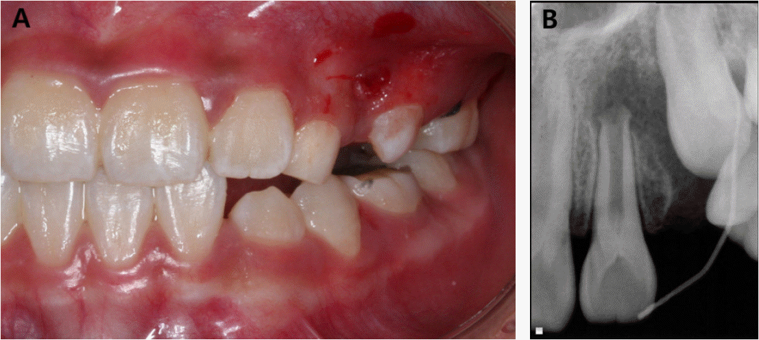
Clinical photograph and periapical radiograph of case 1 at the first visit. (A) Intraoral photograph showing a sinus tract on the buccal gingiva in the distal side of left maxillary deciduous canine. (B) Radiographic image showing an open apex associated with periapical radiolucent lesion of the maxillary left lateral incisor.
When the access cavity was made under rubber dam isolation, the canal was flushed gently with 2.5% sodium hypochlorite (NaOCl) for 10 minutes. Then, a mixture of ciprofloxacin, metronidazole, and cefaclor was placed in the root canal. The access cavity was sealed with sterilized cotton and glass ionomer cement (Fuji IX, GC Corporation, Tokyo, Japan).
About 3 weeks after the antibiotic treatment, the intracanal antibiotic dressing material was removed. After copious irrigation with 2.5% NaOCl, a size #30 precurved K-file was over-instrumented to irritate the apical tissue to create some bleeding into the canal (Fig. 2A). After the canal filled with blood to the level of 3-4 mm below the CEJ, it was left for 15 min to permit clot formation. White MTA (Pro Root MTA, Tulsa Dental, Tulsa, OK, USA) was placed directly over the blood clot (Fig. 2B), followed by a sterilized wet cotton pellet, and glass ionomer cement. A week later, after confirming the tight coronal sealing with MTA (Fig. 2C), the tooth was restored with a dentin-bonded resin composite (FiltekTM Supreme XTE, 3M ESPE, St Paul, MN, USA).
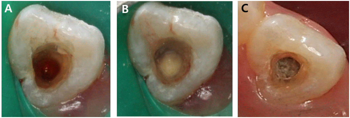
Clinical photographs of case 1. (A) Intracanal bleeding induced from apical tissue. (B) Results following MTA placement. (C) Tight coronal sealing with hardened MTA.
At the 6-month follow-up, the tooth remained asymptomatic and resolution of the apical radiolucency was observed in a periapical radiograph (Fig. 3D). At the 12- and 18-months follow-up, the patient continued to be asymptomatic, and the thickening of the dentinal wall was evident in radiographic examinations (Fig. 3E, 3F). The patient experienced no complications such as tooth discoloration (Fig. 4).

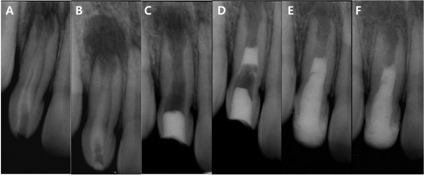
Intraoral radiographs of case 1. (A) Preoperative. (B) Triple antibiotic dressing. (C) Intracanal blood clot formation and coronal sealing with MTA. (D) 6-month follow-up. (E) 12-month follow-up. (F) 18-month follow-up.
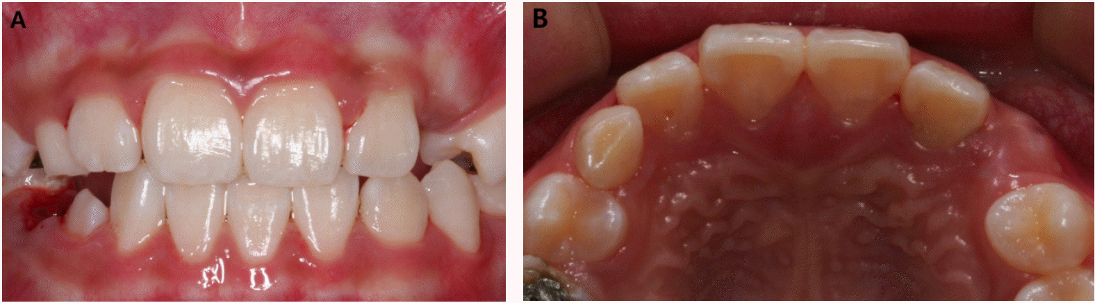
Clinical photographs of case 1. No apparent discoloration of the maxillary left lateral incisor at the 12-month follow-up. (A) Frontal view. (B) Occlusal view.
2. Case 2
A 10-year-old boy was referred from a local dental clinic for evaluation of the right maxillary lateral incisor with a complicated internal structure. He had a history of fluctuant swelling on the labial attached gingiva of right maxillary incisors, and a root canal treatment had already been initiated with systemic antibiotics for the preceding 5 days. When he visited to our clinic for the first time, the access cavity was restored with temporary restorative material and the gingival swelling was relieved (Fig. 5A).
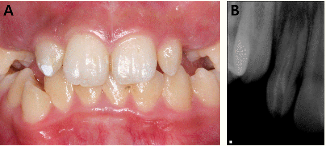
Clinical photograph and periapical radiograph of case 2 at the first visit. (A) Intraoral photograph showing peg shaped maxillary lateral incisors and a temporary restoration in the access cavity of maxillary right lateral incisor. (B) Radiographic image showing an incompletely developed apex with periapical radiolucency and an invagination extending the entire length of the root of maxillary right lateral incisor.
On clinical and radiographic examination, both maxillary lateral incisors were conical in shape. In particular, the right maxillary lateral incisor was an immature tooth with an associated widely open apex and periapical radiolucency. It appeared to have a complex internal structure, suspected to be Oehlers type III dens invaginatus (Fig. 5B).
First, cone-beam computed tomography (CBCT; Alphard VEGA, Asahi Roentgen Ind. Co., Kyoto, Japan) was used to better visualize the complexity of the dens invaginatus (Fig. 6). Calcium hydroxide paste (Ultracal� XS, Ultradent Products Inc., South Jordan, UT, USA) was applied into the infected invagination in isolation, to evaluate whether the infection was associated with the limited space of the invagination or was connected to the pulp canal space. About 4 weeks after the first visit, the tooth was still tender in a percussion test. The internal space of the invagination was supposed to be partially connected with the main root canal space, according to the CBCT images. Thus, the endodontic treatment needed to include both the invagination and the root canal, and it was decided to remove the invagination to redesign the complex internal structure.
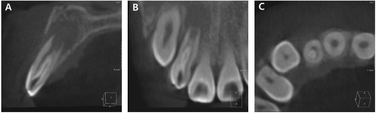
CBCT images. (A) Sagittal view. (B) Coronal view. (C) Axial view.
A surgical operating microscope (OPMI pico Dental Microscope, Carl Zeiss, Oberkochen, Germany) and ultrasonic instruments (CPR tips, Spartan, Fenton, MO, USA) were used for access to the outline of the invagination (Fig. 7A). Once the invagination was separated from the main root trunk, the canal was irrigated gently with 2.5% NaOCl (Fig. 7B). Then, the procedure of antibiotic dressing, intracanal blood clot formation, and coronal sealing with MTA were performed as described for case 1 (Fig. 8C, 8D). A week after the MTA application, the tooth was built-up with a dentin-bonded resin. The maxillary left lateral incisor was also restored for esthetic reasons.
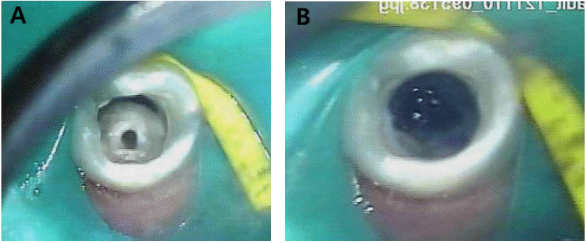
Clinical images captured by a surgical operating microscope. (A) Access to the outline of the invagination. (B) Separating the invagination from the main root trunk.
Intraoral radiographs of case 2. (A) Preoperative. (B) Calcium hydroxide paste applied to the invagination in isolation. (C) Triple antibiotic dressing following complete removal of the invagination. (D) Intracanal blood clot formation and coronal sealing using MTA. (E) 12-month follow-up. (F) 24-month follow-up.
At the 6-month follow-up, the patient reported no further experience of pain or discomfort. Moreover, the radiographs appeared to show complete resolution of the apical radiolucency. However, a bluish discoloration was seen through the resin composite at the cervical portion of the crown (Fig. 9). At the 12- and 24-month follow-ups, the tooth continued to be free of symptoms, and a slightly increased thickness of the dentinal wall seemed apparent, but it was not clearly evident as in case 1 (Fig. 8E, 8F).
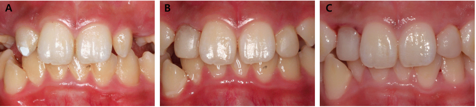
Clinical photographs of case 2. (A) Preoperative. (B) 6-month follow-up. (C) 24-month follow-up.
Ⅲ. Discussion
The dental problems that occur in association with dens invaginatus are related primarily to the degree of bacterial invasion, which can eventually lead to involvement of not only the invagination but also the pulp and/or periodontal ligament. The use of 3-dimensional imaging (CBCT) has helped in identifying the morphology of the complex internal structure clearly so that appropriate treatment can be planned [11,15]. Especially during the diagnostic procedures for the second case, CBCT images were useful in ensuring the extended boundary of the invagination and to establish whether the invagination was clearly separated from the root canal.
In many cases of dens invaginatus, successful outcome depends on the ability to gain access to and disinfect the root canal system despite the complex and unpredictable morphology of the invagination [12]. Several case reports have described successful outcomes following endodontic management of complex cases of dens invaginatus by removing the dens or central hard tissue [15,16]. It may enhance the clinician’s ability to disinfect and seal the canal space, for a more predictable outcome. From this point of view, in the second case of an immature tooth with type III dens invaginatus, it was decided to redesign the internal anatomy. The use of the surgical operating microscope and ultrasonic instrumentation provided better capabilities for a precise and sophisticated approach during the removal of the invagination without compromising the coronal tooth structure.
In the 1960s, Nygaard-östby and coworkers [17-19] suggested the potential of healing on infected, necrotic pulp by a mechanically induced blood clot, and this concept was first called “revascularization”. More recently, there has been a paradigm shift for the treatment of infected immature permanent teeth, from the ‘traditional’ apexification to pulp regeneration [20,21]. Many studies have described that the local application of antibiotics is effective for eradicating common endodontic pathogens, both in vitro and in vivo [22,23]. The use of triple-antibiotic paste (ciprofloxacin, metronidazole, and minocycline), as recommended by Hoshino et al. [22], has been confirmed in many studies [5-7,24,25].
Discoloration of the crown is a common consequence of regenerative procedures. Previous studies have recognized the problem of staining of the tooth structure as a result of using minocycline [22,25]. Thus, there have been trials to eliminate or replace minocycline in the triple-antibiotic paste. The bactericidal efficacy of mixtures of ciprofloxacin and metronidazole with amoxicillin, cefaclor, cefroxadine, fosfomycin, or rokitamycin was compared by Sato et al. [26], and it was found that these combinations could also sterilize endodontic lesions effectively. Thibodeau and Trope [27] and Dabbagh et al. [28] reported no discoloration when using a modified triple-antibiotic paste, in which cefaclor was substituted for minocycline. Hence, in the cases reported here, minocycline was replaced by cefaclor to avoid discoloration. However, a bluish discoloration on the cervical portion of the crown was noticed in the second case. A possible reason may be the MTA used for coronal sealing. Although minocycline has been blamed as the main cause of discoloration, several studies have reported the MTA can also cause discoloration [28,29]. A wider canal space resulting in difficulty in controlling MTA and thinner remaining coronal dentin thickness in the second case versus the first case may have contributed to the different esthetic result.
Additional long-term follow-up will be needed to ensure the treatment efficacy of this procedure in cases of non-vital immature permanent teeth with dens invaginatus. It would be significant to evaluate the risks and benefits in addition to patient preferences and compliance with the treatment plan. Patients and parents should also be informed about possible adverse effects, such as staining of the crown, before treatment initiation.
Ⅳ. Summary
On the basis of the short-term results of these two cases, regenerative endodontic treatment can be effective for managing immature permanent teeth with necrotic pulp tissue, which can lead not only to clinical and radiographic resolution, but also a continuing increase in the thickness of the dentinal walls and apical closure. Considering the anatomical variation in teeth with dens invaginatus, an additional approach, redesigning the complex internal anatomy, may be advisable in some cases to gain better access for effective disinfection and sealing.