유치의 근관 치료 중 차아염소산나트륨의 치근단 유입으로 인한 합병증
Accidental Extrusion of Sodium Hypochlorite during Endodontic Treatment in a Primary Tooth
Article information
Abstract
차아염소산나트륨은 가장 자주 쓰이는 근관 세척제이지만, 그 독성과 부작용에 대해서는 많이 인식되어 있지 않다. 본 증례는 근관 치료 중 차아염소산나트륨이 치근단 조직으로 압출되어 심각한 합병증이 발생하였기에 이를 보고하는 바이다. 만 5세의 남자 환자에서 근관 치료 중 차아염소산나트륨이 치근단 조직으로 유입되어 갑작스런 통증 및 부종, 주위 조직의 피하 출혈 그리고 근관 내의 지속적 출혈을 보였다. 환자는 입원치료를 받았고, 진통제와 항생제를 처방받았다. 근단공이 넓은 미성숙 영구치와 유치, 치근흡수, 천공이 일어난 치아에서는 근관 치료 시 차아염소산나트륨이 압출되지 않도록 더욱 각별한 주의가 필요하다. 합병증이 발생하였을 때에는 적절한 환자의 관리와 약물치료가 필요하다.
Trans Abstract
Although sodium hypochlorite is the most frequently used canal irrigant during endodontic treatment, its complications are not as well recognized as its effectiveness. This report demonstrates that sodium hypochlorite extrusion during endodontic treatment can cause severe complications.
A 5-year-old boy experienced immediate pain and swelling, ecchymosis in surrounding tissues, and profuse bleeding from the root canal during endodontic treatment, because of accidental extrusion of sodium hypochlorite. The patient was hospitalized, and analgesics and antibiotics were prescribed. Accidental extrusion of the irrigating solution occurs more frequently in teeth with immature apices, root resorption, and apical perforations; therefore, caution is needed. When such complications occur, proper management and medications are needed.
Ⅰ. Introduction
The first treatment decision in a young patient with pulpally involved primary molars is whether to retain or extract the teeth. Because premature loss of a primary tooth may cause functional problems, to retain pulpally involved primary teeth, one treatment choice may be pulp therapy [1,2]. However, the complex morphology and irregularities of the root canals of primary teeth negatively affect the success of root canal treatment [3]. Thus, the irrigation procedure for primary teeth is at least as important as that for permanent teeth [4]. Although various solutions have been proposed for root canal irrigation, sodium hypochlorite is the most frequently used irrigant. High concentrations are recommended to degrade protein products present in the root canal. However, these high concentrations can cause damage to the periapical tissue [4-8]. These complications are not as well known as the effectiveness of sodium hypochlorite because of their rarity.
The dental literature contains several case reports on complications during root canal irrigation, including inadvertent injection of sodium hypochlorite into periapical tissues [9-11]. However, only a few cases involving primary teeth [12] or immature permanent teeth have been reported, although there is a higher risk of extravasation of sodium hypochlorite into periapical tissue due to their wider apical foramen [9,13]. This case report describes the clinical features and management of a patient who experienced an accidental extrusion of sodium hypochlorite during endodontic treatment of an upper primary molar.
Ⅱ. Case operation
A 5-year-old boy visited the department of pedodontics of Sun Dental Hospital for treatment of dental caries. Multiple caries were found through clinical (Fig. 1) and radiographic examinations (Fig. 2). In particular, the upper left first primary molar showed root and peripheral bone resorption of the mesiobuccal root in dental periapical radiography. However, the tooth was asymptomatic clinically; negative response to percussion test, no tooth mobility, and no history of pain. Despite the poor prognosis, we decided to attempt a pulpectomy and avoid extraction if possible, because the tooth was clinically asymptomatic and many years were left until the successional replacement of the tooth.
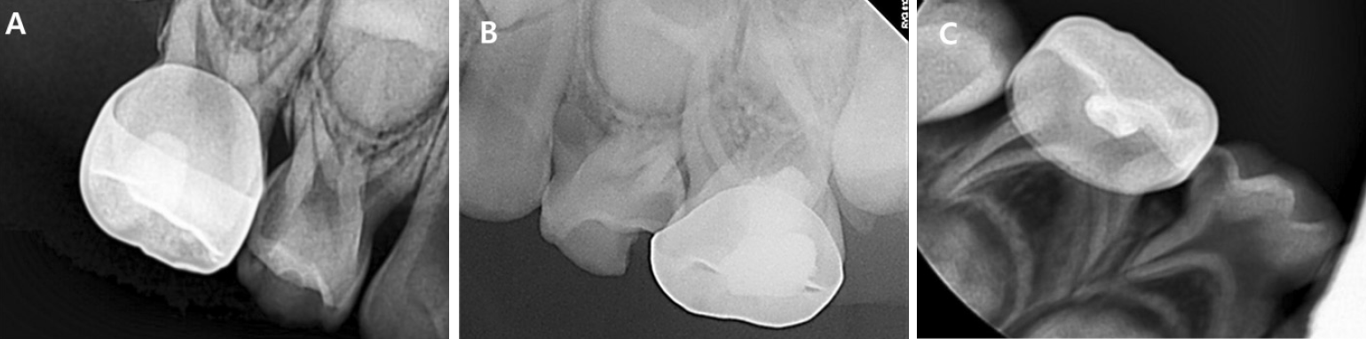
Intraoral periapical radiographs at first visit. (A) Occlusal and proximal caries on #54. (B) Occlusal and proximal caries on #64. Root and peripheral bone resorption can be seen. (C) Proximal caries on #84.
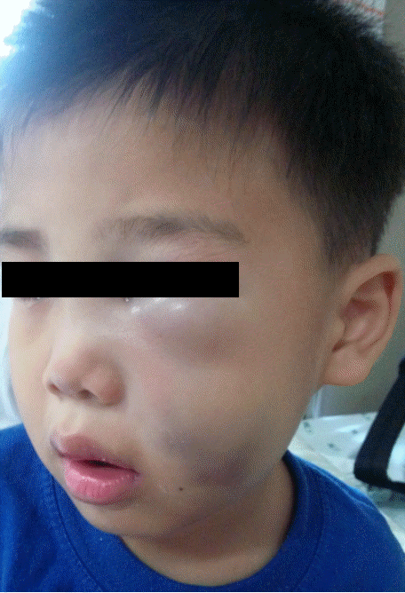
Extraoral photograph a few hours after endodontic treatment of tooth #64.
At the next appointment, root canal treatment of the upper left first primary molar was initiated. Following access cavity preparation under local anesthesia, the pulp tissue was extirpated. Working length was determined by only tactile sense. To aid the initial debridement of the root canal system, the canals were irrigated with 1.2% sodium hypochlorite using 27-guage injection needle and 0.9% saline using 23-guage injection needle. Canal shaping and canal enlargement was performed using #15, #20, #25, and #30 K-files. Canal irrigation was performed between each step.
However, continuous bleeding was seen in the mesiobuccal canal. Attempt to stop the bleeding by placing dry cotton pellets into the canals’orifices was done but the bleeding didn’stop. Therefore, canal filling was suspended. After drying the canal, the access cavity was sealed with cotton and Caviton®(GC Corporation, Tokyo, Japan), and the rubber dam was removed. At this time, mild swelling was visible on the left cheek, and ecchymosis was visible in the left infraorbital area. The possibility of sodium hypochlorite extrusion into the adjacent tissues, probably due to root resorption in the apical area or to an unintentional injection into the foramen, was considered. To prevent secondary infection and relief the pain, antibiotics (amoxicillin clavulanated) and analgesics (ibuprofen) were prescribed. When the patient returned home, the swelling and ecchymosis gradually advanced. In left buccal area, the swelling became more obvious and additional subcutaneous bleeding was visible. Also in left infraorbital area, severe ecchymosis and swelling were found (Fig. 2). The patient went to a nearby dental clinic and the tooth was extracted.
The next day, as the clinical symptoms were aggravated after tooth extraction, the patient revisited our hospital. The patient was immediately referred to an oral surgeon, and was hospitalized for further treatment. At the first day of hospitalization, antibiotics (amoxicillin clavulanated), and analgesics (ketorolac) were administered intravenously. From the second day of hospitalization, the type of antibiotics (cefotiam) was changed but the type of analgesics was the same. The facial swelling and ecchymosis gradually diminished from the second day, the patient was discharged on the fifth day of hospitalization. Although mild swelling was left on the buccal area, there was no pain on palpation of the affected region and the bluish color of the ecchymosis area faded.
A week later, the patient revisited our department for follow up. The swelling and ecchymosis had disappeared without other signs or symptoms (Fig. 3). A band and loop was set for space maintenance at the extraction site (Fig. 4), and restorations of other teeth with caries were carried out. Regular check-ups are being made to observe the development and eruption of the successional permanent tooth; no significant abnormality has yet been found (Fig. 5).

Extraoral photograph 1 week after treatment of sodium hypochlorite complications.
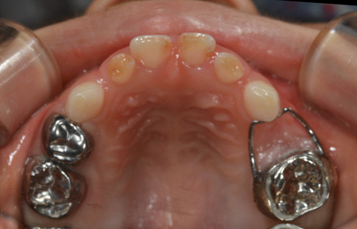
Intraoral photograph after band and loop delivery.
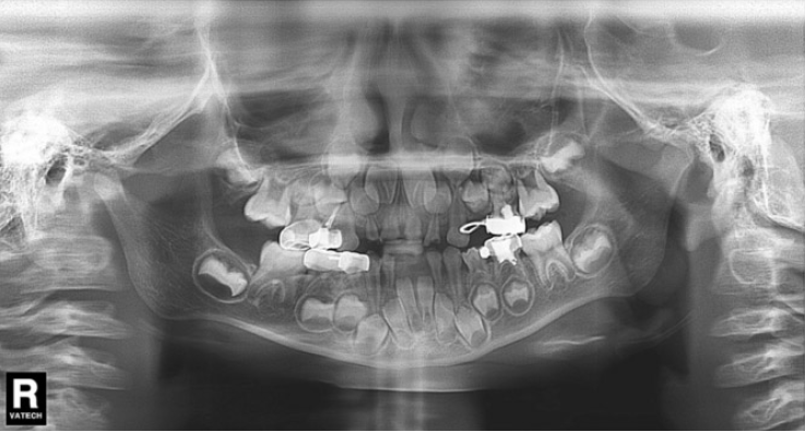
Panoramic view 1 year after treatment.
Ⅲ. Discussion
Sodium hypochlorite is most frequently used as a canal irrigant. There is no general agreement about its optimal concentration; it is used over a range from 0.5% to 5.25%. Its antibacterial and tissue dissolution actions increase with concentration, as does its toxicity [4-8].
Complications of sodium hypochlorite use, such as damage to clothing and surrounding tissues, allergic reactions, chemical burns and tissue necrosis, neurological damage, and upper airway obstruction, may occur with accidental spillage or extrusion beyond the root apex [6,9-11,14]. Several cases similar to the present article have been reported [9-11]. Most complications, including this case, appear to be the result of its accidental injection beyond the root apex, which can cause immediate severe pain and swelling of the surrounding soft tissue, profuse bleeding from the root canal, hemorrhage of the skin and mucosa, secondary infection, and paresthesia [9,10,15]. According to previous reports, inadvertent injection of sodium hypochlorite beyond the apical foramen may occur readily in teeth with perforation, a wide apex, when the apical constriction has been destroyed during root canal preparation, or by resorption [9,13]. In addition, extreme pressure during irrigation or binding of the irrigation needle tip in the root canal may result in delivering large volumes of irrigant to the apical tissues [9].
Various suggestions have been made to prevent the occurrence of such accidents. First, the clinician should check both clinically and radiographically for immature apices, root resorption, apical perforations, and any other condition that may cause extrusion of irrigant from the root canal system into the surrounding tissue before endodontic treatment [16]. When instrumenting the tooth to a large apical preparation size, caution is needed because apical extrusion of the irrigant is more likely to occur regardless of irrigation technique [13]. Careful evaluation of the integrity of individual canals, marking the working length on the irrigation needle, keeping the needle loose in the canal, and not using excessive force on the irrigation syringe are also important [9-11]. Recently, several modifications of the needle-tip design have been introduced to help facilitate effectiveness and minimize safety risks. Closed-end, side-vented needles appear to be significantly safer than open-ended beveled needles [17-20]. Although there is no general agreement on the optimal concentration, the antibacterial effects and toxicity of sodium hypochlorite are dependent on its concentration [4-8]. Thus, increasing the irrigation time with a lower concentration (0.5-1.0%) of sodium hypochlorite may be more suitable for endodontic irrigation to obtain an optimal antimicrobial effect with minimal risk of tissue-irritating injury [15,21].
In the event of accidental extrusion of sodium hypochlorite into the periapical area, there is no standard therapy for complications, probably because they are rare and sporadic [6,9]. However, most studies describe several therapies depending on the character and seriousness of the accident [6,9-11]. If signs and symptoms of complications appear, the dentist should inform the patient about its possible etiology and seriousness. In addition, the patient should be informed that signs and symptoms such as pain and swelling usually disappear in a few weeks, and rarely cause permanent complications, such as neurological damage [22]. Thus, the treatment should focus on minimizing swelling, controlling pain, and preventing secondary infection [11]. Pain control can be achieved with local anesthesia and/or oral analgesics, and the swelling can be treated with cold compresses [6,9-11]. After 1 day, the cold compresses should be replaced by warm compresses to stimulate local microcirculation [9-11]. The need for antihistamines and corticosteroids remains controversial [10]. In serious cases, referral to a maxillofacial department or surgical intervention may be needed [6,11]. Most cases do not need extraction or surgical treatment of the involved tooth, and the root canal treatment can be completed [9]. Irrigation with sterile saline solution or with chlorhexidine gluconate (0.2- 2%) is recommended [8,23]. Nevertheless, in the case of a primary tooth, extraction may be preferred for several reasons, such as preventing secondary infection, uncertain prognosis of endodontic re-treatment, and effects on the successional permanent tooth. In addition, the development and eruption of successional permanent tooth should be observed on an ongoing basis. However, there is no recommended timing of extraction and the effects of extraction during treatment on complications are not clear.
Although sodium hypochlorite is commonly used as an irrigant during endodontic treatment in primary teeth, more research is needed on the degree of sodium hypochlorite inflow in periapical tissues during endodontic treatment, and any long-term effects on the successional permanent tooth.
Ⅳ. Summary
This case report demonstrates that severe complications can occur when sodium hypochlorite is extruded in periapical tissues during endodontic treatment. Although these complications are rare, the dentist should be aware of and have the ability to manage the complications. More caution is needed for teeth with immature apices, root resorption, and apical perforations, to avoid causing extrusion of irrigant from the root canal system into the surrounding tissue during endodontic treatment [9,13]. If complications occur, the treatment should focus on minimizing swelling, controlling pain, and preventing secondary infection [11]. After the treatment of complications, continued observation of the development and eruption of the successional permanent tooth is important.