상악 중절치의 이소맹출로 인해 발생한 자극성 섬유종
Irritation Fibroma Associated with Ectopic Eruption of the Maxillary Incisor
Article information
Abstract
자극성 섬유종은 만성적인 자극 또는 외상에 의해 발생하는 구강 내의 흔한 섬유증식성 병소이다. 자극성 섬유종과 유사한 형태의 다른 양성 또는 악성 연조직종양의 감별진단을 위해서는 병리조직학적 검사가 필요하다.
본 증례는 엔젤만 증후군 환아에서 상악 좌측 중절치의 이소맹출로 인해 자극성 섬유종이 발생하여 미다졸람을 이용한 근육내 진정 하에 절제생검을 시행한 뒤, 병소의 재발 없이 양호한 치유 결과를 나타내고 있기에 보고하는 바이다.
Trans Abstract
Irritation fibroma is a common hyperplastic lesion of the oral mucosa that can occur in response to chronic irritation or trauma.
This report presents an unusual case of irritation fibroma associated with ectopic eruption of the maxillary left central incisor in a patient with Angelman syndrome. Considering the patient’s medical history and cooperative ability, excisional biopsy under intramuscular sedation using midazolam was performed. The patient exhibited successful healing without lesion recurrence.
It is important to perform histopathological examination followed by excisional biopsy, because other benign or malignant tumors may mimic the clinical appearance of an irritation fibroma.
Ⅰ. Introduction
Irritation fibroma is a common intraoral exophytic lesion of the oral cavity. Although it can occur anywhere within the oral cavity, the most common location is within the buccal mucosa along the bite line. The lateral border of the tongue, lips, gingiva and labial mucosa represent other common sites [1].
Irritation fibroma can be caused by chronic irritation or trauma, including habitual lip and cheek biting, illfitting dentures and food impaction [1,2]. Furthermore, a correlation may exist between irritation fibroma and the human papilloma virus [3].
Irritation fibroma lesions are localized and typically appear as dome-shaped or pedunculated nodules of the same color as the surrounding mucosa. The lesion surface may appear white as a result of hyperkeratosis due to continued irritation. Lesion size varies from several millimeters to several centimeters in diameter. However, the majority of fibromas are 2.0 cm or less in diameter. These lesions are associated with no particular symptoms except a feeling of irritation, unless secondary traumatic ulceration of the surface has occurred [4,5].
This report presents an unusual case of irritation fibroma that occurred in the labial mucosa, which was associated with ectopic eruption of the maxillary central incisor in a patient with Angelman syndrome.
Ⅱ. Case Report
A boy aged 7 years and 7 months presented to the Department of Pediatric Dentistry, Yonsei University Dental Hospital with a chief complaint of non-eruption of his front tooth. He had been diagnosed with Angelman syndrome and was taking anticonvulsive medication. Clinical and radiographic examination revealed delayed eruption of the maxillary left central incisor (Fig. 1A). The crown of the maxillary left central incisor was palpated at the vestibular area near the labial frenum. The patient revisited our dental hospital for follow-up at the age of 8 years and 11 months. Radiographic examination demonstrated that his maxillary left central incisor had erupted slightly (Fig. 1B). Swelling of the oral mucosa was observed around the palpated crown; ectopic eruption of the maxillary left central incisor was expected in the future. Eleven months after his second visit, at the age of 9 years and 10 months, the patient’s parents brought him to our dental hospital because they had incidentally observed an unusual mass around the erupting tooth. The maxillary left central incisor was fully erupted, but it was ectopically erupted in the horizontal direction, toward the upper lip (Fig. 1C, Fig. 2A). The swelling of the oral mucosa seen at the last visit became a pedunculated nodule measuring approximately 0.7 × 0.9 × 1.0 cm (Fig. 2A). The lesion was positioned between the labial frenum and mesial surface of the maxillary left central incisor. There was no pain on palpation, but bleeding readily occurred upon stimulation. The patient had difficulty in closing his mouth completely due to the enlarged mass (Fig. 3). Furthermore, heavy plaque was deposited on the labial surface of the maxillary left central incisor because the mass covered part of the crown. The patient’s parents reported difficulty in brushing his front tooth.
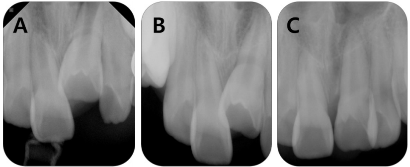
Periapical radiographs of the patient. (A) Periapical radiograph showing delayed eruption of the maxillary left central incisor when the patient was aged 7 years and 7 months. (B) Periapical radiograph taken at 8 years and 11 months, showing that the maxillary left incisor had erupted slightly. (C) Periapical radiograph taken at 9 years and 10 months showing full eruption of the maxillary left central incisor.

Intraoral photographs of the patient. (A) Intraoral photograph showing a pedunculated mass on the labial mucosa. (B) Intraoral photograph taken immediately after the excisional biopsy. (C) Intraoral photo taken at the 8-month check-up visit, showing improved oral hygiene status and spontaneous reduction of labial inclination of the maxillary left central incisor by the force of the upper lip and perioral muscles without recurrence of the lesion.
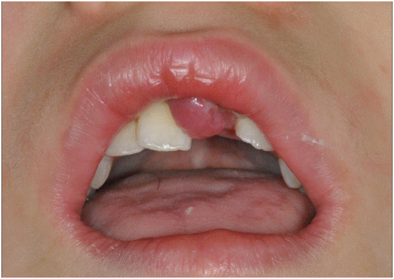
Extraoral photograph showing an enlarged mass.
Considering the patient’s limited cooperative ability, excisional biopsy under intramuscular sedation using midazolam was planned. On the day of the operation, an intramuscular dose of 0.15 mg/kg of midazolam was administered into the upper outer quadrant of the gluteal area. Excisional biopsy was performed under local anesthesia (Fig. 2B). The mass was elevated with a hemostat and excised with 1 to 2 mm peripheral margins. The depth of the excision was at the level of the muscle fascia, such that the edges could be undermined for primary closure. The wound was closed with 4-0 sutures, and the biopsy specimen was immediately fixed in 10% formalin and sent for histological evaluation. Histological evaluation of the sections by light microscopy revealed a nodular mass of fibrous connective tissue covered by hyperparakeratinized stratified squamous epithelium (Fig. 4). The covering epithelium exhibited acanthosis with finger-like elongation of the rete ridges. Dense bundles of collagen fibers, arranged in a circular pattern with vascular hyperplasia and infiltration of inflammatory cells, were observed in the underlying connective tissue. On the basis of clinical and microscopic features, a final diagnosis of irritation fibroma was made.

Histopathological examination of the irritation fibroma. (A) Low-power view showing an exophytic unencapsulated mass of dense fibrous connective tissue. The covering epithelium exhibits acanthosis with finger-like elongation of rete ridges (H-E, ×10). (B) Hyperparakeratinized stratified squamous epithelium with residual nuclei of keratinocytes (H-E, ×600). (C) Dense bundles of collagen fibers arranged in a circular pattern with vascular hyperplasia and infiltration of inflammatory cells (H-E, ×200).
The patient was recalled for follow-ups and there were no signs of recurrence during the 8-month follow-up period (Fig. 2C). Furthermore, the amount of plaque deposited on the labial surface of the maxillary left central incisor decreased markedly commensurate with easier tooth brushing after removal of the mass. Finally, we observed that the labial inclination of the maxillary left central incisor improved spontaneously by the force of the upper lip and perioral muscles. Periodic follow-ups have been scheduled.
Ⅲ. Discussion
The patient described herein had Angelman syndrome, which is caused by a deletion on chromosome 15. The syndrome is characterized by delayed development, mental retardation, seizures, and movement or balance disorders [6,7]. The present patient was taking Topamax® (topiramate) to prevent seizures. Anticonvulsive drugs such as phenytoin, sodium valproate, phenobarbitone, and vigabatrin are known to be associated with gingival overgrowth [8]. The appearance of gingival overgrowth induced by anticonvulsants is generalized, and the marginal gingiva and interdental papilla appear to be the areas predisposed to enlargement [9]. However, with the introduction of a new generation of anticonvulsive drugs including topiramate, lamotrigine, gabapentin, and sulthiame, the possibility of gingival enlargement has been markedly reduced [10]. Therefore, it can be concluded that the irritation fibroma observed in this case was not associated with an anticonvulsive drug. Oral manifestations of Angelman syndrome include mandibular prognathism, a wide mouth, diastema, and a habit of tongue thrusting [11,12]. However, delayed eruption or ectopic eruption of a tooth, as observed in this case, has not been reported previously.
Irritation fibroma is caused by reactive hyperplasia of fibrous connective tissue and is most common in the fourth to sixth decades of life. The male-to-female ratio is almost 1:2 among cases submitted for biopsy [1,13,14], although several reports suggest no gender difference [15,16]. Bouquot and Gundlach [15] reported that of 1,453 oral exophytic lesions found among 23,616 white Americans over 35 years of age, the prevalence of irritation fibroma was 1.2%. Furthermore, irritation fibroma represented 74.5% of all fibrous exophytic lesions. Although there have been numerous epidemiologic studies on the occurrence of irritation fibroma, no studies have assessed this lesion in children and adolescents [17].
It is doubtful that irritation fibroma represents a true neoplasm. True fibroma of the oral mucosa is characterized by an absence of obvious signs of chronic irritation, and sharp demarcation of the lesion from the surrounding tissues by a capsule. However, irritation fibroma occurs in response to local irritation or trauma and is not clearly demarcated from surrounding tissues [18].
Rotaru et al. [19] suggested that TGF-α may play a key role in the fibroblastic proliferation of oral fibroma. Another study suggested that proliferation of fibroblasts and collagen synthesis, due to increased expression of TGF-β1 and imbalance in the expression of MMP-1 and TIMP-1, represent contributory factors to the pathogenesis of irritation fibroma [20].
Previously, microscopic examination of irritation fibroma revealed a nodular mass of fibrous connective tissue covered by stratified squamous epithelium. The epithelium may exhibit hyperkeratosis or ulceration due to chronic irritation. Collagen bundles may be arranged in a radiating, circular, of haphazard fashion. Inflammatory cells, which typically comprise lymphocytes and plasma cells, are frequently observed beneath the epithelial surface [1,2]. As the lesion matures, inflammatory cells and blood vessels observed during the early stage are substituted by fibrous tissue; younger lesions are composed mainly of unpacked collagen, whereas older lesions contain packed, well-organized collagen [21,22]. In the present case, the epithelium was hyperparakeratinized due to chronic and continuous irritation by the ectopically erupting maxillary left central incisor. The epithelium was covered by parakeratin, which contains residual nuclei of keratinocytes. The collagen fibers of the connective tissue observed in this case were arranged in a circular pattern. Toida et al. [23] reported that collagen fibers were arranged in a circular pattern on mobile mucosa, whereas collagen fibers were arranged in a radiating pattern on fixed mucosa.
The differential diagnosis of irritation fibroma should include benign submucosal soft tissue tumors such as true fibroma, giant cell fibroma, peripheral giant cell granuloma, pyogenic granuloma, and lipoma.
Irritation fibroma should be treated by conservative surgical excision because the excessive collagen that characterizes this lesion is permanent and does not decrease spontaneously. Excisional biopsy represents the treatment of choice, and recurrence is extremely rare after removing the lesion and other causes of irritation [1,2]. In our case, because the labial inclination of the maxillary left central incisor improved spontaneously by the force of the upper lip and perioral muscles, it is unlikely that the lesion will recur in the future.
This case report is notable because the irritation fibroma occurred in the labial mucosa, which represents an uncommon location for this type of lesion; furthermore, it was associated with ectopic eruption of the incisor.
Ⅳ. Summary
Irritation fibroma is a reactive hyperplasia of fibrous connective tissue that occurs in response to local irritation or trauma and is characterized by proliferation of collagenous connective tissue and blood vessels. In the case described herein, irritation fibroma occurred in the labial mucosa associated with ectopic eruption of the maxillary left central incisor in a patient with Angelman syndrome. When soft tissue lesions occur in the oral cavity, identifying the causes and performing excisional biopsy are warranted to achieve a definitive diagnosis.