제1대구치 지연 발육의 임상 양상 및 선천성 결손치와의 연관성
Clinical Features and Correlation With Congenital Missing Teeth of Delayed First Permanent Molar
Article information
Abstract
국소적, 전신적 요인을 동반하지 않는 제1대구치의 맹출 지연은 해당 치아의 발육 지연을 동반한다. 본 연구의 목적은 제1대구치의 발육지연의 임상적 양상 및 동반하는 다른 치아의 발달 이상에 관하여 고찰하는 것이다.
국소적 요인이 없이 제1대구치 맹출 지연을 보이는 건강한 어린이 40명의 파노라마방사선 사진 분석을 시행하였다. 2명의 평가자가 Nolla 방법을 이용하여 해당 치아의 발육지연 여부, 해당 분악의 제2대구치 발육 이상 여부 및 제3대구치를 제외한 다른 치아의 선천적 결손 여부에 관하여 조사하였다.
제1대구치의 발육지연은 상악에서 호발하였고, 여성에게서 양측성 이환, 남성에게서 편측성 이환이 많이 관찰되었다. 제1대구치의 발육지연이 있는 환아는 유의하게 높은 선천적 결손치의 유병률을 보였으며, 이환된 분악의 제2대구치는 모두 발육지연 혹은 선천적 결손의 이상을 보였다.
본 연구결과 제1대구치의 발육지연은 다른 치아의 선천적 결손과 밀접한 연관이 있는 것으로 나타났다.
Trans Abstract
Delayed eruption of the first molar, without a generalized or localized cause, is usually associated with delayed development of the affected tooth. The aim of this study was to investigate the clinical features of the first permanent molar showing delayed development and eruption, and its association with developmental anomalies of other teeth.
Panoramic radiographs of 40 healthy children showing delayed development and eruption of first permanent molars were analyzed. The clinical features of affected first molars and developmental anomalies of other teeth (except third molars) were evaluated.
Delayed first molars were more frequent in the maxilla. The incidence of bilateral delayed development of first molars was greater than that of unilateral cases in female patients. In contrast, male patients showed unilateral delayed development of the first molar more frequently.
A higher incidence of congenitally missing teeth was observed in patients with delayed first molar.
In each case, delayed development or congenital absence was observed in the second molar adjacent to the delayed first molar.
Overall, delayed first molar seems to be associated with congenital absence of additional teeth. Understanding the developmental mechanisms of this phenomenon requires further studies.
Ⅰ. Introduction
Delayed tooth eruption is the retarded emergence of a tooth into the oral cavity. There are two methods for estimating the timing of tooth eruption. One method, suggested by Grøn [1], evaluates the extend of dental root development, and denotes the tooth eruption stage as the time point at which 3/4 of the root is formed. The second method estimates the tooth eruption stage based on the chronological age. According to Suri et al. [2], it is defined that delayed eruption occurs when the chronological age is delayed more than two standard deviations from the mean eruption age.
The first permanent molar erupts at the age of 6.12 - 6.39 years [3]. Grover [4] reported that delayed eruption of the first permanent molar is rare compared to other teeth, with an incidence of 0.01% in the general population. Delayed eruption of the first permanent molar in the maxilla and mandible accounted for 5.84% and 2.76%, respectively, of the total delayed eruption in Korean children [5]. Most of the cases of delayed eruption were attributable to local factors, such as early loss of a primary tooth, loss of eruption space, and presence of supernumerary teeth [5].
Delayed eruption of the first permanent molar without systemic or local causes was reported to accompany delayed tooth development or congenital absence of the adjacent second permanent molar [6-9]. Rasmussen [7] named the first permanent molar showing delayed eruption as the “9-year-molar”, because the first permanent molar often erupted at approximately 9 years of age.
In a previous case study, delayed development of the adjacent second molar was also observed in 7 out of 9 cases with delayed eruption of the first permanent molar [8]. In addition, congenital missing of the third molar was observed in all 9 cases in the same study [8].
Nakano et al. [8] proposed that delayed development of the first permanent molar could be resulted from congenital absence of the first permanent molar and mesial migration of second permanent molar. However, the higher rate of congenitally missing teeth cannot be explained in patient with delayed development of the first molar. Furthermore, there are no clinical studies proving the association of congenitally missing teeth with delayed development of the first permanent molar.
The aim of this study was to investigate the clinical aspects of ‘delayed eruption and development of the first permanent molar (DFM)’without systemic or local causes and to demonstrate its association with delayed development or congenital absence of other teeth.
Ⅱ. Materials and Methods
1. Subjects
The study subjects were first selected from those who visited the Department of Pediatric Dentistry, Dental Hospital, Yonsei University between January 2008 and December 2014. The patient selection inclusion criterion was a clinical and radiographical evidence of localized delayed eruption of the first permanent molar. The exclusion criteria were medically compromised patients and patients with localized causes of delayed tooth eruption (such as, odontoma or ectopic impacted teeth). In all cases, clinically delayed eruption accompanied clear delayed development of the first permanent molar, when compared to erupted first permanent molars in other quadrants (Fig. 1). A total of 40 subjects (15 males, 25 females) with DFM were selected; they were aged 6 - 10 years (mean age of 8.62 years) were selected for this study.
Panoramic radiographs of patients with DFM. (A) Unilateral occurrence of DFM and congenital absence of adjacent second permanent molar (closed arrow) in the right maxilla of a male patient (aged 8 years, 2 months). The patient showed congenital absence of the right maxillary second premolar (open arrows). (B) Bilateral occurrence of DFM and congenital absence of adjacent second permanent molar (closed arrow) in the maxilla of a female patient (aged 6 years, 8 months). The patient showed congenital absence of both maxillary second premolars (open arrows).
DFM = delayed eruption and development of the first permanent molar
2. Methods
The panoramic radiographs of the study subjects were examined by two investigators. The location and laterality of DFM, delayed development or congenital absence of the adjacent second permanent molars, and congenital absence of other teeth were examined on panoramic radiographs of the subjects. The dental age of the subjects’ was determined by the calcification stage according to Nolla’s method [10]. Delayed development of teeth was diagnosed when the dental age was delayed by more than 2 standard deviations of the mean calcification stage for Korean children [11]. The interclass correlation coefficient between two examiners was 0.95.
Statistical analysis was performed using Excel 2010 (Microsoft, Redmond, Washington, USA) and R (x64 3.13, R Foundation for Statistical Computing, Vienna, Austria). Statistical significance was determined at a level of p < 0.05. The Fisher’s exact test was used to compare the laterality of DFM according to the gender of the subjects, and the chi-square test was used to assess the differences in the frequency of congenital missing teeth between the DFM affected quadrant and the normal quadrant.
Ⅲ. Results
In this study, 69 cases of DFM were found in 40 subjects; 21 cases were found in 14 male subjects, while 48 were in 26 female subjects.
1. Distribution
DFM was detected in the maxilla of 32 out of 40 (80.0%) subjects, while it was found in the mandible of 5 out of 40 (12.5%) subjects. In total, 7.5% of the patients (3 out of 40) exhibited bimaxillary manifestation of DFM.
2. Laterality
Sixteen subjects (40.0%) showed unilateral DFM in the maxilla (30.0%, 12 subjects) or mandible (10.0%, 4 subjects), 21 subjects (52.5%) showed bilateral involvement of DFM in the maxilla (20 patients, 50.0%) or mandible (1 patient, 2.5%). In 3 subjects (7.5%), DFMs were found in 3 quadrants (Table 1).
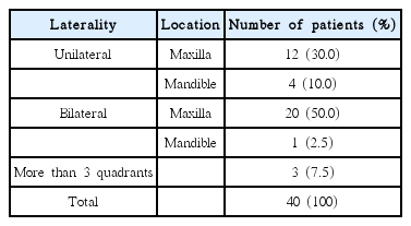
Distribution of DFM in relation to location
Among the 37 subjects (14 males and 23 females), DFM was unilateral in 16 subjects (43.2%, 10 males and 6 females) and bilateral in 21 (56.8%, 4 males and 17 females). Three female subjects with DFM in three quadrants were excluded from this analysis. There was a statistically significant difference in the laterality of DFM in male and female patients. Unilateral manifestation of DFM was significantly frequent in male patients. In contrast, female patients exhibited significantly higher frequency of bilateral presentation of DFM (Fig. 2, 3).
Correlation of bilateral DFM with gender. The incidence of bilateral DFM was significantly higher in female patients than in male patients (Fisher's exact test, * : p < 0.05).
DFM = delayed eruption and development of the first permanent molar
Correlation of unilateral DFM with gender. The incidence of unilateral DFM was significantly higher in male patients than in female patients. (Fisher's exact test, * : p < 0.05).
DFM = delayed eruption and development of the first permanent molar
3. Developmental abnormalities of adjacent second permanent molars.
In 69 cases of DFM, delayed development of the second permanent molar was seen in 39 cases, whereas congenital absence of the adjacent second permanent molar was observed in 30 cases.
4. Positive correlation between DFM and prevalence of congenital missing teeth
Congenital absence of permanent teeth, except the third molar, was evaluated. If the tooth germ was missing in the panoramic views of subjects (aged 6 years and older), the tooth was considered to be congenitally missing.
Based on this criteria, congenital absence of permanent teeth was seen in 23 subjects (57.5%). Among them, 9 were male (60% of all male subjects), while 14 were female (56.0% of all female subjects). The total number of congenital missing teeth was 60, including the congenital absence of the second permanent molar next to the DFM. Distribution of the missing teeth was as follows: 22 (36.7%) upper second molars, 13 (21.7%) lower premolars, 9 (15.0%) upper premolars, 8 (13.3%) lower second molars, and 8 (13.3%) lower incisors (Table 2).

Distribution of congenitally missing teeth in relation to DFM
Among the 60 congenitally missing teeth, 40 were located in the same quadrant as the DFM. The most frequently absent tooth was the second permanent molar (30 teeth, 50.0% of the total missing teeth) in the same quadrant of the DFM.
Regarding the number of congenitally missing teeth, 5 subjects (21.7%) showed only one missing tooth while 18 subjects (78.3%) showed 2 or more missing teeth.
There was a statistically significant correlation between the prevalence of congenitally missing teeth and the DFM-affected quadrants (Fig. 4). The quadrants in which DFM was noted showed a higher prevalence for congenitally missing teeth.
Correlation of congenitally missing teeth with DFM-affected quadrants. The prevalence of congenital missing teeth was significantly higher in DFM-affected quadrants. (Chi-square test, * : p < 0.001)
DFM = delayed eruption and development of the first permanent molar
Ⅳ. Discussion
This study was conducted to investigate the clinical features of DFM on the basis of radiographic analysis of 40 children who showed no underlying general or local factors causing delayed tooth eruption. DFM occurred more frequently in the maxilla (32 subjects, 80%) compared to the mandible. This result is in accordance with previous studies. For instance, Rasmussen [7] reported 22 cases of DFM in 10 subjects, with 18 of them in the maxilla and 4 in the mandible. Nakano et al. [8] reported 9 cases, with 8 occurring in the maxilla and 1 in the mandible. Sano et al. [9] reported 131 cases in the maxilla of 73 subjects. In Korea, Kim et al. [6] reported 7 cases in 4 subjects, with 6 found in the maxilla and only 1 in the mandible. The results from these studies support the conclusion that the incidence of DFM is higher in the maxilla compared to the mandible, and is more frequently seen in female than in male patients (Table 3).
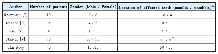
An overview of previous studies about DFM
Regarding the laterality of DFM, the results of this study did not reveal a difference between the incidence of unilateral and bilateral DFM [6,9]. This is in contrast to previous studies, in which it was reported that DFM often occurs bilaterally. However, laterality of occurrence differed according to gender. In female patients, bilateral of DFM was higher than that of unilateral DFM. In contrast, unilateral DFM was more frequent in male patients.
There was significant correlation between the location and prevalence of the DFM in relation to congenitally missing teeth. According to a previous study in Korean children, the incidence of congenitally missing teeth was reported to be 4.7 - 8.8% [12-15]. In the present study, congenital missing teeth were observed in 62.5% of patients (25 out of 40 subjects) who had DFM, and 66.7% of the congenitally missing teeth (40 teeth) were found in the same quadrant as DFM. While the most commonly congenitally missing teeth are the mandiblular lateral incisor (followed by maxilla and mandible second premolar), 50% (30 teeth) of the congenitally missing teeth in the present study involved the second permanent molar in the same quadrant as DFM. In addition, while the rate of absence of single tooth was 43.3 - 53.6% in Korean children [12-14], the proportion of multiple teeth that were congenitally absent (78.3%) was higher than that of single teeth missing (21.7%) in children showing DFM. Nakano et al. [8] reported that 2 of 9 patients with DFM had congenitally absence of the second premolar, as well as second and third permanent molars in the affected quadrant. Kim et al. [6] also reported a case where bilateral DFM accompanied the bilateral congenital absence of lateral incisors. These studies indicate that DFM may not be the result of a localized abnormality, but rather a developmental problem in the molar field where DFM is located.
In a previous study, DFM and the congenital absence of secondary molars were generally interpreted as congenitally absence of the first permanent molar and early eruption of the mesially drifted second permanent molar at the age of nine years old [8]. Nakano et al. [8] interpreted the results based on frequent association of decrease in the size of distolingual cusp, congenital absence of the ipsilateral third molar, and the close proximity of the time of first molar eruption to the normal eruption of the second permanent molar. The decrease in the size of distolingual cusp is a characteristic phenomenon that is observed frequently on the maxillary second molar [16], the incidence of which becomes higher in the absence of a third molar [17]. However, there are limitations in this conjecture in light of previous studies regarding odontogenic development. First, the congenital absence of a tooth has a high correlation with the reduction in tooth size and number of cusps [18-20]. Loss of distolingual cusp observed in DFM may be related to the reduction in tooth size associated with a congenitally missing tooth. Moreover, several studies have reported that congenital absence of a tooth is accompanied by generalized delay in tooth development [21,22]. As congenitally missing teeth are observed in cases of DFM, this phenomenon may occur as a result of a generalized developmental delay or an anomaly in the affected molar field. Although calcification of the third molar varies with the ethnic or geographic variables, the mean age of radiographic appearance of third molar calcification has been reported as 9 years of age based in the Korean children [23,24]. Radiographic examination may be inappropriate for the detection of the third molar because the age of our subjects ranged from 6 to 10 years. Further longitudinal study of our sample group is required to verify the association between DFM and developmental anomaly of the third molar.
Congenitally missing teeth originate from local factors (such as infection [25], trauma [26], drug side effects [27], radiation therapy [28]) and genetic factors [29]. Genetic mutations related to epithelial-mesenchymal interaction lead to agenesis of teeth without a syndrome [30]. Cooperative genetic interaction models for odontogenesis suggest that interaction between mesenchymal cells from neural crest and oral epithelium activates the transcription of several genes including homeobox [31]. Mutations of Msx1 and Pax9, which are the causative genes for congenital absence of teeth, are also involved in general developmental defects of the teeth such as, eruption disturbance [29,30,32]. In our data, all second molars adjacent to DFM were either congenitally absent or showed delayed development. The prevalence of congenitally missing teeth in DFM-affected quadrants was significantly high. This indicates that DFM may be a result of defects in genetic or signal transduction processes involved in tooth development of the affected field, rather than single defects of the first molar. We suspect that genetic mutations affecting molar field development may be the basis of this developmental defect. Considered comprehensively, genetic pathways can be used to explain the etiology and development of DFM.
Ⅴ. Conclusion
The study proved that DFM correlates with the congenital absence of additional teeth, especially in DFM-affected quadrants, with every second permanent molar next to the DFM exhibiting delayed development or being congenitally absent. In conclusion, a close follow-up examination should be performed to assess the temporal development of dentition in patients with DFM. However, a complete understating of the underlying developmental mechanism responsible for development DFM requires further studies.