J Korean Acad Pediatr Dent > Volume 50(1); 2023 > Article
유치에서의 단일 색조 복합레진의 색조 적응력 평가

초록

모든 유치에 알맞은 색조의 복합레진을 수복하는 것은 소아치과 의사에게 큰 어려움이다. 최근 개발된 단일 색조 복합레진은 구조색 현상을 기반으로 주변 치질과 유사한 색상을 나타낼 수 있다. 이 연구는 단일 색조 복합레진의 유치에서의 색조 적응력(Color adjustment potential, CAP)을 일반적인 다색조 복합레진과 비교하여 평가하기 위한 연구이다. 한 개의 단일 색조 복합레진과 두 개의 다색조 복합레진이 연구에 포함되었다. Single, dual 두 종류의 표본이 제작되어 평가되었다. Single 표본은 실험군 레진을 이용하여 제2유구치 인공치를 복제하여 제작하였다. Dual 표본은 발거된 제2유구치 협면에 와동을 형성한 뒤 실험군 레진을 수복하여 제작하였다. 측색계를 이용하여 발거된 치아 및 제작된 표본의 L*, a*, b* 값을 측정하였다. Single, dual 표본에 대한 ΔEab* 값과 CAP 값의 평균을 계산하였으며 Bonferroni 사후 분석을 이용하여 실험군 레진 간의 통계적 유의성을 확인하였다. 실험 결과 단일 색조 복합레진이 다른 복합레진들에 비해 유의하게 큰 ΔESingle 값을 나타내었다(p < 0.0167). 모든 실험군 레진 간 유의한 ΔEDual 값의 차이는 관찰되지 않았다. 단일 색조 복합레진은 다른 다색조 복합레진들과 비교 시 가장 높은 CAP 값을 나타내었다. 단일 색조 복합레진은 제2유구치에서 다른 다색조 복합레진에 비해 준수한 색조 적응력을 나타내었다. 단일 색조 복합레진은 제2유구치에서 색조 선택 과정을 단순화하면서 심미적인 수복물을 제공할 수 있을 것이다.

Abstract

Restoring composite resins with the optimal shades for all primary teeth is a great challenge for pediatric dentists. A newly developed single-shade composite resin can exhibit a color similar to that of the surrounding tooth structure based on the structural color phenomenon. This study aims to evaluate the color adjustment potential (CAP) of a single-shade composite resin compared to conventional multi-shade composite resins in primary teeth. A single-shade composite resin and two conventional multi-shade composite resins were included in this study. Two types of specimens, a single specimen and a dual specimen, were evaluated. For single specimens, duplications of the primary second molar denture teeth were made using experimental composite resins. For dual specimens, cavities were prepared on the buccal surfaces of extracted primary second molars and restored with experimental composite resins. The L*, a*, and b* values were measured using a colorimeter for the extracted teeth and specimens. The mean ΔEab* values for single and dual specimens and CAP were calculated. Bonferroni post-hoc analysis was performed to confirm the statistical significance between the ΔEab* and CAP values of the experimental resins. Among the single specimens, the single-shade composite resin showed significantly higher ΔESingle compared to other composite resins (p < 0.0167). There was no significant difference between ΔEDual for all experimental resins. The single-shade composite resin showed highest CAP compared to other multi-shade composite resins. A single-shade composite resin exhibited the most prominent color adaptability compared to other conventional multi-shade composite resins for primary second molars. A single-shade composite resin can simplify shade matching and provide esthetic outcomes for the restoration of primary second molars.

Introduction

Recently, esthetic demands for dental treatment in children have increased, and esthetic restoration of primary teeth has become more important [1]. Esthetic restorative materials that can be used in primary teeth include glass ionomers, composite resins, and preformed zirconia crowns [2,3]. Among them, the composite resin is most commonly used for the restoration of primary teeth because it has excellent esthetics and allows the preservation of tooth structure [4,5].
Tooth shade varies among individuals. To address this issue, various shades are available for each commercially available composite resin product [6]. However, selecting composite resins that provide the perfect shade for all teeth is a great challenge for clinicians, especially in pediatric patients who require minimal chair time because of behavioral problems [7]. Furthermore, there is a scarcity of research on the color of primary teeth [8,9].
Therefore, clinicians have focused on the “blending effect”, which is the ability of a material to resemble the color of the adjacent tooth structures [10]. The blending effect is affected by the translucency and thickness of tissues or materials [11]. To quantify the physical elements of the blending effect, the concept of color adjustment potential (CAP) was introduced [12]. The CAP can be defined as the proportion of color difference values between two objects observed under two conditions: one enclosed with another and, in isolation [13].
The single-shade composite resin was developed based on the structural color phenomenon, which represents a color through the microstructure of the material that strengthens or weakens different wavelengths [14]. The single-shade composite resin does not contain dyes or pigments, and the specific microstructure formed by the uniform filler particles expresses the color of the material [15,16]. This color, combined with the color reflected from the surrounding tooth structure, exhibits a color similar to that of the adjacent tooth [16]. This material has the advantage of ensuring an esthetic restoration without using different shades of composite resin. Previous studies on artificial teeth [15] and extracted permanent teeth [17] have shown excellent CAP of single-shade composite resins; however, studies on primary teeth are still insufficient.
Therefore, this study aimed to evaluate the color adjustment potential of a single-shade composite resin compared to conventional multi-shade composite resins in primary teeth.

Materials and Methods

This study was approved by the Institutional Review Board (IRB) of Gangneung-Wonju National University Dental Hospital (IRB No.: GWNUDH-IRB2022-A008)

1. Specimen preparation

1) Tooth preparation and experimental resins

Forty extracted human primary second molars, without caries, restorations, or obvious discoloration, were used in this study. All root portions below the cementoenamel junction were removed using a diamond disk. All the remaining organic substances in the teeth were removed, and the teeth were stored in saline at 4°C. The prepared teeth were classified according to the study process (Fig. 1).
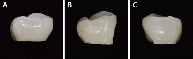
A single-shade composite resin (Omnichroma [Tokuyama Dental, Tokyo, Japan] [OM]) and two multishade composite resins (Filtek Z350XT [3M ESPE, St Paul, MN, USA] [FT] and Estelite Sigma Quick [Tokuyama Dental [ES]) were used in this study (Table 1). The A1 shade of each multi-shade composite resin was selected by a previous study on the color of primary teeth [18]. Two types of specimens, single (Fig. 2) and dual (Fig. 3), were evaluated. For each experimental resin, 10 single and dual specimens were prepared, respectively.

2) Single specimen preparation

For the single specimens, duplications of the primary second molar denture teeth were performed using experimental composite resins. The mold for duplicating the denture teeth was made using silicon impression materials (Exafine Putty Type, GC Co., Tokyo, Japan). Each experimental resin was incrementally filled three times in the manufactured mold, and photopolymerization was performed for a total of 45 seconds, 15 seconds per layer, using a VALO light-emitting diode (Ultradent Products Inc., South Jordan, UT, USA). The single specimens were polished using a series of Sof-lex™ Contouring and Polishing Discs (3M ESPE) under water cooling, according to the manufacturer’s instructions.

3) Dual specimen preparation

For the dual specimens, standardized 4.0 × 4.0 × 1.5 mm cavities were prepared on the center of the buccal surfaces of extracted primary second molars using a #330 carbide bur. The inner surfaces of the cavities were etched with a 37% phosphoric acid gel (Vericom Co., Ltd., Chuncheon, Korea) for 15 seconds. After washing and drying, an 8th generation bonding agent (Scotchbond™ Universal Adhesive, 3M ESPE) was applied evenly inside the cavities and photopolymerized for 15 seconds. Each experimental composite resin was placed into the cavities of the teeth, followed by photopolymerization for 15 seconds and polishing. The prepared specimens were stored for 24 hours in saline at 4°C prior to color measurement.

2. Color measurement

The color parameters of the restorations were measured using a colorimeter (Shade Eye-NCC, Shofu Co., Kyoto, Japan). The colorimeter was calibrated according to the manufacturer’s instructions prior to each measurement. Immediately before the measurement, the remaining solution on the surface of each sample was removed using gauze. The color parameters were measured perpendicular to the center of the restorations or buccal surfaces three times for each specimen, and the average was obtained.
The CIELAB color difference formula was used for color measurements in this study. The color difference (ΔEab*) of each specimen was calculated using the following equation:
ΔEab* = [(ΔL*)2 + (Δa*)2 + (Δb*)2]1/2
where ΔL*, Δa*, and Δb* correspond to lightness, red-green chromaticity, and yellow-blue chromaticity, respectively.
The CAP of each experimental resin was calculated using the following equation.
CAP = 1 - (ΔEDual / ΔESingle)
ΔESingle was calculated as the color difference between the single specimen and the unrestored primary second molar and ΔEDual was calculated as the color difference between the dual specimen and the primary second molar before fabricating the dual specimen. The means and standard deviations were determined for each experimental resin.

3. Statistical analysis

SPSS version 22.0 (SPSS Inc., Chicago, IL, USA) was used to analyze the data. The normality of the data was checked using the Shapiro-Wilk test. The ΔEab* and CAP values of the experimental resins were compared using Bonferroni post-hoc analysis (p < 0.0167).

Results

1. Color difference (ΔEab*)

Among the single specimens, OM showed the highest ΔESingle compared with the other composite resins (Table 2, p < 0.0167). Between the other two multi-shade composite resins, FT showed a significantly smaller ΔESingle than that of ES (Table 2, p < 0.0167). OM showed the smallest ΔEDual, but there was no significant difference among the 3 experimental composite resins (Table 3).

2. Color adjustment potential

OM showed a significantly higher CAP than FT (p < 0.0167). OM also showed higher CAP than ES, but the difference was not statistically significant. There was no significant difference between the CAP for FT and ES.

Discussion

According to previous studies[19-22], the clinical acceptance threshold for ΔEab * ranges from 3.3 to 3.7. Any ΔEab * value higher than these thresholds cannot be considered clinically acceptable [21]. In this study, the average ΔEDual of OM was 3.2, which was lower than that of the other experimental resins; however, no statistically significant difference was observed. Nevertheless, from the results of previous studies, single-shade composite resins are expected to provide good esthetics in the restoration of the primary second molars.
The enamel and dentin of primary teeth are thinner than those of permanent teeth [23]. This provides an environment in which the color of the tooth structure beyond the restoration can be better permeated. Furthermore, the color of the crown is whiter and lighter in the primary teeth compared with that of the permanent teeth [23]. According to a previous study[16] that evaluated the shade matching ability of multiple composite resins in acrylic teeth of various shades, single-shade composite resin showed better shade matching for lighter-shade acrylic teeth. In this study, conducted for primary teeth, OM showed higher CAP values than the two multi-shade composite resins.
In the oral cavity, factors such as the shape or location of teeth and restoration, the dark background of the oral cavity, and surrounding soft tissues affect the shade matching of the restoration [16,24]. Furthermore, according to a study by Kim et al. [18], significant differences in color values were observed between the primary molars and primary incisors in both the maxillary and mandibular teeth. In most cases, the esthetics of anterior tooth restorations are considered critical. Therefore, the CAP of single-shade composite resin in primary incisors was evaluated in the pilot study. However, the enamel and dentin thicknesses of the primary incisors were too low to obtain an adequate cavity depth. Future in vitro studies on primary incisors should be designed to overcome these limitations. Moreover, the shade matching of single-shade composite resin for primary incisors should be clinically evaluated.
One of the limitations of this study was the lack of visual evaluation by the observers. The visual method is a subjective assessment, but the visual judgment of color matching is a decisive factor in patient acceptance [25]. Furthermore, in this study, color measurement was performed only once after 24 hours of specimen fabrication. Long-term studies are needed to evaluate the color stability of single-shade composite resins in comparison with conventional multi-shade resins.

Conclusion

Within the limitations of this current study, the results showed that the single-shade composite resin exhibited the most prominent color adaptability for primary second molars compared with the conventional multi-shade composite resins. Single-shade composite resins can simplify shade matching and provide better esthetic outcomes for the restoration of primary second molars.

NOTES

Conflict of Interest

The authors have no potential conflicts of interest to disclose.

Fig 1.
Schematic diagram of the study process.
jkapd-50-1-113f1.jpg
Fig 2.
Single specimens of each experimental composite resin. A denture tooth was replicated using experimental composite resins. (A) Omnichroma, (B) Filtek Z350XT, (C) Estelite Sigma Quick.
jkapd-50-1-113f2.jpg
Fig 3.
Dual specimens of each experimental composite resin. Extracted primary second molars were restored with experimental composite resins. (A) Omnichroma, (B) Filtek Z350XT, (C) Estelite Sigma Quick.
jkapd-50-1-113f3.jpg
Table 1.
Experimental composite resins used in this study
Material Manufacturer Filler content Filler type Monomer Shade
Wt% Vol%
Omnichroma (OM) Tokuyama Dental, Tokyo, Japan 79.0 68.0 Uniform sized supranano spherical filler UDMA TEGDMA -
Filtek Z350XT (FT) 3M ESPE, St Paul, MN, USA 72.5 55.6 Nanofil Bis-GMA UDMA A1B
TEGDMA Bis-EMA
Estelite Sigma Quick (ES) Tokuyama Dental, Tokyo, Japan 82.0 71.0 Nanofil Bis-GMA TEGDMA A1

UDMA: Urethane dimethacrylate; TEGDMA: Triethylene glycol dimethacrylate; Bis-GMA: Bisphenol A-glycidyl methacrylate; Bis-EMA: Ethoxylated bisphenol A glycol dimethacrylate.

Table 2.
ΔESingle of the experimental composite resins
Material (n=10) Mean (SD) p value
ΔESingle Omnichroma 10.2 (1.1)
Filtek Z350XT 4.1 (0.6) < 0.0001
Estelite Sigma Quick 6.4 (4.8) 0.001

p value from the Bonferroni’s post-hoc test.

ΔESingle: The color difference between the single specimen and the unrestored primary second molar; SD: standard deviation.

Table 3.
ΔEDual of the experimental composite resins
Material (n = 10) Mean (SD) p value
ΔEDual Omnichroma 3.2 (1.3)
Filtek Z350XT 4.0 (2.7) 1.000
Estelite Sigma Quick 5.2 (2.4) 0.833

p value from the Bonferroni’s post-hoc test.

ΔEDual: The color difference between the dual specimen and the primary second molar before restoration; SD: standard deviation.

Table 4.
The color adjustment potential of the experimental composite resins
Material (n = 10) Mean (SD) p value
CAP Omnichroma 0.69 (0.1)
Filtek Z350XT 0.01 (0.7) 0.006
Estelite Sigma Quick 0.17 (0.4) 0.043

p value from the Bonferroni’s post-hoc test.

CAP: Color adjustment potential; SD: standard deviation.

References

1. Salami A, Walia T, Bashiri R : Comparison of Parental Satisfaction with Three Tooth-Colored Full-Coronal Restorations in Primary Maxillary Incisors. J Clin Pediatr Dent, 39:423-428, 2015.
crossref pmid pdf
2. Chisini LA, Collares K, Cademartori MG, de Oliveira LJC, Conde MCM, Demarco FF, Correa MB : Restorations in primary teeth: a systematic review on survival and reasons for failures. Int J Paediatr Dent, 28:123-139, 2018.
crossref pmid pdf
3. Ashima G, Sarabjot KB, Gauba K, Mittal HC : Zirconia Crowns for Rehabilitation of Decayed Primary Incisors: An Esthetic Alternative. J Clin Pediatr Dent, 39:18-22, 2014.
crossref pmid pdf
4. Lynch CD, Frazier KB, McConnell RJ, Blum IR, Wilson NH : Minimally invasive management of dental caries: Contemporary teaching of posterior resin-based composite placement in U.S. and Canadian dental schools. J Am Dent Assoc, 142:612-620, 2011.
pmid
5. Pani SC, Saffan AA, AlHobail S, Salem FB, AlFuraih A, AlTamimi M : Esthetic Concerns and Acceptability of Treatment Modalities in Primary Teeth: A Comparison between Children and Their Parents. Int J Dent, 2016:3163904, 2016.
crossref pmid pmc pdf
6. Park SK, Lee YK : Shade distribution of commercial resin composites and color difference with shade guide tabs. Am J Dent, 20:335-9, 2007.
pmid
7. Aminabadi NA, Farahani RM : The Efficacy of a Modified Omega Wire Extension for the Treatment of Severely Damaged Primary Anterior Teeth. J Clin Pediatr Dent, 33:283-288, 2009.
crossref pmid pdf
8. Balakrishnan A, Sudhan M : The Ideal Shade of Composite for Primary Dentition. J Pharm Sci Res, 9:35-36, 2017.
9. Muhamad AH, Aspasia S : The Color of Primary Teeth: A Literature Review. Dentistry, 2:144, 2012.
crossref
10. Paravina RD, Westland S, Kimura M, Powers JM, Imai FH : Color interaction of dental materials: Blending effect of layered composites. Dent Mater, 22:903-908, 2006.
crossref pmid
11. Paravina RD, Westland S, Imai FH, Kimura M, Powers JM : Evaluation of blending effect of composites related to restoration size. Dent Mater, 22:299-307, 2006.
crossref pmid
12. Paravina RD, Westland S, Johnston WM, Powers JM : Color Adjustment Potential of Resin Composites. J Dent Res, 87:499-503, 2008.
crossref pmid pdf
13. Durand LB, Ruiz-López J, Perez BG, Ionescu AM, Carrillo-Pérez F, Ghinea R, Perez MM : Color, lightness, chroma, hue, and translucency adjustment potential of resin composites using CIEDE2000 color difference formula. J Esthet Restor Dent, 33:836-843, 2021.
crossref pmid pdf
14. Sharma N, Samant PS : Omnichroma: The See-It -To-Believe -It Technology. EAS J Dent Oral Med, 3:100-104, 2021.
15. Sanchez NP, Power JM, Paravina RD : Instrumental and visual evaluation of the color adjustment potential of resin composite. J Esthet Restor Dent, 31:465-470, 2019.
crossref pmid pdf
16. Iyer RS, Babani VR, Yaman P, Dennison J : Color match using instrumental and visual methods for single, group, and multi-shade composite resins. J Esthet Restor Dent, 33:394-400, 2021.
crossref pmid pdf
17. Kobayashi S, Nakajima M, Furusawa K, Tichy A, Hosaka K, Tagami J : Color adjustment potential of single-shade resin composite to various-shade human teeth: Effect of structural color phenomenon. Dent Mater J, 40:1033-1040, 2021.
crossref pmid
18. Kim J, Paravina RD, Chen JW : In Vivo Evaluation of Color of Primary Teeth. Pediatr Dent, 29:383-386, 2007.
pmid
19. An JS, Son HH, Qadeer S, Ju SW, Ahn JS : The influence of a continuous increase in thickness of opaqueshade composite resin on masking ability and translucency. Acta Odontol Scand, 71:120-129, 2013.
crossref pmid
20. Kim SJ, Son HH, Cho BH, Lee IB, Um CM : Translucency and masking ability of various opaque-shade composite resins. J Dent, 37:102-107, 2009.
crossref pmid
21. Miotti LL, Santos IS, Nicoloso GF, Pozzobon RT, Susin AH, Durand LB : The Use of Resin Composite Layering Technique to Mask Discolored Background: A CIELAB/CIEDE2000 Analysis. Oper Dent, 42:165-174, 2017.
crossref pmid pdf
22. Zhao IS, Chu S, Yu OY, Mei ML, Chu CH, Lo EC : Effect of silver diamine fluoride and potassium iodide on shear bond strength of glass ionomer cements to caries-affected dentine. Int Dent J, 69:341-347, 2019.
crossref pmid pdf
23. Cleghorn BM, Boorberg NB, Christie WH : Primary human teeth and their root canal systems. Endod Topics, 23:6-33, 2012.
crossref
24. Ismail EH, Paravina RD : Color adjustment potential of resin composites: Optical illusion or physical reality, a comprehensive overview. J Esthet Restor Dent, 34:42-54, 2022.
crossref pmid pdf
25. Trifkovic B, Powers JM, Paravina RD : Color adjustment potential of resin composites. Clin Oral Invest, 22:1601-1607, 2018.
crossref pdf


ABOUT
BROWSE ARTICLES
EDITORIAL POLICY
FOR CONTRIBUTORS
Editorial Office
Seoul National University, Dental Hospital, B1-166 101, Daehak-ro, Jongno-gu, Seoul 03080, Republic of Korea
Tel: +82-70-4145-8875    Fax: +82-2-745-8875    E-mail: info@kapd.org                

Copyright © 2024 by Korean Academy of Pediatric Dentistry.

Developed in M2PI

Close layer
prev next