


Introduction
Composite resins are commonly used in restorative treatments for not only adults but also children and adolescents because of their aesthetic appeal, excellent bonding properties, and minimally invasive cavity preparation requirements. Following the introduction of resin adhesive techniques by Buonocore in 1955 [1], restorative treatment by bonding composite resins to tooth structures has become possible. The bonding of enamel and composite resin demonstrates strong bond strength and stability, and the dentin bonding system has achieved sufficient bond strength for clinical use through the incorporation of a 3-step etch-and-rinse adhesive system, which involves an etchant, primer, and adhesive [2].
However, composite resin restorations are techniquesensitive and require a significant amount of time because of the multiple clinical steps for bonding [3-6]. Particularly in pediatric patients, lack of cooperation can result in inadequate moisture control, which can reduce bond strength, increase microleakage, and raise failure rates [7]. In an attempt to simplify the bonding process, self-adhesive flowable composites, which combine the advantages of self-etch adhesive technology with flowable composites, were developed. This type of composite resin has the benefits of a simplified application process without the need for separate steps of etching, priming, and adhesion and reduced chair time [8]. The composition of the self-adhesive flowable composite is similar to that of other flowable composites except that it includes acidic monomers that can chemically bond to the tooth structure [9-11]. Several self-adhesive flowable composites have been developed and introduced to the market, and their performance has been thoroughly evaluated [10-14].
Recently, a self-adhesive giomer (SAG) was developed and commercialized. The giomer is a type of composite resin that contains a surface pre-reacted glass ionomer (S-PRG) filler. S-PRG fillers can release and recharge six different ions: fluoride, strontium, sodium, aluminum, silicate, and borate [15], inhibiting plaque formation and bacterial growth [16,17]. SAG incorporates a phosphonic acid monomer in resin components, which enables bonding to the tooth structure [9] so SAG can be applied directly to the cavity without any bonding process like other self-adhesive flowable composites.
For clinical use, restorative materials must have sufficient bond strength and marginal sealing. Microleakage after restoration can lead to postoperative sensitivity, bacterial penetration, and restoration failure. The longevity and success of restorations can be ensured by achieving complete marginal sealing, which is considered a fundamental goal of composite resin restorations. [18,19]. However, the performance of the SAG has not yet been evaluated in the literature.
Therefore, the purpose of this study was to evaluate the shear bond strength and microleakage of the SAG compared with the conventional flowable composite in enamel and dentin. The null hypotheses are: (1) there is no difference in shear bond strength between the SAG and conventional flowable composite in enamel and dentin; (2) there is no difference in microleakage between the SAG and conventional flowable composite.
Materials and Methods
This study was conducted with the approval of the Institutional Review Board of Gangneung-Wonju National University Dental Hospital (GWNUDH-IRB2022-A010).
In this study, a total of 104 healthy human premolars extracted for orthodontic purposes within the last 6 months were used. The premolars had intact buccal surfaces and no caries, restorations, fractures, or cracks. The teeth were collected immediately after extraction. The periodontal ligaments and other contaminants on the surface were removed with a scaler and stored in a saline solution at 4.0℃ until the study. The saline solution was replaced every month. SAG was used for the study group, and a conventional flowable composite with a 2-step etch-and-rinse adhesive system (CF) was used for the control group. Table 1 contains a list of all materials used in this study.
1. Shear bond strength
The root was sectioned at the cemento-enamel junction of the extracted premolar, and the pulp of the crown was removed. Exposing the buccal surface, the crown was embedded in a self-polymerizing acrylic resin, measuring 20.0 mm × 20.0 mm × 20.0 mm, and it was stored in a cold saline solution to disperse the heat generated during the resin curing. The enamel specimens were prepared using a specimen grinder (RB 209 MINIPOL, R&B Inc., Daejeon, Korea) under water cooling with 320 grit and 600 grit silicon carbide abrasive papers to expose a flat and uniform enamel surface. For dentin specimens, a water-cooled model trimmer was used to expose sufficient dentin to place the composite resin specimens with an inner diameter of 3.0 mm. The dentin surfaces were then polished using a specimen grinder (RB 209 MINIPOL, R&B Inc.) under water cooling with 320 grit and 600 grit abrasive papers to expose a flat and uniform surface. A total of 26 enamel specimens and 26 dentin specimens were randomly assigned to the study and control groups, with 13 enamel and 13 dentin specimens in each group.
The enamel and dentin surfaces were restored with the flowable composites of each group following the manufacturer’s instructions (Table 2), using a Teflon mold with an inner diameter of 3.0 mm and a height of 3.0 mm. Polymerization was performed by minimizing the distance between the light source and the composite resin using a light-emitting diode light-curing unit (Valo, Ultradent Products Inc., South Jordan, UT, USA) with an intensity of 1,400 mW/cm2. The bonded specimens were stored in distilled water at 37.0℃ for 24 hours.
A universal testing machine (RB-306, R&B Inc.) was used to measure the shear bond strength by applying a shear force of 500 kgf at a crosshead speed of 1.0 mm/min until fracture occurred. The maximum load at the moment of fracture was determined with RB 306 Helio X software and converted to shear bond strength per unit area.
R (MPa) = F (N) / A (mm2)
After measuring the shear bond strength, the surfaces of the specimens were observed using a stereomicroscope (LEICA M320, Leica Microsystems, Wetzlar, Germany) at 8× and 12.5× magnifications to evaluate the failure mode. The failure mode was divided into 3 categories: cohesive failure occurring within the adhesive or tooth structure; adhesive failure at the adhesive-tooth interface; and mixed failure, which is a combination of the previous 2 failure modes.
2. Microleakage
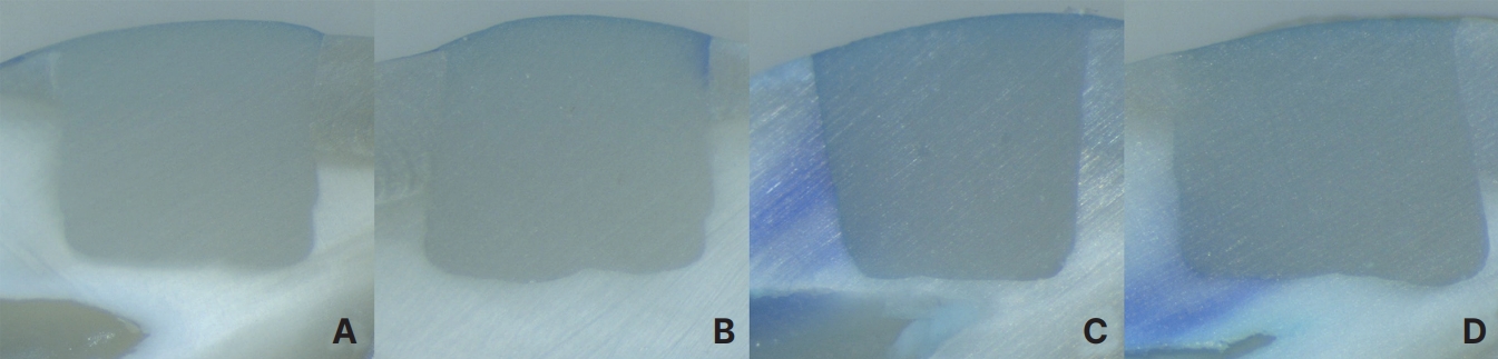
A cavity of 5.0 mm in width, 3.0 mm in height, and 3.0 mm in depth was prepared on the buccal surface of the extracted premolars using a high-speed handpiece with #330 carbide burs (Komet/Gebr. Brasseler GmbH & Co. KG, Lemgo, Germany). The bur was replaced every five tooth preparations. The size of the cavity was verified using a Williams periodontal probe. A total of 52 premolars were randomly assigned to the study and control groups, with 26 teeth in each group. The cavities were restored with the respective flowable composites following the manufacturer’s instructions (Table 2). The restorations were polished using Sof-Lex disks (3M ESPE, St. Paul, MN, USA) under water cooling. The tooth apex was sealed with composite resin to prevent dye penetration through the apical foramen. The restored teeth were stored in distilled water at 37.0℃ for 24 hours. After storage, thermal cycling was performed using a thermal cyclic tester (R&B Inc.), immersing the teeth for 30 seconds each in water baths at 5.0℃ and 55.0℃ for a total of 1,500 cycles. Nail varnish was applied twice to the entire tooth surface, excluding the restoration surface and the surrounding 1.0 mm. All the prepared specimens were immersed in a 2% methylene blue solution and stored for 24 hours. After rinsing under running water, the specimens were cut into sections along the long axis of the tooth, passing through the center of the restoration, using a low-speed diamond saw under water cooling. The penetration of the dye solution along the cavity walls on both sectioned surfaces was observed using a stereomicroscope (LEICA M320, Leica Microsystems) at 20× magnification, and the larger score was recorded based on the criteria listed in Table 3 from a previous study [20].
3. Statistical analysis
All statistical analyses were performed with SPSS 25.0 (IBM, Armonk, NY, USA). Shear bond strength data were tested for normal distribution using the Shapiro-Wilk test. The shear bond strengths of all groups satisfied normality. One-way analysis of variance and Tukey’s honestly significant difference test were used to compare the mean shear bond strengths of the enamel and dentin surfaces among the 4 groups. In addition, a 2-way analysis of variance was carried out to observe the significance of the interaction between the type of restoration and the type of tooth substrate on the mean values of shear bond strength. Fisher’s exact test was used to evaluate the failure modes. The microleakage scores were statistically analyzed using the Mann-Whitney U test. Statistical significance was set at α = 0.05.
Results
1. Shear bond strength
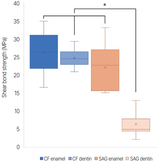
No statistically significant difference between the SAG and the conventional flowable composite was observed in the shear bond strength on enamel (p= 0.091). However, on dentin, the shear bond strength of the SAG was significantly lower than that of the conventional flowable composite (p= 0.000, Table 4, Fig. 1). The shear bond strength was significantly related to the type of restoration and tooth substrate, and the interaction was found to be significant.
The distribution of failure modes for each group is shown in Fig. 2. A significant difference was observed in the distribution of the failure modes among the groups (p < 0.001). In the enamel of SAG and conventional flowable composite and in the dentin of conventional flowable composite, mixed failure was predominant and adhesive failure was not observed. However, in the SAG on dentin, adhesive failure was observed (46.15%), whereas cohesive failure did not occur.
Discussion
Several studies have shown that current self-adhesive flowable composites bond less effectively to tooth substrates than conventional flowable composites that use multistep etch-and-rinse or self-etch adhesives [10-14]. Nevertheless, this study was conducted with the expectation that a new SAG may demonstrate bonding effectiveness similar to that of conventional flowable composites using an existing adhesive system. The results of this study indicate that the shear bond strength of the SAG did not differ significantly from that of the conventional flowable composite in enamel, although it was significantly lower in dentin. Microleakage was significantly greater in the SAG. Therefore, the null hypotheses were rejected.
A 2-step etch-and-rinse adhesive was selected as the control because of its excellent performance, as demonstrated in long-term laboratory and clinical studies [21-23]. An important factor in evaluating the performance of a restorative system is comparing the system of interest to an adhesive system that has been used for a long time, in the same laboratory environment [24-26]. The elastic modulus of the composite resin significantly affected the bond strength [27]. Therefore, a flowable composite resin, rather than a conventional composite resin, was selected to be used in combination with the 2-step etch-and-rinse adhesive for the control group. Products from the same manufacturer were used to minimize the risk of side effects due to unknown interactions between the materials [28].
In a systematic review and meta-analysis of the bond strength of self-adhesive flowable composite resins by David et al. [29], 3 existing self-adhesive flowable composites demonstrated significantly lower bonding performance than conventional composite resins with an adhesive system, regardless of the substrate evaluated, storage time, or type of dentition. In this study, the SAG demonstrated comparable bond strength to the conventional flowable composite in enamel, but significantly lower bond strength in dentin.
Ikemura et al. [9] developed phosphonic acid monomers with a hydrophilic moiety. The SAG incorporates these acidic monomers into the resin components. It is speculated that the ionized phosphonic acid group of phosphonic acid monomers chemically interacts as ligand monomers with the calcium cation of hydroxyapatite in teeth, and this chemical interaction provides adhesion between the SAG and tooth structure. Enamel is composed of over 95wt% carbonated hydroxyapatite [30], while dentin consists of 70% hydroxyapatite, 20% organic matrix, and 10% water on a weight basis [31]. Since the composition ratio of hydroxyapatite in enamel is much higher, the bonding of SAG to enamel could be much better than that of dentin. Also, the chemical interactions could sufficiently occur in enamel, leading to a bond strength comparable to that of conventional flowable composites.
The SAG, as a flowable composite resin, has a higher viscosity and lower wettability than independent adhesive systems. While the SAG does not contain solvents, the 2-step etch-and-rinse adhesive contains ethanol and water as solvents. As a result, the SAG cannot deeply penetrate the dentinal tubules or the space between collagen fibers, making it difficult to achieve sufficient micromechanical interlocking and, thus, compromising the bonding effectiveness and sealing ability within the tooth structure. On the contrary, alcohol included as a solvent in the 2-step etch-and-rinse adhesive increases penetration into the dentinal tubules. Moisture in the dentinal tubules tends to attract alcohol components, dragging the resin along with them. Then, moisture and alcohol evaporate, leaving the resin components behind, resulting in increased bond strength [32]. However, in the case of a highly viscous SAG, the first layer of the SAG may act as a semipermeable membrane, preventing the diffusion of moisture. Bumrungruan and Sakoolnamarka [11] observed the dentin-resin interface using scanning electron microscopy and confirmed the presence of gaps without a distinct hybrid layer at the interface of the selfadhesive flowable composite samples. Davidson et al. [33] stated that a minimum of 17 - 20 MPa bond strength is required to withstand the polymerization shrinkage of the composite resin, and Munksgaard et al. [34] claimed that a bond strength of at least 17.6 MPa is required. In this study, the mean shear bond strength value of the SAG was 22.07 MPa in enamel and 6.42 MPa in dentin, indicating insufficient bond strength in dentin.
A self-adhesive composite resin containing hydrophilic monomers exhibits considerable hygroscopic expansion when immersed in water, and significant dehydration shrinkage during water desorption [35]. It also exhibited appreciable solubility during water sorption, and visible cracks indicating obvious degradation and solubility after desorption [36]. The SAG contains hydrophilic phosphonic acid monomers, and these points may explain the greater microleakage in the SAG after thermal cycling.
The limitation of this study is that it was conducted in a laboratory setting. The oral environment is dynamic in terms of temperature, moisture, pH, and occlusal forces, and it is impossible to fully replicate it in a laboratory. Questions may be raised regarding the ability of shortterm laboratory studies to predict clinical results, and further in vivo studies evaluating the clinical performance of the SAG are necessary.
In addition, research on other characteristics that can be used to evaluate the clinical success of the SAG, such as wear resistance, is required. Because SAGs bond directly to the tooth structure without an intervening bonding layer, they may release various ions, including fluoride, more directly to the tooth structure. If SAG’s better ion release ability is confirmed through additional studies, it can be beneficial in terms of resistance to demineralization and promotion of remineralization, making it a significant advantage for restorative materials and supporting SAG’s clinical use.
Conclusion
Within the limitations of this in vitro study, SAG exhibited a bond strength comparable to that of conventional flowable composites using a 2-step etch-and-rinse adhesive in enamel. SAG is considered useful for restoring small cavities at the enamel level of pediatric patients by reducing chair time with the advantage of a simple bonding process. However, as the study was conducted in a laboratory setting, further research in a clinical environment is deemed necessary.

 PDF Links
 PubReader
 ePub Link
 Full text via DOI
 Download Citation
 Print
Print


